EasySep™ Direct Human Monocyte Isolation Kit

Immunomagnetic negative isolation of untouched human monocytes directly from whole blood
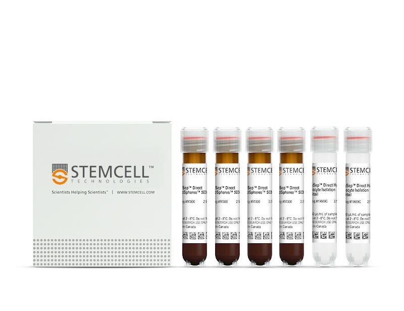
EasySep™ Direct Human Monocyte Isolation Kit

Immunomagnetic negative isolation of untouched human monocytes directly from whole blood

From: 587 USD
Catalog #
(Select a product)
Immunomagnetic negative isolation of untouched human monocytes directly from whole blood
Add to Wish List

Product Advantages


  • 99.9% RBC depletion without the need for density gradient centrifugation, sedimentation, or lysis

  • Up to 92% purity of isolated cells

  • Fast, easy-to-use and column-free

  • Isolated cells are untouched

What's Included

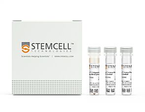
  • EasySep™ Direct Human Monocyte Isolation Kit (Catalog #19669)
    • EasySep™ Direct Human Monocyte Isolation Cocktail, 2 x 2.5 mL
    • EasySep™ Direct RapidSpheres™, 4 x 2.5 mL
  • RoboSep™ Human Monocyte Isolation Kit with Filter Tips (Catalog #19669RF)
    • EasySep™ Direct Human Monocyte Isolation Cocktail, 2 x 2.5 mL
    • EasySep™ Direct RapidSpheres™, 4 x 2.5 mL
    • RoboSep™ Buffer (Catalog #20104)
    • RoboSep™ Filter Tips (Catalog #20125) x 2
Products for Your Protocol
To see all required products for your protocol, please consult the Protocols and Documentation.

Overview

Easily and efficiently isolate highly purified human CD14+ monocytes directly from human whole blood samples by immunomagnetic negative selection, with the EasySep™ Direct Human Monocyte Isolation Kit. Widely used in published research for more than 20 years, EasySep™ combines the specificity of monoclonal antibodies with the simplicity of a column-free magnetic system.

In this EasySep™ negative selection procedure, unwanted cells are labeled with antibody complexes and magnetic particles called EasySep™ Direct RapidSpheres™. The following unwanted cells are targeted for removal: granulocytes, T cells, B cells, NK cells, dendritic cells, platelets, and erythroid cells. The magnetically labeled cells are then separated from the untouched desired CD14+ monocytes by using an EasySep™ magnet and simply pouring or pipetting the desired cells into a new tube. Following magnetic cell isolation, the desired CD14+ monoctes are ready for downstream applications such as flow cytometry, culture, or DNA/RNA extraction.

Learn more about how immunomagnetic EasySep™ technology works or how to fully automate immunomagnetic cell isolation with RoboSep™ to save time and increase laboratory throughput. Explore additional products optimized for your workflow, including those for cell characterization, cryopreservation, and more.

Magnet Compatibility
• EasySep™ Magnet (Catalog #18000)
• “The Big Easy” EasySep™ Magnet (Catalog #18001)
• Easy 50 EasySep™ Magnet (Catalog #18002)
• EasyEights™ EasySep™ Magnet (Catalog #18103)
• RoboSep™-S (Catalog #21000)
Subtype
Cell Isolation Kits
Cell Type
Monocytes, Myeloid Cells
Species
Human
Sample Source
Whole Blood
Selection Method
Negative
Application
Cell Isolation
Brand
EasySep, RoboSep
Area of Interest
Drug Discovery and Toxicity Testing, Immunology

Data Figures

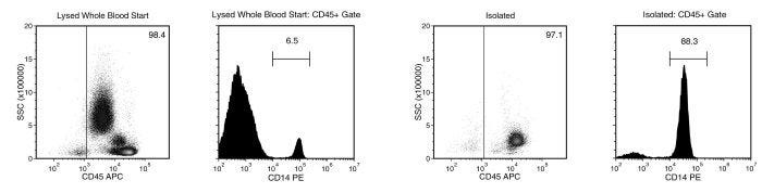
Monocyte separation using EasySep™ Direct Human Monocyte Isolation Kit

Figure 1. Typical EasySep™ Direct Human Monocyte Isolation Profile

Starting with human whole blood from normal healthy donors, the typical monocyte (CD14+) content of the non-lysed final Isolated fraction is 82.2 ± 8.4% (gated on CD45) or 79.0 ± 10.1% (not gated on CD45). In the example above, the monocyte (CD14+) content of the lysed whole blood start sample and the non-lysed final isolated fraction is 6.5% and 88.3% (gated on CD45), respectively, or 6.4% and 85.7% (not gated on CD45), respectively. The starting frequency of monocytes in the non-lysed whole blood start sample above is approximately 0.007% (data not shown).

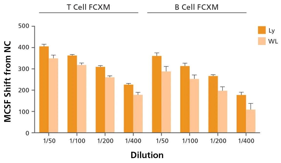
Use of Highly Enriched Lymphocytes Isolated with EasySep™ Direct Improves Donor Specific Antibody DSA Detection Compared to Whole Leukocyte Cell Preparations

Figure 2. Use of Highly Enriched Lymphocytes Isolated with EasySep™ Direct Improves Donor Specific Antibody DSA Detection Compared to Whole Leukocyte Cell Preparations

Lymphocytes (Ly), neutrophils (Nu), and monocytes (Mo) were isolated from volunteer donors (n=5) using EasySep™ Direct Human Total Lymphocyte Isolation Kit (Catalog #19655), Human Neutrophil Isolation Kit (Catalog #19666), and Human Monocyte Isolation Kit (Catalog #19669). Whole leukocyte (WL) preparations were obtained by adding Ly, Nu, and Mo cells in equal proportions. WL (low lymphocyte purity) and Ly (high lymphocyte purity) preparations were treated with pronase and then used to perform the FCXM assay against negative control sera or several dilutions of positive control sera. The median channel fluorescence shifts (MCFS) were generated by using the negative control sera samples as a baseline. The MCF shifts between WL and Ly were then compared. Each column with error bars represents the mean ± SEM (n = 5 donors). Data kindly provided by Dr. Robert Liwski.

Protocols and Documentation

Find supporting information and directions for use in the Product Information Sheet or explore additional protocols below.

Document Type
Product Name
Catalog #
Lot #
Language
Catalog #
19669RF
Lot #
All
Language
English
Catalog #
19669
Lot #
All
Language
English
Document Type
Safety Data Sheet 1
Catalog #
19669RF
Lot #
All
Language
English
Document Type
Safety Data Sheet 2
Catalog #
19669RF
Lot #
All
Language
English
Document Type
Safety Data Sheet 3
Catalog #
19669RF
Lot #
All
Language
English
Document Type
Safety Data Sheet 1
Catalog #
19669
Lot #
All
Language
English
Document Type
Safety Data Sheet 2
Catalog #
19669
Lot #
All
Language
English

Applications

This product is designed for use in the following research area(s) as part of the highlighted workflow stage(s). Explore these workflows to learn more about the other products we offer to support each research area.

Resources and Publications

Frequently Asked Questions

Can EasySep™ be used for either positive or negative selection?

Yes. The EasySep™ kits use either a negative selection approach by targeting and removing unwanted cells or a positive selection approach targeting desired cells. Depletion kits are also available for the removal of cells with a specific undesired marker (e.g. GlyA).

How does the separation work?

Magnetic particles are crosslinked to cells using Tetrameric Antibody Complexes (TAC). When placed in the EasySep™ Magnet, labeled cells migrate to the wall of the tube. The unlabeled cells are then poured off into a separate fraction.

Which columns do I use?

The EasySep™ procedure is column-free. That's right - no columns!

How can I analyze the purity of my enriched sample?

The Product Information Sheet provided with each EasySep™ kit contains detailed staining information.

Can EasySep™ separations be automated?

Yes. RoboSep™, the fully automated cell separator, automates all EasySep™ labeling and cell separation steps.

Can EasySep™ be used to isolate rare cells?

Yes. We recommend a cell concentration of 2x10⁸ cells/mL and a minimum working volume of 100 µL. Samples containing 2x10⁷ cells or fewer should be suspended in 100 µL of buffer.

Are the EasySep™ magnetic particles FACS-compatible?

Yes, the EasySep™ particles are flow cytometry-compatible, as they are very uniform in size and about 5000X smaller than other commercially available magnetic beads used with column-free systems.

Can the EasySep™ magnetic particles be removed after enrichment?

No, but due to the small size of these particles, they will not interfere with downstream applications.

Can I alter the separation time in the magnet?

Yes; however, this may impact the kit's performance. The provided EasySep™ protocols have already been optimized to balance purity, recovery and time spent on the isolation.

For positive selection, can I perform more than 3 separations to increase purity?

Yes, the purity of targeted cells will increase with additional rounds of separations; however, cell recovery will decrease.

How does the binding of the EasySep™ magnetic particle affect the cells? is the function of positively selected cells altered by the bound particles?

Hundreds of publications have used cells selected with EasySep™ positive selection kits for functional studies. Our in-house experiments also confirm that selected cells are not functionally altered by the EasySep™ magnetic particles.

If particle binding is a key concern, we offer two options for negative selection. The EasySep™ negative selection kits can isolate untouched cells with comparable purities, while RosetteSep™ can isolate untouched cells directly from whole blood without using particles or magnets.

Publications (16)

Dynamic expression of complement receptor immunoglobulin (CRIg) on monocytes and its role in phagocytosis and killing of Staphylococcus aureus. K. Perveen et al. Journal of biomedical science 2026 Mar

Abstract

BACKGROUND: The complement receptor immunoglobulin (CRIg), a key microbial pathogen phagocytosis-promoting receptor, responsible for intravascular clearance of bacteria, is purported to be expressed selectively on tissue-fixed macrophages such as Kupffer cells. However, recently it has been reported that neutrophils can also express functional CRIg following activation by inflammatory mediators. Monocytes have been reported not to express CRIg under non-activated conditions. Thus, investigations were undertaken to examine whether blood monocytes express CRIg under cell activation conditions and its role in anti-microbial immunity. METHODS: Monocytes CRIg expression in whole human and mouse blood or peripheral blood mononuclear cells and purified monocytes using density gradient centrifugation or an affinity purification kit was examined using PE/FITC-labelled anti-CRIg monoclonal antibody and flow cytometry. Characterization of CRIg isoforms in monocytes was determined by the detection of CRIg mRNA transcripts and protein using RT-PCR and Western blot, respectively. Gene-edited CRIg- and CD18- monocytic THP-1 cell lines were generated to assess the role of CRIg and CD18 in cell adhesion, phagocytosis, and microbial killing. Functional assays were performed using Staphylococcus aureus as a model pathogen. RESULTS: CRIg was constitutively expressed, dynamically, on the surface of human and mouse blood monocytes. All three human monocyte subpopulations expressed CRIg, equally. The inability to demonstrate expression on monocytes cell surface by previous studies can be explained by its lability during blood storage and loss during monocyte isolation steps. Interestingly of the monocyte subpopulations only the classical and intermediate but not the non-classical showed a loss of CRIg expression. The data showed that loss from the surface was most likely due to relocation of the receptor intracellularly. Monocytes expressed 6 different CRIg mRNA transcripts and immunoreactive isoforms. Using CRIg- and CD18- THP-1 monocytic cells, we found that both CRIg and CD18 (CR3/CR4) were critical for cell adhesion, but for phagocytosis and killing of S. aureus, either receptor was independently effective. CONCLUSION: The data provide compelling evidence that monocytes express functional CRIg, relevant to the cells' anti-microbial role of the 'wandering' phagocyte and consolidate a view that CRIg is widely expressed in our phagocytic cell system, similar to the classical complement receptors CR3 and CR4.
Consequences of the Novel ALS-Associated KIF5A Variant c.2993-6C > A for Exon 27 Splicing and Axonal Transport of SFPQ G. A. Rouleau et al. Neurology: Genetics 2026 Mar

Abstract

Background and Objectives: Recent studies have identified variants in the kinesin family member 5A (KIF5A) gene that predispose to amyotrophic lateral sclerosis (ALS). These ALS-linked KIF5A variants lead to the exclusion of exon 27, resulting in the production of a mutated protein with an altered C-terminal region (KIF5A ΔExon27). Through whole genome sequencing, we identified a novel KIF5A intronic variant, rs1057522322 (c.2993-6C > A; chr12:57582596C > A, GRCh38.p14), in a family segregating ALS. Our goal is to investigate the effect of this variant on exon 27 splicing and to assess its functional consequences on KIF5A-mediated cargo transport. Methods: Induced pluripotent stem cells (iPSCs) were generated from siblings with and without the c.2993-6C > A variant. RT-PCR was performed on RNA extracted from iPSC-derived neurons to assess exon 27 splicing. Functional studies were conducted on iPSC-derived motor neurons (MNs). Results: RT-PCR confirmed that the c.2993-6C > A variant induced exon 27 skipping in KIF5A. Immunofluorescent staining showed that KIF5A ΔExon27 abolished the axonal interaction with splicing factor proline- and glutamine-rich, a cargo specifically transported by KIF5A. Under stress conditions, MNs carrying the c.2993-6C > A variant exhibited TDP-43 proteinopathy. Discussion: KIF5A intronic variant c.2993-6C > A could be a risk factor for ALS. KIF5A ΔExon27 impairs KIF5A-mediated cargo transport and contributes to ALS pathogenesis in a TDP-43–dependent manner.
Stress Hyperglycemia Drives CD4+ T Cell PANoptosis and Postoperative Organ Injury via Monocyte-Derived Succinate. S. Zhao et al. Advanced science (Weinheim, Baden-Wurttemberg, Germany) 2026 Aug

Abstract

Perioperative stress hyperglycemia is a transient but frequent metabolic disturbance strongly linked to postoperative organ injury and mortality; however, the immunometabolic mechanisms driving this association remain largely undefined. In a two-center cohort of patients undergoing total aortic arch replacement, we identify stress hyperglycemia as an independent determinant of poor postoperative outcomes that associates strongly with CD4+ T cell loss. Hyperglycemia induces inflammatory PANoptosis in CD4+ T cells from patients in response to surgical trauma. This results from elevated glucose driving the accumulation and release of succinate from monocytes, which subsequently acts on CD4+ T cells to compromise mitochondrial integrity and activate ZBP1-mediated PANoptosis. Our findings define a monocyte-T cell metabolic signaling axis that transduces hyperglycemic stress via elevated succinate to adaptive immune cell death and reveal potential therapeutic targets to prevent postoperative immune dysfunction and organ injury, especially for patients with hyperglycemic comorbidities.