Make more informed purchasing decisions with our new product availability and delivery estimate feature, now available on all product pages, in your cart, and during checkout.
Sign In
New to STEMCELL?
Register for an account to quickly and easily purchase products online and for one-click access to all educational content.
New look, same high quality and support! You may notice that your instrument or reagent packaging looks slightly different from images displayed on the website, or from previous orders. We are updating our look but rest assured, the products themselves and how you should use them have not changed. Learn more
Request Pricing
Thank you for your interest in this product.
Please provide us with your contact information and your local representative
will contact you with a customized quote. Where appropriate, they can also assist you with a(n):
Estimated delivery time for your area
Product sample or exclusive offer
In-lab demonstration
By submitting this form, you are providing your consent to STEMCELL Technologies Canada Inc. and its subsidiaries and affiliates (“STEMCELL”) to collect and use your information, and send you newsletters and emails in accordance with our privacy policy. Please contact us with any questions that you may have. You can unsubscribe or change your email preferences at any time.
Compatible antibodies for purity assessment of isolated cells
What Our Scientist Says
RBC lysis and other preprocessing steps can complicate cell isolation procedures. We developed this kit so you can keep things simple and quickly isolate neutrophils directly from whole blood.
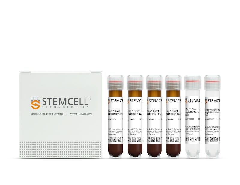
Easily and efficiently isolate highly purified human neutrophils directly from human whole blood samples by immunomagnetic negative selection, with the EasySep™ Direct Human Neutrophil Isolation Kit. Widely used in published research for more than 20 years, EasySep™ combines the specificity of monoclonal antibodies with the simplicity of a column-free magnetic system.
In this EasySep™ negative selection procedure, unwanted cells are labeled with antibody complexes and magnetic particles called EasySep™ Direct RapidSpheres™. The following unwanted cells are targeted for removal: basophils, eosinophils, T cells, B cells, monocytes, NK cells, and erythroid cells. The magnetically labeled cells are then separated from the untouched desired neutrophils by using an EasySep™ magnet and simply pouring or pipetting the desired cells into a new tube. Following magnetic cell isolation, the desired neutrophils are ready for downstream applications such as flow cytometry, culture, or DNA/RNA extraction.
This product replaces the EasySep™ Human Neutrophil Isolation Kit (Catalog #19257) for even faster cell isolations.
Learn more about how immunomagnetic EasySep™ technology works or how to fully automate immunomagnetic cell isolation with RoboSep™ to save time and increase laboratory throughput. Explore additional products optimized for your workflow, including those for cell characterization, cryopreservation, and more.
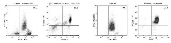
Figure 1. Typical EasySep™ Direct Human Neutrophil Isolation Profile
Starting with human whole blood from normal healthy donors, the typical neutrophil (CD66b+CD16+) content of the non-lysed final isolated fraction Is 97.3 ± 1.4% (gated on CD45) or 94.0 ± 3.7% (not gated on CD45). In the example above, the neutrophil (CD66b+CD16+) content of the lysed whole blood start sample and the non-lysed final isolated fraction is 50.6% and 97.2% (gated on CD45), respectively, or 43.6% and 95.9% (not gated on CD45), respectively. The starting frequency of neutrophils in the non-lysed whole blood start sample above is 0.04% (data not shown).
Figure 2. Typical EasySep™ Direct Protocol
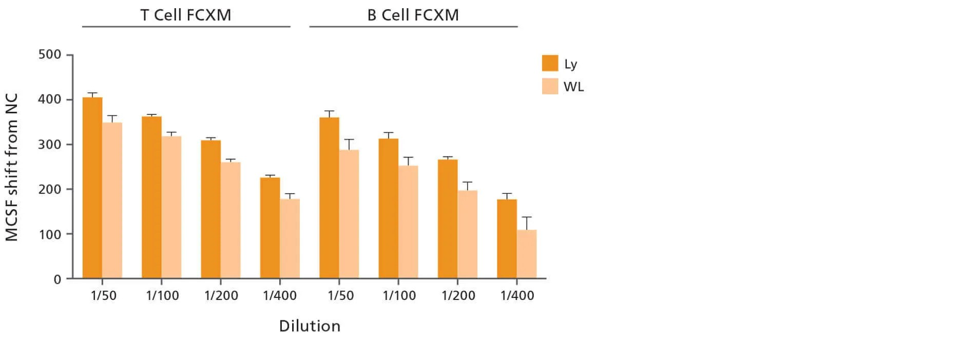
Figure 3. Use of Highly Enriched Lymphocytes Isolated with EasySep™ Direct Improves DSA Detection Compared to Whole Leukocyte Cell Preparations
Lymphocytes (Ly), neutrophils (Nu), and monocytes (Mo) were isolated from volunteer donors (n = 5) using EasySep™ Direct Catalog #19655, #19666, and #19669, respectively. Whole leukocyte (WL) preparations were obtained by adding Ly, Nu, and Mo cells in equal propotions. WL (low lymphocyte purity) and Ly (high ymphocyte purity) preparations were treated with pronase and then used to perform the flow cytometry cross-match (FCXM) assay against negative control sera or several dilutions of positive control sera. The median channel fluorescence shifts (MCFS) were generated by using the negative control sera samples as a baseline. The MCF shifts between WL and Ly were then compared. Each column with error bars represents the mean ± SEM (n = 5 donors). Data kindly provided by Dr. Robert Liwski.
This product is designed for use in the following research area(s) as part
of the highlighted workflow stage(s). Explore these workflows to learn more about the other products we
offer to support each research area.
Can EasySep™ be used for either positive or negative selection?
Yes. The EasySep™ kits use either a negative selection approach by targeting and removing unwanted cells or a positive selection approach targeting desired cells. Depletion kits are also available for the removal of cells with a specific undesired marker (e.g. GlyA).
How does the separation work?
Magnetic particles are crosslinked to cells using Tetrameric Antibody Complexes (TAC). When placed in the EasySep™ Magnet, labeled cells migrate to the wall of the tube. The unlabeled cells are then poured off into a separate fraction.
Which columns do I use?
The EasySep™ procedure is column-free. That's right - no columns!
How can I analyze the purity of my enriched sample?
The Product Information Sheet provided with each EasySep™ kit contains detailed staining information.
Can EasySep™ separations be automated?
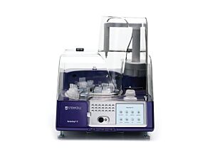
Yes. RoboSep™, the fully automated cell separator, automates all EasySep™ labeling and cell separation steps.
Can EasySep™ be used to isolate rare cells?
Yes. We recommend a cell concentration of 2x10⁸ cells/mL and a minimum working volume of 100 µL. Samples containing 2x10⁷ cells or fewer should be suspended in 100 µL of buffer.
Are the EasySep™ magnetic particles FACS-compatible?
Yes, the EasySep™ particles are flow cytometry-compatible, as they are very uniform in size and about 5000X smaller than other commercially available magnetic beads used with column-free systems.
Can the EasySep™ magnetic particles be removed after enrichment?
No, but due to the small size of these particles, they will not interfere with downstream applications.
Can I alter the separation time in the magnet?
Yes; however, this may impact the kit's performance. The provided EasySep™ protocols have already been optimized to balance purity, recovery and time spent on the isolation.
For positive selection, can I perform more than 3 separations to increase purity?
Yes, the purity of targeted cells will increase with additional rounds of separations; however, cell recovery will decrease.
How does the binding of the EasySep™ magnetic particle affect the cells? is the function of positively selected cells altered by the bound particles?
Hundreds of publications have used cells selected with EasySep™ positive selection kits for functional studies. Our in-house experiments also confirm that selected cells are not functionally altered by the EasySep™ magnetic particles.
If particle binding is a key concern, we offer two options for negative selection. The EasySep™ negative selection kits can isolate untouched cells with comparable purities, while RosetteSep™ can isolate untouched cells directly from whole blood without using particles or magnets.
NETosis and thrombosis in vaccine-induced immune thrombotic thrombocytopenia.
H. H. L. Leung et al.
Nature communications 2022 sep
Abstract
Vaccine-induced immune thrombotic thrombocytopenia (VITT) is a rare yet serious adverse effect of the adenoviral vector vaccines ChAdOx1 nCoV-19 (AstraZeneca) and Ad26.COV2.S (Janssen) against COVID-19. The mechanisms involved in clot formation and thrombocytopenia in VITT are yet to be fully determined. Here we show neutrophils undergoing NETosis and confirm expression markers of NETs in VITT patients. VITT antibodies directly stimulate neutrophils to release NETs and induce thrombus formation containing abundant platelets, neutrophils, fibrin, extracellular DNA and citrullinated histone H3 in a flow microfluidics system and in vivo. Inhibition of NETosis prevents VITT-induced thrombosis in mice but not thrombocytopenia. In contrast, in vivo blockage of Fc$\gamma$RIIa abrogates both thrombosis and thrombocytopenia suggesting these are distinct processes. Our findings indicate that anti-PF4 antibodies activate blood cells via Fc$\gamma$RIIa and are responsible for thrombosis and thrombocytopenia in VITT. Future development of NETosis and Fc$\gamma$RIIa inhibitors are needed to treat VITT and similar immune thrombotic thrombocytopenia conditions more effectively, leading to better patient outcomes.
Neutrophil degranulation and severely impaired extracellular trap formation at the basis of susceptibility to infections of hemodialysis patients.
S. Talal et al.
BMC medicine 2022 oct
Abstract
BACKGROUND Chronic kidney disease patients are at increased risk of mortality with cardiovascular diseases and infections as the two leading causes of death for end-stage kidney disease treated with hemodialysis (HD). Mortality from bacterial infections in HD patients is estimated to be 100-1000 times higher than in the healthy population. METHODS We comprehensively characterized highly pure circulating neutrophils from HD and healthy donors. RESULTS Protein levels and transcriptome of HD patients' neutrophils indicated massive neutrophil degranulation with a dramatic reduction in reactive oxygen species (ROS) production during an oxidative burst and defective oxidative cellular signaling. Moreover, HD neutrophils exhibit severely impaired ability to generate extracellular NET formation (NETosis) in NADPH oxidase-dependent or independent pathways, reflecting their loss of capacity to kill extracellular bacteria. Ectopic hydrogen peroxidase (H2O2) or recombinant human SOD-1 (rSOD-1) partly restores and improves the extent of HD dysfunctional neutrophil NET formation. CONCLUSIONS Our report is one of the first singular examples of severe and chronic impairment of NET formation leading to substantial clinical susceptibility to bacteremia that most likely results from the metabolic and environmental milieu typical to HD patients and not by common human genetic deficiencies. In this manner, aberrant gene expression and differential exocytosis of distinct granule populations could reflect the chronic defect in neutrophil functionality and their diminished ability to induce NETosis. Therefore, our findings suggest that targeting NETosis in HD patients may reduce infections, minimize their severity, and decrease the mortality rate from infections in this patient population.
Peripheral blood mononuclear cell phenotype and function are maintained after overnight shipping of whole blood.
R. K. Johnson et al.
Scientific reports 2022 nov
Abstract
Same day processing of biospecimens such as blood is not always feasible, which presents a challenge for research programs seeking to study a broad population or to characterize patients with rare diseases. Recruiting sites may not be equipped to process blood samples and variability in timing and technique employed to isolate peripheral blood mononuclear cells (PBMCs) at local sites may compromise reproducibility across patients. One solution is to send whole blood collected by routine phlebotomy via overnight courier to the testing site under ambient conditions. Determining the impact of shipping on subsequent leukocyte responses is a necessary prerequisite to any experimental analysis derived from transported samples. To this end, whole blood was collected from healthy control subjects and processed fresh or at 6, 24 and 48 h after collection and handling under modeled shipping conditions. At endpoint, whole blood was assessed via a complete blood count with differential and immunophenotyped using a standardized panel of antibodies [HLADR, CD66b, CD3, CD14, CD16]. PBMCs and neutrophils were isolated from whole blood and subjected to ex vivo stimulation with lipopolysaccharide and heat-killed Staphylococcus aureus. Stimulated release of cytokines and chemokines was assessed by cytometric bead array. RNA was also isolated from PBMCs to analyze transcriptional changes induced by shipping. The complete blood count with differential revealed that most parameters were maintained in shipped blood held for 24 h at ambient temperature. Immunophenotyping indicated preservation of cellular profiles at 24 h, although with broadening of some populations and a decrease in CD16 intensity on classical monocytes. At the transcriptional level, RNAseq analysis identified upregulation of a transcription factor module associated with inflammation in unstimulated PBMCs derived from whole blood shipped overnight. However, these changes were limited in both scale and number of impacted genes. Ex vivo stimulation of PBMCs further revealed preservation of functional responses in cells isolated from shipped blood held for 24 h at ambient temperature. However, neutrophil responses were largely abrogated by this time. By 48 h neither cell population responded within normal parameters. These findings indicate that robust immunophenotyping and PBMC stimulated response profiles are maintained in whole blood shipped overnight and processed within 24 h of collection, yielding results that are representative of those obtained from the sample immediately following venipuncture. This methodology is feasible for many patient recruitment sites to implement and allows for sophisticated immunological analysis of patient populations derived from large geographic areas. With regard to rare disease research, this meets a universal need to enroll patients in sufficient numbers for immunoprofiling and discovery of underlying pathogenic mechanisms.
Mouse monoclonal IgM antibody against human, chimpanzee CD66b
Item added to your cart
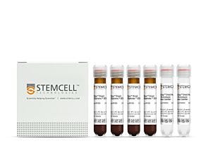
EasySep™ Direct Human Neutrophil Isolation Kit
New look, same high quality and support! You may notice that your instrument or reagent packaging looks slightly different from images displayed on the website, or from previous orders. We are updating our look but rest assured, the products themselves and how you should use them have not changed. Learn more
Quality Statement:
PRODUCTS ARE FOR RESEARCH USE ONLY AND NOT INTENDED FOR HUMAN OR ANIMAL DIAGNOSTIC OR THERAPEUTIC USES UNLESS OTHERWISE STATED. FOR ADDITIONAL INFORMATION ON QUALITY AT STEMCELL, REFER TO WWW.STEMCELL.COM/COMPLIANCE.

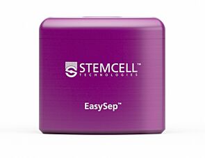 EasySep™ Magnet