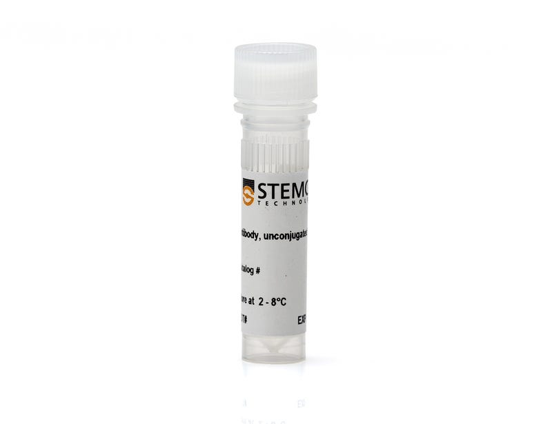
Anti-Oligodendrocyte Marker O4 Antibody, Clone 81

Mouse monoclonal IgM antibody against human, mouse, rat oligodendrocyte marker O4

Anti-Oligodendrocyte Marker O4 Antibody, Clone 81

Mouse monoclonal IgM antibody against human, mouse, rat oligodendrocyte marker O4

From: 834 USD
Catalog #
(Select a product)
Mouse monoclonal IgM antibody against human, mouse, rat oligodendrocyte marker O4
Add to Wish List

Overview

The 81 antibody clone reacts with the O4 antigen, a sulfated galactocerebroside (sulfatide). O-Antigens are primarily expressed on the extracellular leaflet of the myelin plasma membrane of differentiating and mature oligodendrocytes in the central nervous system, as well as on Schwann cells in the peripheral nervous system. O4 is formed during postnatal development and is a marker for cell bodies and processes of oligodendrocytes types I and II. O4 is expressed in pro-oligodendrocytes, however, it is not found on O-2A-progenitor cells. Abnormal expression of O4 has been associated with various disorders, including neurofibromatosis and schizophrenia.
Subtype
Primary Antibodies
Target Antigen
Oligodendrocyte Marker O4
Alternative Names
3-O-sulfogalactosylceramide, SM4, Sulfatide
Reactive Species
Chicken, Human, Mouse, Rat
Conjugation
Unconjugated
Host Species
Mouse
Cell Type
Neural Cells, PSC-Derived, Neural Stem and Progenitor Cells
Species
Human, Mouse, Other, Rat
Application
Flow Cytometry, Immunocytochemistry, Immunofluorescence, Immunohistochemistry
Area of Interest
Neuroscience
Clone
81
Isotype
IgM

Protocols and Documentation

Find supporting information and directions for use in the Product Information Sheet or explore additional protocols below.

Document Type
Product Name
Catalog #
Lot #
Language
Catalog #
60053
Lot #
All
Language
English
Document Type
Safety Data Sheet
Catalog #
60053
Lot #
All
Language
English

Applications

This product is designed for use in the following research area(s) as part of the highlighted workflow stage(s). Explore these workflows to learn more about the other products we offer to support each research area.

Resources and Publications

Publications (1)

Modeling neuronal defects associated with a lysosomal disorder using patient-derived induced pluripotent stem cells. Lemonnier T et al. Human Molecular Genetics 2011 SEP

Abstract

By providing access to affected neurons, human induced pluripotent stem cells (iPSc) offer a unique opportunity to model human neurodegenerative diseases. We generated human iPSc from the skin fibroblasts of children with mucopolysaccharidosis type IIIB. In this fatal lysosomal storage disease, defective α-N-acetylglucosaminidase interrupts the degradation of heparan sulfate (HS) proteoglycans and induces cell disorders predominating in the central nervous system, causing relentless progression toward severe mental retardation. Partially digested proteoglycans, which affect fibroblast growth factor signaling, accumulated in patient cells. They impaired isolation of emerging iPSc unless exogenous supply of the missing enzyme cleared storage and restored cell proliferation. After several passages, patient iPSc starved of an exogenous enzyme continued to proliferate in the presence of fibroblast growth factor despite HS accumulation. Survival and neural differentiation of patient iPSc were comparable with unaffected controls. Whereas cell pathology was modest in floating neurosphere cultures, undifferentiated patient iPSc and their neuronal progeny expressed cell disorders consisting of storage vesicles and severe disorganization of Golgi ribbons associated with modified expression of the Golgi matrix protein GM130. Gene expression profiling in neural stem cells pointed to alterations of extracellular matrix constituents and cell-matrix interactions, whereas genes associated with lysosome or Golgi apparatus functions were downregulated. Taken together, these results suggest defective responses of patient undifferentiated stem cells and neurons to environmental cues, which possibly affect Golgi organization, cell migration and neuritogenesis. This could have potential consequences on post-natal neurological development, once HS proteoglycan accumulation becomes prominent in the affected child brain.