STEMdiff™ Monocyte Differentiation Supplement (100X)

Supplement for the differention to CD14+ monocytes

STEMdiff™ Monocyte Differentiation Supplement (100X)

Supplement for the differention to CD14+ monocytes

From: 210 USD
Catalog #
(Select a product)
Supplement for the differention to CD14+ monocytes
Add to Wish List

Product Advantages


  • Robust differentiation to monocytes

What's Included

  • STEMdiff™ Monocyte Differentiation Supplement (100X), 1 mL

Overview

Use STEMdiff™ Monocyte Differentiation Supplement (100X) to support the differention of human pluripotent stem cells (hPSCs) to CD14+ monocytes.

Available either individually or as part of the STEMdiff™ Monocyte Kit, the supplement is intended to be combined with StemSpan™ SFEM II to prepare the Monocyte Differentiation Medium. This medium can be used during the STEMdiff™ monocyte differentiation protocol to promote the production of CD14+ monocytes.

For detailed information on the STEMdiff™ monocyte differentiation protocol, please explore the STEMdiff™ Monocyte Kit Product Information Sheet.
Subtype
Supplements
Cell Type
Dendritic Cells, Macrophages, Monocytes, Myeloid Cells, Pluripotent Stem Cells
Species
Human
Application
Cell Culture, Differentiation
Brand
STEMdiff
Area of Interest
Disease Modeling, Immunology, Stem Cell Biology

Data Figures

Differentiation of hPSC-Derived CD34+ Cells into CD14+ Monocytes

Figure 1. Monocyte Differentiation Protocol

One day prior to differentiation, human pluripotent stem cell (hPSC) colonies are harvested and seeded as small aggregates (100 - 200 μm in diameter) at 10 - 20 aggregates/cm2 in mTeSR™1, TeSR™-E8™, or mTeSR™ Plus media. After one day, the medium is replaced with Medium A (STEMdiff™ Hematopoietic Basal Medium + Supplement A) to induce mesodermal specification (stage 1). On day 3, the medium is changed to Medium B (STEMdiff™ Hematopoietic Basal Medium + Supplement B) to promote hematopoietic specification (stage 2). On day 7, the medium is replaced with Monocyte Differentiation Medium (StemSpan™ SFEM II + STEMdiff™ Monocyte Differentiation Supplement) to promote the production of CD14+ monocytes (stage 3). Monocyte Differentiation Medium is used for all medium changes for the remaining culture period. CD14+ cells can be detected in suspension starting after day 14, and their frequency gradually increases until day 17 - 23. CD14+ cells can be harvested directly from the culture supernatant during medium changes.

hPSC-Derived CD14+ Monocyte Characterization, Frequency and Yield

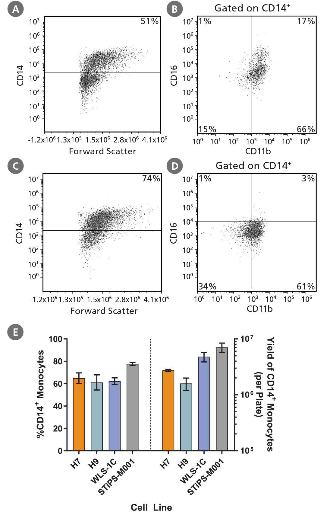
Figure 2. Robust and Efficient Generation of CD14⁺ Monocytes Using STEMdiff™ Monocyte Kit

hPSCs were differentiated to monocytes using the 2D culture system described in Figure 1. Between days 17 and 23, cells were harvested every 2 - 3 days and analyzed by flow cytometry for CD14 expression. Representative flow cytometry plots are shown for (A, B) iPS (WLS-1C)-derived cells and (C, D) ES (H9)-derived cells. (E) The average frequency of viable CD14+ monocytes at the peak harvest was 61 - 78%. The average yield of CD14+ monocytes produced per 6-well plate at the peak harvest was between 1.6 x 10^6 and 7.1 x 10^6 cells. Data are shown as mean ± SEM (n = 3 - 14).

Characterization and Phagocytosis Analysis of Macrophages

Figure 3. STEMdiff™ Monocyte Kit Generates Monocytes That Are Capable of Differentiation to Macrophages

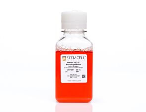
hPSC-derived monocytes were harvested after 21 days of culture. These were then differentiated to macrophages using ImmunoCult™-SF Macrophage Medium (Catalog #10961) with 100 ng/mL M-CSF for 4 days. Macrophages were then incubated for an additional 2 days with either 10 ng/mL of LPS and 50 ng/mL of IFN-γ, or 10 ng/mL IL-4, to become polarized to M1 or M2a macrophages, respectively. Representative flow cytometry plots of (A) M1 and (B) M2a macrophages produced from the WLS-1C iPS cell line are shown. (C) To measure phagocytosis, PSC-derived M2a macrophages and peripheral blood (PB) monocyte-derived M2a macrophages (primary M2a macrophages), were incubated with pHrodo™ Red Zymosan A BioParticles® Conjugate and incubated at 37°C for 8 hours. Images were acquired using the IncuCyte® ZOOM every 30 minutes and analyzed for internalization of pHrodo™ Red Zymosan A BioParticles® (measured as red object/mm2). hPSC-derived and primary M2a macrophages show similar phagocytic activity.

Characterization of Dendritic Cells

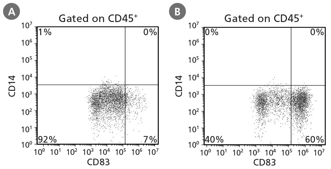
Figure 4. STEMdiff™ Monocyte Kit Generates Monocytes That Can Be Differentiated to Dendritic Cells

hPSCs were differentiated into monocytes, harvested after 21 days, and differentiated to dendritic cells using ImmunoCult™ Dendritic Cell Culture Kit (Catalog #10985). Half of the dendritic cells were harvested on day 7 and examined for CD14 and CD83 expression to identify CD14⁻CD83⁻/lo immature dendritic cells. The remaining dendritic cells were activated for 2 days and assessed for the presence of CD14⁻CD83⁺ mature dendritic cells at day 7. Representative cultures initiated with ES (H9) cells are shown for production of (A) immature dendritic cells and (B) mature dendritic cells.

Protocols and Documentation

Find supporting information and directions for use in the Product Information Sheet or explore additional protocols below.

Document Type
Product Name
Catalog #
Lot #
Language
Catalog #
05324
Lot #
All
Language
English
Document Type
Safety Data Sheet
Catalog #
05324
Lot #
All
Language
English

Applications

This product is designed for use in the following research area(s) as part of the highlighted workflow stage(s). Explore these workflows to learn more about the other products we offer to support each research area.