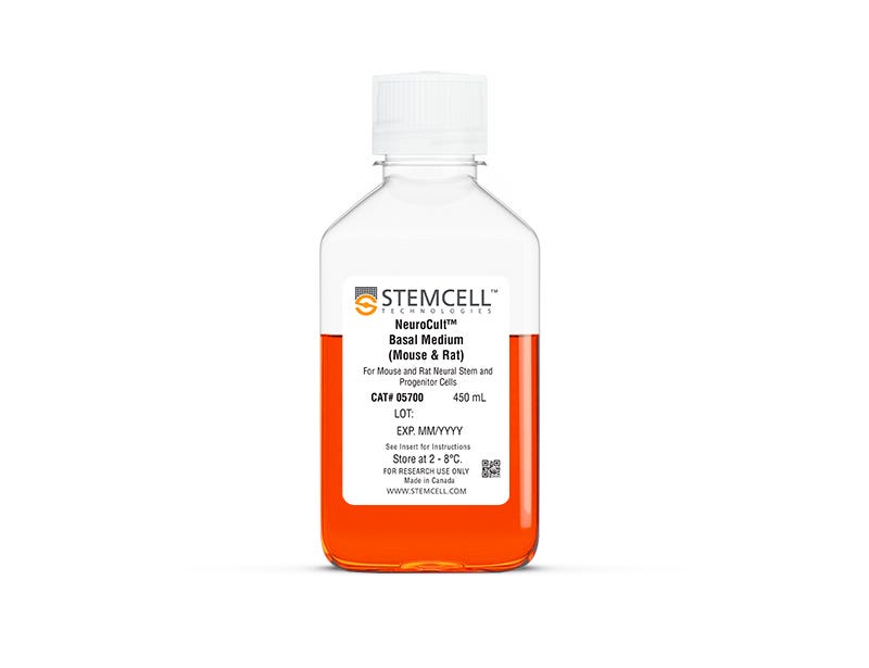
NeuroCult™ Basal Medium (Mouse & Rat)

Basal medium for culture of mouse and rat neural stem and progenitor cells

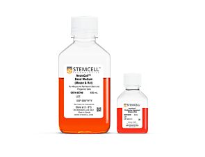
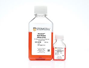
NeuroCult™ Basal Medium (Mouse & Rat)

Basal medium for culture of mouse and rat neural stem and progenitor cells

From: 112 USD
Catalog #
(Select a product)
Basal medium for culture of mouse and rat neural stem and progenitor cells
Add to Wish List
Products for Your Protocol
To see all required products for your protocol, please consult the Protocols and Documentation.

Overview

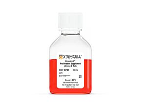
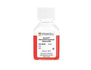
NeuroCult™ Basal Medium (Mouse & Rat) is a standardized serum-free basal medium designed to be supplemented with NeuroCult™ Proliferation Supplement (Mouse & Rat; Catalog #05701) and appropriate cytokines for the in vitro culture and expansion of mouse and rat neural stem and progenitor cells. NeuroCult™ Basal Medium (Mouse & Rat) can also be supplemented with NeuroCult™ Differentiation Supplement (Mouse & Rat; Catalog #05703) for the differentiation of mouse and rat neural stem and progenitor cells into neurons, astrocytes, and oligodendrocytes. NeuroCult™ Basal Medium (Mouse & Rat) is a component of NeuroCult™ Proliferation Kit (Mouse & Rat; Catalog #05702) and NeuroCult™ Differentiation Kit (Mouse & Rat; Catalog #05704).

NOTE: When preparing Complete NeuroCult™ Proliferation Medium, addition of Human Recombinant EGF (Catalog #78006.1) is required. When culturing cells obtained from adult mouse or rat, Human Recombinant bFGF (Catalog #78003.1) and Heparin Solution (Catalog #07980) are also required.
Subtype
Basal Media, Specialized Media
Cell Type
Brain Tumor Stem Cells, Neural Stem and Progenitor Cells
Species
Mouse, Rat
Application
Cell Culture, Colony Assay, Differentiation, Expansion, Functional Assay, Spheroid Culture
Brand
NeuroCult
Area of Interest
Cancer, Disease Modeling, Neuroscience, Stem Cell Biology
Formulation Category
Serum-Free

Protocols and Documentation

Find supporting information and directions for use in the Product Information Sheet or explore additional protocols below.

Document Type
Product Name
Catalog #
Lot #
Language
Catalog #
05700
Lot #
All
Language
English
Document Type
Technical Manual
Catalog #
05700
Lot #
All
Language
English
Document Type
Safety Data Sheet
Catalog #
05700
Lot #
All
Language
English

Applications

This product is designed for use in the following research area(s) as part of the highlighted workflow stage(s). Explore these workflows to learn more about the other products we offer to support each research area.

Resources and Publications

Publications (128)

Recombinant insulin-like growth factor binding protein-4 inhibits proliferation and promotes differentiation of neural progenitor cells Niu H et al. Neuroscience Letters 2017 MAR

Abstract

Insulin-like growth factor (IGF) is involved in regulating many processes during neural development, and IGF binding protein-4 (IGFBP4) functions as a modulator of IGF actions or in an IGF-independent manner (e.g., via inhibiting Wnt/β-catenin signaling). In the present study, neural progenitor cells (NPCs) were isolated from the forebrain of newborn mice to investigate effects of IGFBP4 on the proliferation and differentiation of NPCs. The proliferation of NPCs was evaluated using Cell Counting Kit-8 (CCK-8) after treatment with or without IGFBP4 as well as blockers of IGF-IR and β-catenin. Phosphorylation levels of Akt, Erk1, 2 and p38 were analyzed by Western blotting. The differentiation of NPCs was evaluated using immunofluorescence and Western blotting. It was shown that exogenous IGFBP4 significantly inhibited the proliferation of NPCs and it did not induce a more pronounced inhibition of cell proliferation after blockade of IGF-IR but it did after antagonism of β-catenin. Akt phosphorylation was significantly decreased and phosphorylation levels of Erk1, 2 and p38 were not significantly changed in IGFBP4-treated NPCs. Excessive IGFBP4 significantly promoted NPCs to differentiate into astrocytes and neurons. These data suggested that exogenous IGFBP4 inhibits proliferation and promotes differentiation of neural progenitor cells mainly through IGF-IR signaling pathway.
Cathepsin B Improves ß-Amyloidosis and Learning and Memory in Models of Alzheimer's Disease. Embury CM et al. Journal of neuroimmune pharmacology : the official journal of the Society on NeuroImmune Pharmacology 2017 JUN

Abstract

Amyloid-ß (Aß) precursor protein (APP) metabolism engages neuronal endolysosomal pathways for Aß processing and secretion. In Alzheimer's disease (AD), dysregulation of APP leads to excess Aß and neuronal dysfunction; suggesting that neuronal APP/Aß trafficking can be targeted for therapeutic gain. Cathepsin B (CatB) is a lysosomal cysteine protease that can lower Aß levels. However, whether CatB-modulation of Aß improves learning and memory function deficits in AD is not known. To this end, progenitor neurons were infected with recombinant adenovirus expressing CatB and recovered cell lysates subjected to proteomic analyses. The results demonstrated Lamp1 deregulation and linkages between CatB and the neuronal phagosome network. Hippocampal injections of adeno-associated virus expressing CatB reduced Aß levels, increased Lamp1 and improved learning and memory. The findings were associated with the emergence of c-fos + cells. The results support the idea that CatB can speed Aß metabolism through lysosomal pathways and as such reduce AD-associated memory deficits.
L2hgdh deficiency accumulates L-2-hydroxyglutarate with progressive leukoencephalopathy and neurodegeneration Ma S et al. Molecular and Cellular Biology 2017 JAN

Abstract

L-2-hydroxyglutarate aciduria (L-2-HGA) is an autosomal recessive neurometabolic disorder caused by a mutation in the L-2-hydroxyglutarate dehydrogenase ( L2HGDH ) gene. In this study, we generated L2hgdh knockout (KO) mice and observed a robust increase of 2-hydroxyglutarate (L-2-HG) levels in multiple tissues. The highest levels of L-2-HG were observed in the brain and testis with a corresponding increase in histone methylation in these tissues. L2hgdh KO mice exhibit white matter abnormalities, extensive gliosis, microglia-mediated neuroinflammation, and an expansion of oligodendrocyte progenitor cells (OPCs). Moreover, L2hgdh deficiency leads to impaired adult hippocampal neurogenesis and late-onset neurodegeneration in mouse brains. Our data provide in vivo evidence that L2hgdh mutation leads to L-2-HG accumulation, leukoencephalopathy, and neurodegeneration in mice, thus offering new insights into the pathophysiology of L-2-HGA in humans.