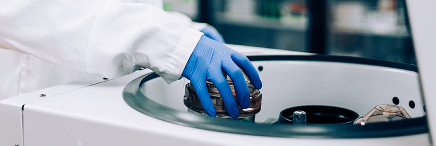
How to Process a Leukopak for Downstream Cell Isolation
How to process a leukopak for isolating cells using manual or automated immunomagnetic and column-free EasySep™ cell isolation kits
The protocol below outlines how to process a leukopak for isolating cells using manual or automated immunomagnetic and column-free EasySep™ cell isolation kits. Following immunomagnetic cell separation, the untouched cells are immediately ready for downstream analysis. Explore our wide range of EasySep™ kits for isolating your cells of interest.
Materials
- Leukopak (e.g. Catalog #70500)*
- Bag extractor stand
- Transfer set spike
- Transfer set tubing
- Sterile container(s)
- 50 mL polyproylene conical tubes (e.g. Falcon® Conical Tubes, 50 mL, Catalog #38010)
Protocol
Before You Begin: Ensure that you handle the leukopak and its contents aseptically within a biosafety cabinet.
Part I: Transfer Leukopak Contents to a Sterile Container
- To transfer the contents of the leukopak to a sterile container, place the leukopak in an extractor stand, open the port on the bag, and insert the transfer set spike.
- Close the plate of the extractor stand to apply pressure to the bag.
-
Open the valve and dispense the leukopak contents via the transfer set tubing into a sterile container. Alternatively, if you don’t have an extractor stand, apply pressure to the bag or drain by gravity.
Note: Wash the leukopak bag with some of the buffer used for dilution to help recover remaining cells in the bag.
Part II: Prepare Leukopak Contents
Notes:
- Before You Begin: It is recommended to perform an initial cell count of the leukopak prior to any processing or washing steps. This number can then be compared to the cell count after the washing steps and used to calculate cell recovery.
- If you are performing downstream cell isolation using an EasySep™ kit, consult the provided Product Information Sheet (PIS) to determine if red blood cell (RBC) lysis and additional platelet removal steps are required. If RBC lysis and additional platelet removal steps are NOT mentioned or required, wash the leukopak contents as outlined in Option 1. If the EasySep™ selection kit requires performing RBC lysis and platelet removal, refer to Option 2.
- Due to donor-to-donor variability, the content of RBCs and platelets in the leukopak may vary. After completing the washing steps in Option 1, an additional RBC lysis step and additional washes to remove platelets may be required (Option 2).
Option 1: Wash Leukopak Contents
- After the contents of the leukopak have been transferred to a sterile container, add an equivalent volume of buffer (PBS + 2% FBS recommended) to the container.
- Aliquot the diluted leukopak contents into the appropriate tubes for centrifugation. We recommend using 50 mL conical tubes for centrifugation.
- Centrifuge the cells at 300 x g for 10 minutes at room temperature (15 - 25ºC) with the brake ON.
- Carefully remove the supernatant without disturbing the cell pellet.
- Resuspend cells by adding recommended medium to the sample. The cells are ready to be counted.
-
If desired, pool all the cells from the different tubes into one single tube as follows:
a. Resuspend cells in a small volume of recommended medium.
b. Transfer resuspended cells from individual 50 mL tubes into one 50 mL tube. Make sure to rinse the tubes with recommended medium to recover as many cells as possible.
Option 2: RBC Lysis and Platelet Removal
- After the contents of the leukopak have been transferred to a sterile container, add Ammonium Chloride Solution to the sample at a volume:volume ratio of 4:1. Mix by gently inverting the tube.
- Incubate on ice for 15 minutes.
- Centrifuge the sample at 300 x g for 10 min at room temperature (15 - 25ºC) with the brake ON.
- Remove the supernatant and resuspend the cells.
-
Perform a wash step to remove platelets, as follows:
a. Top up the tube with PBS + 2% FBS. Centrifuge at 120 - 130 x g for 10 minutes at room temperature with the brake OFF.
b. Remove the supernatant.
c. Resuspend the cells in a small volume of recommended medium. - If additional platelet removal is required, perform one to two additional washing steps.
Part III: Count Cells and Prepare Cell Suspension for Cell Separation
- Perform a cell count using either Trypan Blue (Catalog #07050) or 3% Acetic Acid with Methylene Blue (Catalog #07060) and a hemocytometer. See this protocol on How to Count Cells with a Hemocytometer for cell counting instructions.
-
Calculate the Total Volume Required to resuspend the cells at the desired concentration, using the following equation:
Total Volume Required = Cell Concentration in Sample x Sample Volume / Desired Final Cell ConcentrationExample:
Desired final cell concentration = 5 x 10⁷ cells/mL in recommended medium
Cell concentration in sample = 18,250,000 cells/mL
Sample volume = 40 mL
(18,250,000 cells/mL x 40 mL) / 5 x 10⁷ cells/mL = 14.6 mL
The total volume required is 14.6 mL. - After determining the volume required to obtain the desired cell concentration, centrifuge cells at 300 x g for 10 minutes at room temperature (15 - 25ºC) with the brake ON.
-
Remove the supernatant and resuspend the cells in the desired medium with the appropriate volume to obtain the desired cell concentration.
Note: For downstream manual isolation with EasySep™ or automated cell isolation with RoboSep™ instruments, resuspend cells in EasySep™ Buffer, RoboSep™ Buffer, or PBS containing 2% FBS and 1 mM EDTA. Medium should be free of Ca++ and Mg++.
Part IV: Cell Isolation Using EasySep™ or RoboSep™
Option 1: Manual Cell Isolation
- Follow the instructions provided in the Product Information Sheet of the particular EasySep™ kit.For efficient isolation of immune cells from leukopaks, we recommend using EasySep™ magnets that allow for large-volume processing.
- Easy 50 EasySep™ Magnet (Catalog #18002) can process up to 40 mL and 4 x 10⁹ cells.
- Easy 250 EasySep™ Magnet (Catalog #100-0821) can process up to 225 mL and 1.25 x 10¹⁰ cells.
- Compare across the different EasySep™ Magnets.
Option 2: Automated Cell Isolation Using RoboSep™-S
- Add the cell suspension at the indicated cell concentration in the EasySep™ Product Information Sheet to a 14 mL (17 x 95 mm) polystyrene round-bottom tube.
- Select the protocol on RoboSep™-S and follow the onscreen prompts to load samples, reagents, and tips.
- Press ‘Run’ to start the protocol.
- After the run has been completed, unload the carousel. Isolated cells are ready for use.


Request Pricing
Thank you for your interest in this product. Please provide us with your contact information and your local representative will contact you with a customized quote. Where appropriate, they can also assist you with a(n):
Estimated delivery time for your area
Product sample or exclusive offer
In-lab demonstration
By submitting this form, you are providing your consent to STEMCELL Technologies Canada Inc. and its subsidiaries and affiliates (“STEMCELL”) to collect and use your information, and send you newsletters and emails in accordance with our privacy policy. Please contact us with any questions that you may have. You can unsubscribe or change your email preferences at any time.


