ImmunoCult™ Human CD3/CD28/CD2 T Cell Activator

Human T cell activation and expansion reagent

ImmunoCult™ Human CD3/CD28/CD2 T Cell Activator

Human T cell activation and expansion reagent
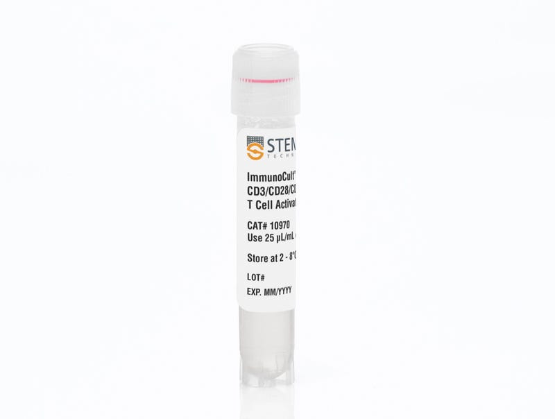
From: 209 USD
Catalog #
(Select a product)
Human T cell activation and expansion reagent
Add to Wish List

Product Advantages


  • Robust activation and expansion of human T cells without the use of magnetic beads, feeder cells, or antigen

  • Provides a gentle activation stimulus that maintains high viability of activated and expanded T cells

  • Highly stable, filter-sterilized soluble reagent

Overview

Achieve robust activation and expansion of T cells in the absence of magnetic beads, feeder cells, or antigens.

This product’s gentle activation stimulus ensures a high viability of activated T cells, which can be further expanded in ImmunoCult™-XF T Cell Expansion Medium (Catalog #10981) or other media for culturing human T cells. ImmunoCult™ Human CD3/CD28/CD2 T Cell Activator consists of soluble antibody complexes that bind to and cross-link CD3, CD28, and CD2 cell surface ligands, thereby providing the required primary and co-stimulatory signals for T cell activation.

This product is designed for research applications. If you require reagents suitable for use in cell therapy manufacturing, ImmunoCult™ Human CD3/CD28/CD2 T Cell Activators (Catalog #100-0785) are produced under relevant GMPs.
Contains
• Anti-human CD3 monospecific antibody complex
• Anti-human CD28 monospecific antibody complex
• Anti-human CD2 monospecific antibody complex
Subtype
Supplements
Cell Type
T Cells, T Cells, CD4+, T Cells, CD8+
Species
Human
Application
Activation, Cell Culture, Expansion
Brand
ImmunoCult
Area of Interest
Immunology, Cell Therapy Development

Data Figures

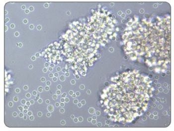
Activated Morphology of Human T Cells Stimulated With ImmunoCult™ Human CD3/CD28/CD2 T Cell Activator

Figure 1. Activated Morphology of Human T Cells Stimulated With ImmunoCult™ Human CD3/CD28/CD2 T Cell Activator

Image of human T cells isolated using the EasySep™ Human T Cell Isolation Kit (Catalog #17951), stimulated with ImmunoCult™ Human CD3/CD28/CD2 T Cell Activator, and cultured in ImmunoCult™-XF T Cell Expansion Medium (Catalog #10981).

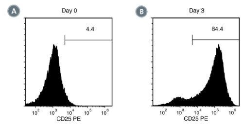
Activation of EasySep™ Isolated Human T Cells Stimulated With ImmunoCult™ Human CD3/CD28/CD2 T Cell Activator

Figure 2. Activation of EasySep™ Isolated Human T Cells Stimulated With ImmunoCult™ Human CD3/CD28/CD2 T Cell Activator

EasySep™-isolated human T cells were stimulated with ImmunoCult™ Human CD3/CD28/CD2 T Cell Activator and cultured in ImmunoCult™-XF T Cell Expansion Medium. Activation of viable CD3+ T cells was assessed by CD25 expression using flow cytometry. On day 0, the frequency of CD25 positive cells was (A) 5.6 ± 2.4% (mean ± SD). Following 3 days of culture, the frequency of CD25 positive cells was (B) 88.8 ± 3.2% (mean ± SD) when stimulated with ImmunoCult™ Human CD3/CD28/CD2 T Cell Activator.

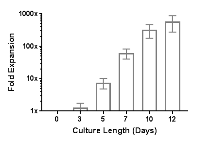
Robust Human T Cell Expansion with ImmunoCult™ Human CD3/CD28/CD2 T Cell Activator

Figure 3. Robust Human T Cell Expansion with ImmunoCult™ Human CD3/CD28/CD2 T Cell Activator

EasySep™-isolated human T cells were expanded over 12 days with ImmunoCult™ Human CD3/CD28/CD2 T Cell Activator in ImmunoCult™-XF T Cell Expansion Medium supplemented with Human Recombinant IL-2. On day 0, 1 x 10^6 EasySep™-isolated human T cells were stimulated with 25 μL of ImmunoCult™ Human CD3/CD28/CD2 T Cell Activator in ImmunoCult™-XF T Cell Expansion Medium supplemented with 10 ng/mL Human Recombinant IL-2. On days 3, 5, 7, and 10, viable cells were counted and fresh medium supplemented with IL-2 was added. No additional ImmunoCult™ Human CD3/CD28/CD2 T Cell Activator was added during the 12-day culture period (mean ± SD in 6 experiments with 3 donors).

Table 1. Comparison of ImmunoCult™ Human T Cell Activation Reagents

Table comparing ImmunoCult™ Human T Cell Activation Reagents

Protocols and Documentation

Find supporting information and directions for use in the Product Information Sheet or explore additional protocols below.

Document Type
Product Name
Catalog #
Lot #
Language
Catalog #
10990, 10970
Lot #
All
Language
English
Document Type
Safety Data Sheet
Catalog #
10990, 10970
Lot #
All
Language
English

Applications

This product is designed for use in the following research area(s) as part of the highlighted workflow stage(s). Explore these workflows to learn more about the other products we offer to support each research area.

Resources and Publications

Educational Materials (22)

Publications (43)

Targeting the ATX-LPA Axis Overcomes TKI Resistance and Immunosuppression in Renal Cell Carcinoma via Dual Inhibition of AKT/mTOR and TBK1/IRF3 Pathways. J. Luo et al. Advanced science (Weinheim, Baden-Wurttemberg, Germany) 2026 Jun

Abstract

BACKGROUND: Therapeutic resistance limits durable survival in advanced/metastatic renal cell carcinoma (RCC) treated with first-line tyrosine kinase inhibitor (TKI) plus immune checkpoint inhibitor (ICI). We sought to define key resistance drivers and actionable targets. METHODS: Integrated RNA sequencing of cabozantinib-resistant RCC cells, lipid metabolomics, and PD-L1 correlation analyses identified ENPP2 as a candidate driver. Its role in TKI resistance and survival signaling was validated by apoptosis, CCK-8, and colony formation assays in vitro and by nude-mouse xenograft models in vivo. ELISA, flow cytometry and tumor cell-T-cell co-culture assays were used to dissect ENPP2-dependent CD8+ T-cell dysfunction. The therapeutic benefit of pharmacologic ATX inhibition combined with standard TKI-ICI regimens was tested in RCC patient-derived xenograft models. RESULTS: The ATX-LPA axis conferred TKI resistance via constitutive AKT/mTOR activation and promoted immune evasion by upregulating PD-L1 through TBK1/IRF3 signaling, thereby impairing intratumoral CD8+ T-cell function. ENPP2 enhanced PD-L1 transcription by facilitating IRF3 nuclear translocation and its direct recruitment to the CD274 promoter. ATX inhibition improved the antitumor efficacy of TKI-ICI therapy in preclinical models. CONCLUSIONS: Targeting the ATX-LPA axis represents a promising strategy to overcome resistance to current TKI-ICI combinations.
Butyrate enhances tumor immune evasion by increasing PD-L1 abundance. X. Sun et al. International journal of biological sciences 2026 Jul

Abstract

As a short-chain fatty acid and histone deacetylase (HDAC) inhibitor, butyrate possesses extensive immunomodulatory properties. Here, we uncover a novel mechanism wherein butyrate markedly upregulates the expression of the immune checkpoint PD-L1 across diverse tumor cell types. Mechanistically, epigenetic analyses revealed that butyrate drives PD-L1 transcriptional activation through dual synergistic pathways: increasing histone H3 acetylation (H3Ac) at the PD-L1 promoter via HDAC inhibition, and enhancing histone H3K4me3 modification by upregulating transglutaminase 2 (TGM2). Combining butyrate or high-fiber diets with anti-PD-L1 antibodies profoundly suppressed tumor growth and bolstered anti-tumor immunity in both subcutaneous and AOM-DSS-induced colorectal cancer (CRC) murine models. Ultimately, our results highlight butyrate and dietary fiber supplementation as promising sensitizing strategies to augment the efficacy of PD-L1 blockade in patients with low baseline PD-L1 expression.
Combined transcriptomic and lipidomic analysis reveals enhanced lipogenesis in memory Tregs upon TCR activation. Y. Sato ImmunoHorizons 2026 Jul

Abstract

Regulatory T cells (Tregs) maintain immune homeostasis in vivo. Similar to conventional T cells (Tconvs), Tregs are divided into naïve and memory cells. Tregs have unique metabolic properties, including enhanced oxidative phosphorylation. The lipidomic profiles of human Tregs have been studied previously; however, those of naïve and memory Tregs have not yet been consistently compared. Thus, in the present study, we used a combined transcriptomic and lipidomic analysis to assess the metabolic features of human naïve and memory Tregs upon activation. Using transcriptomic analysis, we identified distinct gene expression profiles in naïve and memory Tregs compared with those in Tconvs. Upon TCR stimulation, memory Tregs showed a lipidomic profile distinct from that of memory Tconvs, whereas the lipidomic profiles of naïve Tregs were similar to those of naïve Tconvs. Furthermore, upon TCR stimulation, memory Tregs expressed triglycerides (TGs) that were more enriched in monounsaturated fatty acids and polyunsaturated fatty acids (PUFAs) than those of memory Tconvs. However, memory Tconvs also had higher PUFA-TG levels than naïve Tconvs and Tregs after TCR stimulation, although their levels remained lower than those in memory Tregs. These findings suggest PUFA-TGs could be a marker of memory Tregs. In turn, higher frequency of memory cells within the Treg population may also contribute to the observed enrichment of PUFA-TGs in Tregs upon TCR activation. Our study demonstrated unique transcriptomic and lipidomic profiles and enhanced lipogenesis in memory Tregs upon TCR stimulation.