Enzyme-Free Passaging of Human Pluripotent Stem Cells Using Gentle Cell Dissociation Reagent
How to passage ES and iPS cells cultured in mTeSR™ Plus using GCDR
Cell passaging, also known as subculturing or cell splitting, is a technique used to promote growth in culture. Cultured cells are transferred to fresh growth medium using enzymatic or non-enzymatic methods, depending on the cell culture medium and matrix being used. For example, human embryonic stem (ES) and induced pluripotent stem (iPS) cells maintained in mTeSR™ Plus on Vitronectin XF™ are recommended to be passaged with non-enzymatic reagents. Gentle Cell Dissociation Reagent (GCDR) is an enzyme-free reagent for passaging human ES and iPS cells as aggregates with manual scraping to generate cell aggregates. This manual process enables selection of differentiated areas if desired.
The following protocol describes how to passage ES and iPS cells cultured in mTeSR™ Plus using GCDR. These instructions are for passaging cells from one well of a 6-well plate. If using other cultureware, adjust volumes accordingly. Please consult your Product Information Sheet for recommendations on suitable passaging reagents and methods for your cell culture system.
Materials
- Cell culture matrix (e.g. Vitronectin XF™, Catalog #07180)
- mTeSR™ Plus (Catalog #05825)
- Note: This protocol can also be used for ES and iPS cells cultured in mTeSR™1 (Catalog #85850) or TeSR™-E8™ (Catalog #05990)
- 6-well plate (e.g. Falcon® 6-Well Flat-Bottom Plate, Tissue Culture-Treated, Catalog #38016)
- Pipette tips (e.g. Corning® Filtered Pipette Tips, Catalog #38034)
- Pipettor (e.g. Corning® Lambda™ Plus Pipettor, Catalog #38060)
Protocol
This procedure has been optimized for use with hPSC maintenance reagents and multiple embryonic stem (ES) and induced pluripotent stem (iPS) cell lines. For upstream protocols and source materials, please see the mTeSR™ Plus Technical Manual and the Product Information Sheet for STEMCELL’s highly quality-controlled Healthy Control Human iPSC Line, Female, SCTi003-A.
Before You Begin: Use sterile technique when coating cultureware. Non-tissue culture-treated cultureware is required for use with Vitronectin XF™.
- At least 1 hour before passaging, coat new plates with either Vitronectin XF™ or Corning® Matrigel® (see section 4.2 of the Technical Manual: Maintenance of Human Pluripotent Stem Cells in mTeSR™ Plus).
- Aliquot sufficient complete mTeSR™ Plus and warm to room temperature (15 - 25°C).
Note: Do not warm mTeSR™ Plus in a 37°C water bath. - Use a microscope to visually identify regions of differentiation. Mark these using a felt tip or lens marker on the bottom of the plate.
- Remove regions of differentiation by scraping with a pipette tip or by aspiration. Avoid having the culture plate out of the incubator for more than 15 minutes at a time.
Note: Selection may not be required if differentiation is < 5%. Selection should not exceed 20% of the well if the culture is of high quality. For representative pictures of regions of differentiation see Figure 1.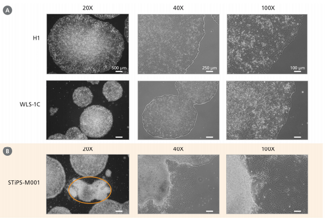
Figure 1. Morphology of Human ES and iPS Cells Cultured in mTeSR™ Plus
(A) Undifferentiated human ES cells (H1) on Corning® Matrigel® and iPS cells (WLS-1C) on Vitronectin XF™ at the optimal time of passaging. (B) Area of spontaneous differentiation (orange circle) between undifferentiated STiPS-M001 colonies. Magnifications: 20X, 40X, and 100X.
- Aspirate medium from the well and add 1 mL of GCDR.
- Incubate at room temperature. Refer to Table 1 for recommended incubation times and Figure 4 for a representative example of the desired appearance following incubation.
Table 1. GCDR Incubation Times for Cultures Plated on Different Matrices
MatrixIncubation Time with GCDRVitronectin XF™8 - 12 minutesCorning® Matrigel®8 - 10 minutesNote: Incubation times may vary when using different cell lines or other non-enzymatic cell passaging reagents; dissociation should be monitored under the microscope until the optimal time is determined. - Aspirate the GCDR, then add 1 mL of mTeSR™ Plus. Gently detach the colonies by scraping with a serological glass pipette or a cell scraper.
Note: Take care to minimize the breakup of colonies. - Transfer the detached cell aggregates to a 15 mL conical tube.
Optional: Rinse the well with an additional 1 mL of mTeSR™ Plus to collect remaining cell aggregates.
Note: Centrifugation of cell aggregates is not required. - Carefully pipette the cell aggregate mixture up and down to break up the aggregates as needed. Refer to Table 2 for suggestions on how to break up cell aggregates grown on different types of matrices. A uniform suspension of aggregates approximately 50 - 200 µm in size is optimal; do not create a single-cell suspension (for more information, see section 8.0 of the Technical Manual: Maintenance of Human Pluripotent Stem Cells in mTeSR™ Plus).
Table 2. Suggested Methods for Breaking Up Cell Aggregates
MatrixPipette TypeNumber of Times to Pipette Up and DownVitronectin XF™1 mL pipettor1 - 2Corning® Matrigel®2 mL serological pipette2 - 5 - Plate the cell aggregate mixture at the desired density onto coated wells containing mTeSR™ Plus. If the colonies are at an optimal density, the cultures can be split every 4 - 7 days using 1 in 10 to 1 in 50 splits (i.e. cell aggregates from 1 well can be plated in 10 - 50 wells). If the colonies are too dense or too sparse, at the next time of passaging adjust the split ratio accordingly (see section 6.4 of the Technical Manual: Maintenance of Human Pluripotent Stem Cells in mTeSR™ Plus).
Note: Work quickly to transfer cell aggregates into new cultureware to maximize viability and attachment. - Place the plate in a 37°C incubator. Move the plate in several quick, short, back-and-forth and side-to-side motions to distribute the cell aggregates. Do not disturb the plate for 24 hours.
Note: Uneven distribution of aggregates may result in increased differentiation of human ES and iPS cells. - Perform medium changes as needed using mTeSR™ Plus and visually assess cultures to monitor growth until the next passaging time. Medium can be changed daily or every other day. To skip two consecutive days of feeding, add twice the volume of medium.
Request Pricing
Thank you for your interest in this product. Please provide us with your contact information and your local representative will contact you with a customized quote. Where appropriate, they can also assist you with a(n):
Estimated delivery time for your area
Product sample or exclusive offer
In-lab demonstration


