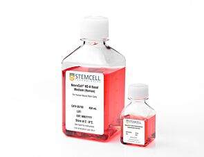
NeuroCult™-XF Proliferation Medium

Xeno-free medium for expansion of human neural stem and progenitor cells

NeuroCult™-XF Proliferation Medium

Xeno-free medium for expansion of human neural stem and progenitor cells

From: 393 USD
Catalog #
(Select a product)
Xeno-free medium for expansion of human neural stem and progenitor cells
Add to Wish List

What's Included

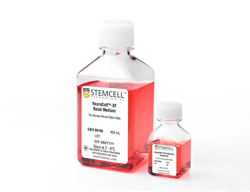
  • NeuroCult™-XF Basal Medium, 450 mL
  • NeuroCult™-XF Proliferation Supplement, 50 mL
Products for Your Protocol
To see all required products for your protocol, please consult the Protocols and Documentation.

Overview

NeuroCult™-XF Proliferation Medium is a standardized, xeno-free, serum-free medium for the culture of human neural stem and progenitor cells from normal tissues or tumor samples, in the neurosphere or adherent monolayer system. When supplemented with appropriate cytokines, NeuroCult™-XF Proliferation Medium is optimized to maintain human neural stem cells in culture for extended periods of time without the loss of their self-renewal, proliferation or differentiation potential.

NOTE: Addition of Carrier-Free rh EGF (Catalog #78006) and rh bFGF (Catalog #78003) is required. Heparin (Catalog #07980) can also be added, however, the heparin solution contains non-human animal-derived components and can be omitted if a complete xeno-free system is desired.
Subtype
Specialized Media
Cell Type
Brain Tumor Stem Cells, Neural Stem and Progenitor Cells
Species
Human
Application
Cell Culture, Colony Assay, Expansion, Functional Assay, Spheroid Culture
Brand
NeuroCult
Area of Interest
Cancer, Disease Modeling, Drug Discovery and Toxicity Testing, Neuroscience, Stem Cell Biology, Transplantation Research
Formulation Category
Serum-Free, Xeno-Free

Data Figures

NSCs cultured in NeuroCult™-ACF Proliferation Medium maintain multi-lineage potential in long-term cultures

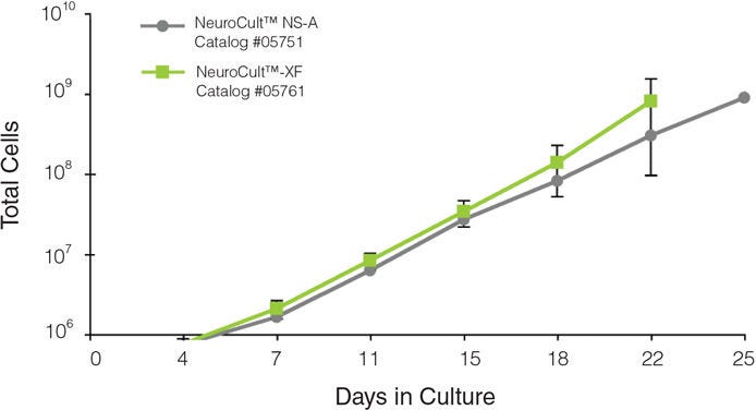
Figure 1. Neurospheres Derived from Human Fetal CNS Tissue can be Efficiently Generated for Multiple Passages in NeuroCult™-XF Proliferation Medium (n = 3)

Total cell expansion obtained with NeuroCult™-XF is comparable to that obtained with NeuroCult™ NS-A Proliferation Medium (Human).

Neurospheres derived from human fetal CNS tissue can be efficiently generated for multiple passages in NeuroCult™-ACF Proliferation Medium

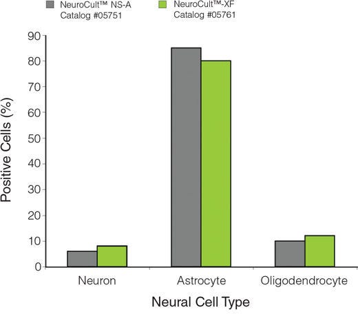
Figure 2. NSCs Cultured in NeuroCult™-XF Proliferation Medium Maintain Multi-Lineage Differentiation Potential in Long-Term Cultures

The graph indicates the percentage of neurons (class III ß-tubulin-positive), astrocytes (GFAP-positive) and oligodendrocytes (O4-positive) generated following differentiation of NSCs previously cultured in NeuroCult™-XF or NeuroCult™ NS-A Proliferation Medium (Human). The differentiation potential of NSCs cultured in NeuroCult™-XF is comparable to NSCs cultured in NeuroCult™ NS-A Proliferation Medium (Human).

Protocols and Documentation

Find supporting information and directions for use in the Product Information Sheet or explore additional protocols below.

Document Type
Product Name
Catalog #
Lot #
Language
Catalog #
05761
Lot #
All
Language
English
Document Type
Technical Manual
Catalog #
05761
Lot #
All
Language
English
Document Type
Safety Data Sheet 1
Catalog #
05761
Lot #
All
Language
English
Document Type
Safety Data Sheet 2
Catalog #
05761
Lot #
All
Language
English

Applications

This product is designed for use in the following research area(s) as part of the highlighted workflow stage(s). Explore these workflows to learn more about the other products we offer to support each research area.

Resources and Publications

Publications (3)

The Use of Autologous Mesenchymal Stem Cells for Cell Therapy of Patients with Amyotrophic Lateral Sclerosis in Belarus. Rushkevich YN et al. Bulletin of experimental biology and medicine 2015 AUG

Abstract

We studied a new method of treatment of amyotrophic lateral sclerosis with autologous mesenchymal stem cells. Autologous mesenchymal stem cells were injected intravenously (intact cells) or via lumbar puncture (cells committed to neuronal differentiation). Evaluation of the results of cell therapy after 12-month follow-up revealed slowing down of the disease progression in 10 patients in comparison with the control group consisting of 15 patients. The cell therapy was safe for the patients.
Chemically defined serum-free and xeno-free media for multiple cell lineages. Usta S et al. Annals of translational medicine 2014 OCT

Abstract

Cell culture is one of the most common methods used to recapitulate a human disease environment in a laboratory setting. Cell culture techniques are used to grow and maintain cells of various types including those derived from primary tissues, such as stem cells and cancer tumors. However, a major confounding factor with cell culture is the use of serum and animal (xeno) products in the media. The addition of animal products introduces batch and lot variations that lead to experimental variability, confounds studies with therapeutic outcomes for cultured cells, and represents a major cost associated with cell culture. Here we report a commercially available serum-free, albumin-free, and xeno free (XF) media (Neuro-Pure(TM)) that is more cost-effective than other commercial medias. Neuro-Pure was used to maintain and differentiate various cells of neuronal lineages, fibroblasts, as well as specific cancer cell lines; without the use of contaminants such serum, albumin, and animal products. Neuro-Pure allows for a controlled and reproducible cell culture environment that is applicable to translational medicine and general tissue culture.
Hypoxia induces a phase transition within a kinase signaling network in cancer cells Wei W et al. Proceedings of the National Academy of Sciences 2013 APR

Abstract

Hypoxia is a near-universal feature of cancer, promoting glycolysis, cellular proliferation, and angiogenesis. The molecular mechanisms of hypoxic signaling have been intensively studied, but the impact of changes in oxygen partial pressure (pO2) on the state of signaling networks is less clear. In a glioblastoma multiforme (GBM) cancer cell model, we examined the response of signaling networks to targeted pathway inhibition between 21% and 1% pO2. We used a microchip technology that facilitates quantification of a panel of functional proteins from statistical numbers of single cells. We find that near 1.5% pO2, the signaling network associated with mammalian target of rapamycin (mTOR) complex 1 (mTORC1)--a critical component of hypoxic signaling and a compelling cancer drug target--is deregulated in a manner such that it will be unresponsive to mTOR kinase inhibitors near 1.5% pO2, but will respond at higher or lower pO2 values. These predictions were validated through experiments on bulk GBM cell line cultures and on neurosphere cultures of a human-origin GBM xenograft tumor. We attempt to understand this behavior through the use of a quantitative version of Le Chatelier's principle, as well as through a steady-state kinetic model of protein interactions, both of which indicate that hypoxia can influence mTORC1 signaling as a switch. The Le Chatelier approach also indicates that this switch may be thought of as a type of phase transition. Our analysis indicates that certain biologically complex cell behaviors may be understood using fundamental, thermodynamics-motivated principles.