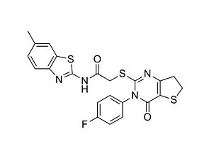
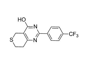
IWP-2

WNT pathway inhibitor; Inhibits Porcupine

IWP-2

WNT pathway inhibitor; Inhibits Porcupine

From: 69 USD
Catalog #
(Select a product)
WNT pathway inhibitor; Inhibits Porcupine
Add to Wish List

Overview

IWP-2 inhibits the WNT pathway (IC₅₀ = 27 nM) at the level of the pathway activator Porcupine. Porcupine is a membrane-bound acyltransferase that palmitoylates WNT proteins, which leads to WNT secretion and signaling capability. (Chen et al., Willert et al.)

DIFFERENTIATION
· Suppresses self-renewal of mouse embryonic stem (ES) cells and supports their conversion to epiblast-like stem cells (ten Berge et al.).
· Inhibits maintenance and proliferation of mouse Lgr5+ intestinal and cochlear epithelial stem cells, demonstrating the importance of WNT signaling in these processes (Chai et al., Farin et al.).
· Promotes cardiomyocyte differentiation from human pluripotent stem cells (Lian et al., Minami et al.).
Cell Type
Cardiomyocytes, PSC-Derived, Intestinal Cells, Pluripotent Stem Cells
Species
Human, Mouse, Non-Human Primate, Other, Rat
Application
Differentiation
Area of Interest
Epithelial Cell Biology, Stem Cell Biology
CAS Number
686770-61-6
Chemical Formula
C₂₂H₁₈N₄O₂S₃
Purity
≥ 95%
Pathway
WNT
Target
Porcupine

Protocols and Documentation

Find supporting information and directions for use in the Product Information Sheet or explore additional protocols below.

Document Type
Product Name
Catalog #
Lot #
Language
Product Name
IWP-2
Catalog #
72122, 72124
Lot #
Lot# 1000035788 or higher for 72122 | Lot# 1000027797 or higher for 72124
Language
English
Document Type
Safety Data Sheet
Product Name
IWP-2
Catalog #
72122, 72124
Lot #
All
Language
English

Applications

This product is designed for use in the following research area(s) as part of the highlighted workflow stage(s). Explore these workflows to learn more about the other products we offer to support each research area.

Resources and Publications

Publications (7)

Directed cardiomyocyte differentiation from human pluripotent stem cells by modulating Wnt/β-catenin signaling under fully defined conditions. Lian X et al. Nature protocols 2013 JAN

Abstract

The protocol described here efficiently directs human pluripotent stem cells (hPSCs) to functional cardiomyocytes in a completely defined, growth factor- and serum-free system by temporal modulation of regulators of canonical Wnt signaling. Appropriate temporal application of a glycogen synthase kinase 3 (GSK3) inhibitor combined with the expression of β-catenin shRNA or a chemical Wnt inhibitor is sufficient to produce a high yield (0.8-1.3 million cardiomyocytes per cm(2)) of virtually pure (80-98%) functional cardiomyocytes in 14 d from multiple hPSC lines without cell sorting or selection. Qualitative (immunostaining) and quantitative (flow cytometry) characterization of differentiated cells is described to assess the expression of cardiac transcription factors and myofilament proteins. Flow cytometry of BrdU incorporation or Ki67 expression in conjunction with cardiac sarcomere myosin protein expression can be used to determine the proliferative capacity of hPSC-derived cardiomyocytes. Functional human cardiomyocytes differentiated via these protocols may constitute a potential cell source for heart disease modeling, drug screening and cell-based therapeutic applications.
A small molecule that promotes cardiac differentiation of human pluripotent stem cells under defined, cytokine- and xeno-free conditions. Minami I et al. Cell reports 2012 NOV

Abstract

Human pluripotent stem cells (hPSCs), including embryonic stem cells and induced pluripotent stem cells, are potentially useful in regenerative therapies for heart disease. For medical applications, clinical-grade cardiac cells must be produced from hPSCs in a defined, cost-effective manner. Cell-based screening led to the discovery of KY02111, a small molecule that promotes differentiation of hPSCs to cardiomyocytes. Although the direct target of KY02111 remains unknown, results of the present study suggest that KY02111 promotes differentiation by inhibiting WNT signaling in hPSCs but in a manner that is distinct from that of previously studied WNT inhibitors. Combined use of KY02111 and WNT signaling modulators produced robust cardiac differentiation of hPSCs in a xeno-free, defined medium, devoid of serum and any kind of recombinant cytokines and hormones, such as BMP4, Activin A, or insulin. The methodology has potential as a means for the practical production of human cardiomyocytes for regeneration therapies.
Wnt signaling induces proliferation of sensory precursors in the postnatal mouse cochlea. Chai R et al. Proceedings of the National Academy of Sciences of the United States of America 2012 MAY

Abstract

Inner ear hair cells are specialized sensory cells essential for auditory function. Previous studies have shown that the sensory epithelium is postmitotic, but it harbors cells that can behave as progenitor cells in vitro, including the ability to form new hair cells. Lgr5, a Wnt target gene, marks distinct supporting cell types in the neonatal cochlea. Here, we tested the hypothesis that Lgr5(+) cells are Wnt-responsive sensory precursor cells. In contrast to their quiescent in vivo behavior, Lgr5(+) cells isolated by flow cytometry from neonatal Lgr5(EGFP-CreERT2/+) mice proliferated and formed clonal colonies. After 10 d in culture, new sensory cells formed and displayed specific hair cell markers (myo7a, calretinin, parvalbumin, myo6) and stereocilia-like structures expressing F-actin and espin. In comparison with other supporting cells, Lgr5(+) cells were enriched precursors to myo7a(+) cells, most of which formed without mitotic division. Treatment with Wnt agonists increased proliferation and colony-formation capacity. Conversely, small-molecule inhibitors of Wnt signaling suppressed proliferation without compromising the myo7a(+) cells formed by direct differentiation. In vivo lineage tracing supported the idea that Lgr5(+) cells give rise to myo7a(+) hair cells in the neonatal Lgr5(EGFP-CreERT2/+) cochlea. In addition, overexpression of β-catenin initiated proliferation and led to transient expansion of Lgr5(+) cells within the cochlear sensory epithelium. These results suggest that Lgr5 marks sensory precursors and that Wnt signaling can promote their proliferation and provide mechanistic insights into Wnt-responsive progenitor cells during sensory organ development.