EasySep™ Dead Cell Removal (Annexin V) Kit

Immunomagnetic depletion of apoptotic (Annexin V+) cells
EasySep™ Dead Cell Removal (Annexin V) Kit

Immunomagnetic depletion of apoptotic (Annexin V+) cells

From: 505 USD
Catalog #
(Select a product)
Immunomagnetic depletion of apoptotic (Annexin V+) cells
Add to Wish List

Product Advantages


  • Fast and easy-to-use

  • No columns required

  • Compatible across EasySep™, "The Big Easy", and EasyEights™ platforms

What's Included

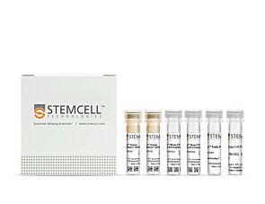
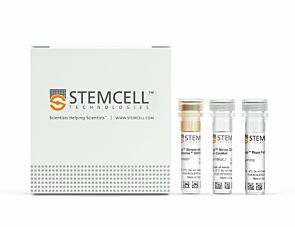
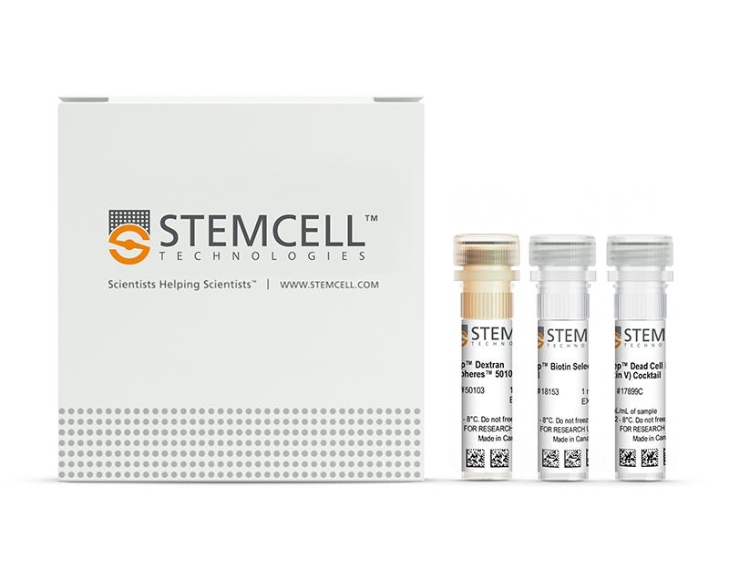
  • EasySep™ Dead Cell Removal (Annexin V) Kit (Catalog #17899)
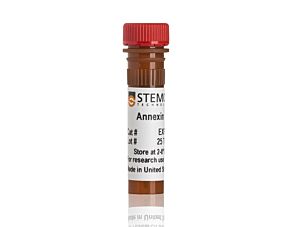
    • EasySep™ Dead Cell Removal (Annexin V) Cocktail, 0.5 mL
    • EasySep ™ Biotin Selection Cocktail, 1 mL
    • EasySep™ Dextran RapidSpheres™ 50103, 1 mL
 

Overview

Efficiently deplete apoptotic (Annexin V+) cells from cell culture or tissue preparation samples by immunomagnetic negative selection, with the EasySep™ Dead Cell Removal (Annexin V) Kit. Widely used in published research for more than 20 years, EasySep™ combines the specificity of monoclonal antibodies with the simplicity of a column-free magnetic system.

This straightforward, optimized EasySep™ procedure involves labeling cells with antibody complexes recognizing Annexin V and magnetic particles. Labeled cells are separated from untouched cells using an EasySep™ magnet and by simply pouring or pipetting off the unlabeled cells. The Annexin V+ cells remain in the tube. Following magnetic cell isolation, desired cells are ready for downstream applications. Annexin V binds phosphatidylserine on the outer leaflet of the cell membrane during apoptosis.

Learn more about how immunomagnetic EasySep™ technology works. Explore additional products optimized for your workflow, including culture media, supplements, antibodies, and more.
Magnet Compatibility
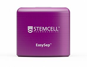
• EasySep™ Magnet (Catalog #18000), or
• “The Big Easy” EasySep™ Magnet (Catalog #18001), or
• EasyEights™ EasySep™ Magnet (Catalog #18103)
 
Subtype
Cell Isolation Kits
Cell Type
Other
Species
Human, Mouse, Non-Human Primate, Other, Rat
Sample Source
Other
Selection Method
Depletion
Brand
EasySep
Area of Interest
Immunology

Data Figures

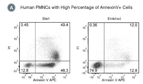
Figure 1. Typical Profile for Dead Cell Removal from Human PMNCs Using EasySep™ Dead Cell Removal (Annexin V) Kit

Starting with human polymorphonuclear cells (PMNCs) cultured overnight, the live cell content (AnnexinV-/PI-) of the enriched fraction is typically 69.7± 12.5% (mean ± SD), using the purple EasySep™ Magnet. In the above example, the percentages of live cells in the start and final enriched fractions are 12.8% and 74.9%, respectively.

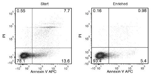
Figure 2. Typical Profile for Dead Cell Removal from Mouse Splenocytes Using EasySep™ Dead Cell Removal (Annexin V) Kit

Starting with 24- to 48-hour-old mouse splenocytes, the live cell content of the enriched fraction is typically 79.8 ± 11.4% (mean ± SD), using the purple EasySep™ Magnet. In the above example, the percentages of live cells in the start and final enriched fractions are 78.1% and 93.4%, respectively.

Protocols and Documentation

Find supporting information and directions for use in the Product Information Sheet or explore additional protocols below.

Document Type
Product Name
Catalog #
Lot #
Language
Catalog #
17899
Lot #
All
Language
English
Document Type
Safety Data Sheet 1
Catalog #
17899
Lot #
All
Language
English
Document Type
Safety Data Sheet 2
Catalog #
17899
Lot #
All
Language
English
Document Type
Safety Data Sheet 3
Catalog #
17899
Lot #
All
Language
English

Resources and Publications

Publications (26)

Optimized patient-derived lung cancer organoids recapitulating the immune landscape for precision therapy evaluation H. Xing et al. Cellular Oncology (Dordrecht, Netherlands) 2026 Jan

Abstract

Purpose: Current lung cancer organoid models often fail to replicate the complex tumor immune microenvironment, reducing their predictive value for immunotherapy and radiotherapy. Therefore, it is crucial to establish an optimized lung cancer organoid model which could recapitulate the tumor immune microenvironment, enabling more accurate evaluation of therapeutic responses. Methods: We developed an optimized air-liquid interface (ALI) culture method to generate patient-derived lung cancer organoids (ALI-LUOs) from 19 lung cancer samples. The tumor microenvironment, including immune and stromal components, was characterized using immunofluorescence, flow cytometry, and single-cell RNA sequencing. The organoids were further used to assess responses to αPD-1 therapy and radiotherapy. Results: The optimized method significantly improved organoid formation efficiency while preserving immune cell viability for up to 30 days. Immune and fibroblast populations were confirmed by immunofluorescence and flow cytometry. Single-cell RNA sequencing demonstrated that ALI-LUOs accurately replicate the tumor immune landscape. Key tumor immunity pathways such as cGAS-STING could be captured by ALI-LUOs. Importantly, ALI-LUOs modeled clinical responses to immune checkpoint inhibitors and radiotherapy with high fidelity. Conclusions: The ALI-LUOs, developed through an optimized culture method, faithfully capture the key characteristics of lung cancer, including its immunosuppressive tumor microenvironment. Our findings highlight this modified ALI-LUOs as a valuable preclinical platform for evaluating antitumor immunity and refining lung cancer treatments.
ArchetypeShift: An R Package Integrating KEGG-Informed Pathway Analysis and IPA-Derived Functional Predictions for Validation of Single-Cell Archetypes. A. Wilson et al. Bioinformatics and biology insights 2026 Aug

Abstract

Functional archetype analysis of single-cell RNA-sequencing (scRNA-seq) data is important because clustering of cell types is often nuanced and inexact. Intermediate phenotypes exist that are difficult to account for in these analyses, particularly in the setting of infant development. Further, different cell types work together to achieve biological functions, thereby broadly supporting tissue function at homeostasis and through environmental challenges via phenotypic plasticity. Currently, the process of assigning archetypes to cell clusters is labor-intensive because annotation requires manual upload of expression data to multiple programs. ArchetypeShift is an R-based pipeline that integrates existing archetypal analysis methods with annotation by Ingenuity Pathway Analysis (IPA) and Kyoto Encyclopedia of Genes and Genomes (KEGG), graphics visualization, and trajectory analysis for scRNA-seq data. Generating IPA- and/or KEGG-informed dot plots, UMAPs (uniform manifold approximation and projections) of archetype weights, archetype maps, heatmaps defining the top genes for each archetype program, and trajectory analysis graphics, ArchetypeShift streamlines the biological interpretation of archetype programs within a unified and efficient analytical framework. The source code for ArchetypeShift is available on GitHub (https://github.com/Neo-NEC-Lab/ArchetypeShift).
Human microglia differentially respond to β‐amyloid, tau, and combined Alzheimer's disease pathologies in vivo M. Coburn et al. Alzheimer's & Dementia 2025 Nov

Abstract

AbstractINTRODUCTIONRecent studies have identified important species‐dependent differences in the response of microglia to β‐amyloid (Aβ) pathology. Yet, whether human microglia also interact differently with the pathognomonic combination of amyloid and tau pathologies that occur in Alzheimer's disease (AD) remains unclear.METHODSWe generated a xenotolerant mouse model of AD that develops both plaque and tangle pathologies, transplanted stem cell‐derived microglial progenitors and examined the interactions between human microglia and AD pathologies with scRNA sequencing, immunohistochemistry, and in vitro modeling.RESULTSThe combined amyloid and tau pathologies induced robust type‐I interferon and proinflammatory cytokine responses, as well as an increased adoption of a distinct “rod” morphology in human microglia. The rod morphology could be induced with type‐I interferon treatment in vitro.DISCUSSIONWe provide new insights into human microglial responses to combined AD pathologies and a novel platform to investigate and manipulate human microglia in vivo.Highlights Amyloid pathology promotes the rapid development of neurofibrillary tangles and neuronal loss in a novel chimeric model of AD.Combined Alzheimer's disease pathologies lead to an expansion of disease‐associated microglia (DAM) and exacerbate Interferon‐responsive and cytokine/chemokine‐enriched states in xenotransplanted human microglia.The combination of amyloid and tau promotes the development of a distinctive rod microglial phenotype that closely correlates with tau pathology and neurodegeneration.Rod morphology and transcriptional changes can be modeled in vitro by treatment of induced pluripotent stem cells (iPSC) ‐microglia with type‐I interferons.