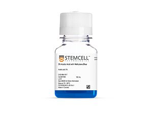
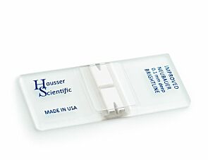
Before performing cell isolation using EasySep™, consult the product information sheet (PIS) to determine whether red blood cell (RBC) lysis is required for your sample type. RBC lysis should only be performed if indicated in the PIS. It is often recommended for blood samples; however, RBC lysis is not recommended for mouse splenocytes as it may reduce cell recovery. For the most accurate cell recovery calculation, we recommend performing total nucleated cell (TNC) count.
Thank you for your interest in this product.
Please provide us with your contact information and your local representative
will contact you with a customized quote. Where appropriate, they can also assist you with a(n):
Estimated delivery time for your area
Product sample or exclusive offer
In-lab demonstration
By submitting this form, you are providing your consent to STEMCELL Technologies Canada Inc. and its subsidiaries and affiliates (“STEMCELL”) to collect and use your information, and send you newsletters and emails in accordance with our privacy policy. Please contact us with any questions that you may have. You can unsubscribe or change your email preferences at any time.
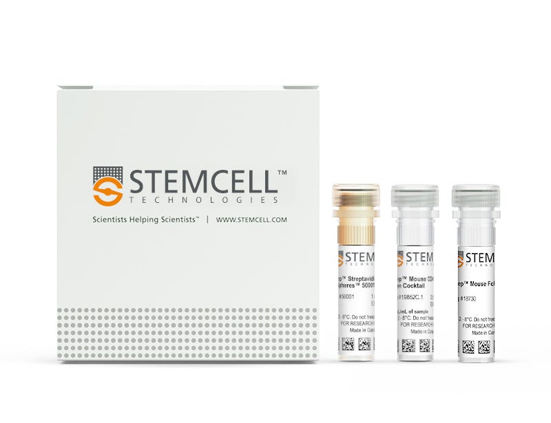
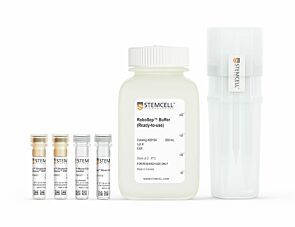
Easily and efficiently isolate highly purified mouse CD4+ T cells from single-cell suspensions of splenocytes or other tissues by immunomagnetic negative selection, with the EasySep™ Mouse CD4+ T Cell Isolation Kit. Widely used in published research for more than 20 years, EasySep™ combines the specificity of monoclonal antibodies with the simplicity of a column-free magnetic system.
In this EasySep™ negative selection procedure, unwanted cells are labeled with antibody complexes and magnetic particles. Unwanted cells expressing the following markers are targeted for removal: CD11b, CD45R, Ter119, CD8a, CD49b, CD19, CD11c, TCRgd and CD24. The magnetically labeled cells are then separated from the untouched desired mouse CD4+ T cells by using an EasySep™ magnet and simply pouring or pipetting the desired cells into a new tube. Following magnetic cell isolation in as little as 15 minutes, the desired CD4+ T cells are ready for downstream applications such as flow cytometry, culture, and cell-based experiments.
Learn more about how immunomagnetic EasySep™ technology works or how to fully automate immunomagnetic cell isolation with RoboSep™. Explore additional products optimized for your workflow, including culture media, supplements, antibodies, and more.
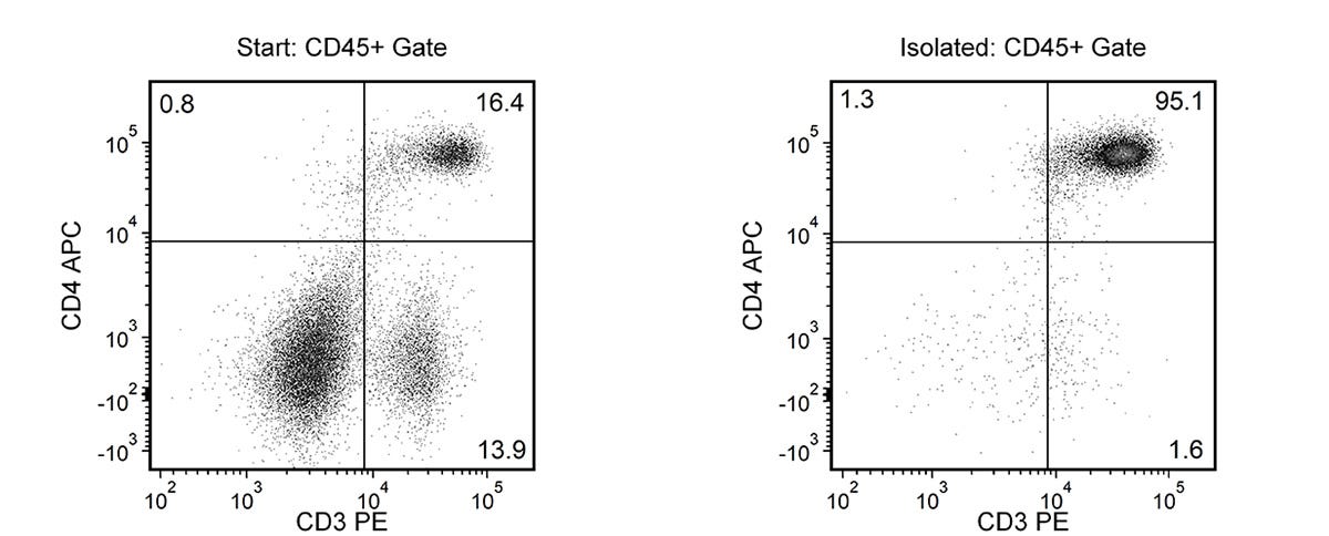
Figure 1. Typical EasySep™ Mouse CD4+ T Cell Isolation Profile
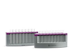
Starting with mouse splenocytes, the CD4+ T cell content (CD3+CD4+) of the isolated fraction is 95.4 ± 3% (mean ± SD), using the purple EasySep™ magnet.
This product is designed for use in the following research area(s) as part
of the highlighted workflow stage(s). Explore these workflows to learn more about the other products we
offer to support each research area.
Can EasySep™ Streptavidin RapidSpheres™ be used for either positive or negative selection?
Currently, EasySep™ Streptavidin RapidSphere™ kits are only available for negative selection and work by targeting and removing unwanted cells.
How does the separation work?
Streptavidin RapidSphere™ magnetic particles are crosslinked to unwanted cells using biotinylated antibodies. When placed in the EasySep™ Magnet, labeled cells migrate to the wall of the tube. The unlabeled cells are then poured off into a new tube.
Which columns do I use?
The EasySep™ procedure is column-free. That's right - no columns!
How can I analyze the purity of my enriched sample?
The Product Information Sheet provided with each EasySep™ kit contains detailed staining information.
Can EasySep™ Streptavidin RapidSphere™ separations be automated?
Yes. RoboSep™, the fully automated cell separator, automates all EasySep™ labeling and cell separation steps.
Are cells isolated using EasySep™ RapidSphere™ products FACS-compatible?
Yes. Desired cells are unlabeled and ready to use in downstream applications, such as FACS analysis.
Can I alter the separation time in the magnet?
Yes; however, this may impact the kit's performance. The provided EasySep™ protocols have already been optimized to balance purity, recovery and time spent on the isolation.
IL-2 mutein promotes antigen-specific transplant acceptance in mice through expansion of ST2+ regulatory T cells.
Y. Ganchiku et al.
Nature communications 2026 Jun
Abstract
Although transplantation is the preferred treatment for end-stage organ disease, long-term outcomes are limited by immunosuppressive drug toxicity and immune-mediated injury. Selective in vivo expansion of regulatory T cells (Tregs) using interleukin-2 (IL-2) analogs has emerged as a strategy to induce antigen-specific transplant tolerance with fewer side effects. Herein, we investigate the therapeutic efficacy of an IL-2 mutein molecule (mIL-2) with enhanced receptor specificity and extended half-life in murine models of solid organ transplantation. mIL-2 therapy significantly improves allograft survival in an antigen-specific manner, accompanied by increased Treg activation, decreased effector T cell activation and reduced donor-specific antibody production. Transcriptional profiling reveals expansion of Tregs expressing the suppression of tumorigenicity 2 (ST2) Tregs with heightened activation status and suppressive function. Accordingly, the mIL-2-induced long-term allograft survival is abrogated in Treg-specific ST2 knockout graft recipients, underscoring the critical role of ST2+ Tregs. These findings identify mIL-2 as an approach to promote long-term transplant tolerance while reducing reliance on conventional immunosuppression.
Nanobioconjugate Trispecific Antibody Augments Antitumor Immunity of Triple Negative Breast Cancer.
Q. Chang and L. Liu
International journal of nanomedicine 2026 Jun
Abstract
INTRODUCTION: Triple-negative breast cancer (TNBC) is characterized as the most unfavorable prognosis of the breast cancer subtype. Chemotherapy is currently the primary treatment owing to a persistent lack of effective alternative medicine. To fulfill this unaddressed clinical requirement, we utilized the PGLU-Fc-III-4C MsAb platform to develop a nanobioconjugate trispecific antibody (TsAb; CD3×CD137×CD276) that targeted CD3, CD137, and CD276, aiming to restrict the growth of TNBC tumors (4T1 model) and provide a novel therapeutic strategy. METHODS: The capacity for binding to target cells and anti-tumor effects of TsAb in vitro were evaluated. In vivo antitumor efficacy and biosafety were further assessed in the 4T1 subcutaneous tumor model. Anti-tumor immune responses induced by TsAb on tumor-infiltrating CD8+ T cells were monitored. RESULTS: The TsAb effectively bound and facilitated interactions between 4T1 tumor cells and T cells, significantly boosting the anti-tumor effect. TsAb promoted the expansion of tumor and splenic T lymphocytes and facilitated the recruitment of splenic and blood T lymphocytes to tumor tissues. Compared with the PGLU-Fc-III-4C-IgG treatment group, the TsAb treatment group had varying degrees of increase in CD8+ tissue-resident memory T cells (TRM), central memory T cells (TCM), and terminal effector memory T cells (TEM) in tumor tissues. The TsAb treatment group exhibited a significant increase of PD-1+ CD8+ T cells and TCF1-Tim-3+ CD8+ terminally exhausted T cells in tumor tissues. The safety profile demonstrated no obvious systemic toxicity. DISCUSSION: Briefly, TsAb mediated CD8+ T cell activation, proliferation, and terminal differentiation, accompanied by increasing cytokine production to eliminate the tumor. Meanwhile, no obvious systemic toxicity was observed. In general, the CD3×CD137×CD276 nanobioconjugate trispecific antibody provides a promising immunotherapeutic approach via regulating CD8+ T cell immune response for the treatment of triple-negative breast cancer.
Rapid and Uniform NHS-Ester-Based Membrane Protein Labeling of Live Mammalian Cells
A. Burgess et al.
Bio-protocol 2025 Oct
Abstract
Rapid and uniform labeling of plasma membrane proteins is essential for high-resolution imaging of dynamic membrane topologies and intercellular communication in live mammalian cells. Existing strategies for labeling live cell membranes, such as fluorescent fusion proteins, enzyme-mediated tags, metabolic bioorthogonal labeling, and lipophilic dyes, face trade-offs in the requirement of genetic manipulation, the presence of non-uniform labeling, the need for extensive preparation times, and limited choices of fluorophores. Here, we present a streamlined protocol that leverages N-hydroxysuccinimide (NHS)-ester chemistry to achieve rapid (≤5 min), covalent conjugation of synthetic small-molecule dyes to surface-exposed primary amines, enabling pan-membrane-protein labeling. This workflow covers dye stock preparation, labeling for suspension and adherent cells, multiplex live-cell imaging, fusion protein co-staining (including insulin-triggered receptor endocytosis), 3D membrane visualization, and in vivo assays for visualizing membrane-derived material transfers between donor and recipient cells using a lymphoma T-cell mouse model. This high-density labeling approach is compatible with various cell types across diverse imaging platforms. Its speed, versatility, and stability make it a broadly applicable tool for studying plasma membrane dynamics and intercellular membrane trafficking. Key features • Rapid high-density membrane labeling with small-molecule fluorescent dyes.• Enables live-cell multiplexed imaging, amenable to primary cells and cells expressing fluorescent fusion proteins, and supports in vivo studies of membrane-associated cell–cell communications.• Compatible with various fluorescence imaging modalities.
Before performing cell isolation using EasySep™, consult the product information sheet (PIS) to determine whether red blood cell (RBC) lysis is required for your sample type. RBC lysis should only be performed if indicated in the PIS. It is often recommended for blood samples; however, RBC lysis is not recommended for mouse splenocytes as it may reduce cell recovery. For the most accurate cell recovery calculation, we recommend performing total nucleated cell (TNC) count.
PRODUCTS ARE FOR RESEARCH USE ONLY AND NOT INTENDED FOR HUMAN OR ANIMAL DIAGNOSTIC OR THERAPEUTIC USES UNLESS OTHERWISE STATED. FOR ADDITIONAL INFORMATION ON QUALITY AT STEMCELL, REFER TO WWW.STEMCELL.COM/COMPLIANCE.