EasySep™ Mouse CD8+ T Cell Isolation Kit

Immunomagnetic negative isolation of untouched mouse CD8+ T cells
Before performing cell isolation using EasySep™, consult the product information sheet (PIS) to determine whether red blood cell (RBC) lysis is required for your sample type. RBC lysis should only be performed if indicated in the PIS. It is often recommended for blood samples; however, RBC lysis is not recommended for mouse splenocytes as it may reduce cell recovery. For the most accurate cell recovery calculation, we recommend performing total nucleated cell (TNC) count.

See How to Calculate Cell Recovery & Measure Purity After Cell Isolation >

EasySep™ Mouse CD8+ T Cell Isolation Kit

Immunomagnetic negative isolation of untouched mouse CD8+ T cells

From: 806 USD
Catalog #
(Select a product)
Immunomagnetic negative isolation of untouched mouse CD8+ T cells
Add to Wish List

Product Advantages


  • Fast and easy-to-use

  • Up to 95% purity

  • No columns required

  • Untouched, viable cells

What's Included

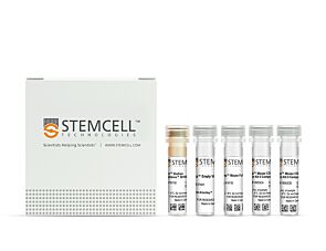
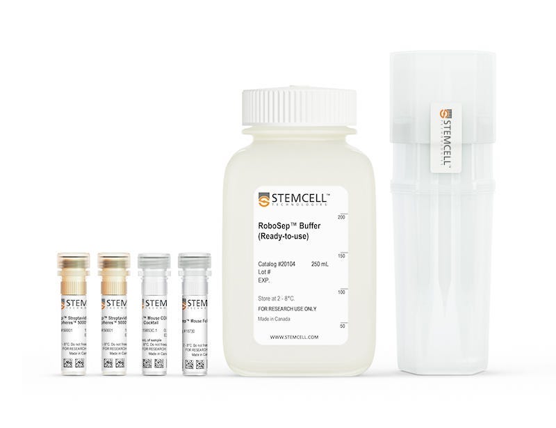
  • EasySep™ Mouse CD8+ T Cell Isolation Kit (Catalog #19853)
    • EasySep™ Mouse CD8+ T Cell Isolation Cocktail, 0.5 mL
    • EasySep™ Streptavidin RapidSpheres™ 50001, 2 x 1 mL
    • EasySep™ FcR Mouse Blocker, 0.2 mL
  • RoboSep™ Mouse CD8+ T Cell Isolation Kit (Catalog #19853RF)
    • EasySep™ Mouse CD8+ T Cell Isolation Cocktail, 0.5 mL
    • EasySep™ Streptavidin RapidSpheres™ 50001, 2 x 1 mL
    • EasySep™ FcR Mouse Blocker, 0.2 mL
    • RoboSep™ Buffer (Catalog #20104)
    • RoboSep™ Filter Tips (Catalog #20125)
Products for Your Protocol
To see all required products for your protocol, please consult the Protocols and Documentation.

Overview

Easily and efficiently isolate highly purified mouse CD8+ T cells from single-cell suspensions of splenocytes or other tissues by immunomagnetic negative selection, with the EasySep™ Mouse CD8+ T Cell Isolation Kit. Widely used in published research for more than 20 years, EasySep™ combines the specificity of monoclonal antibodies with the simplicity of a column-free magnetic system.

In this EasySep™ negative selection procedure, unwanted cells are labeled with antibody complexes and magnetic particles. Unwanted cells expressing the following markers are targeted for removal: CD11b, CD45R, Ter119, CD4, CD49b, CD19, CD11c, TCRgd and CD24. The magnetically labeled cells are then separated from the untouched desired mouse CD8+ T cells by using an EasySep™ magnet and simply pouring or pipetting the desired cells into a new tube. Following magnetic cell isolation in as little as 17.5 minutes, the desired CD8+ T cells are ready for downstream applications such as flow cytometry, culture, and cell-based experiments.

This product replaces the EasySep™ Mouse CD8+ T Cell Enrichment Kit (Catalog #19753) for even faster cell isolations.

Learn more about how immunomagnetic EasySep™ technology works or how to fully automate immunomagnetic cell isolation with RoboSep™. Explore additional products optimized for your workflow, including culture media, supplements, antibodies, and more.
Magnet Compatibility
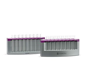
• EasySep™ Magnet (Catalog #18000)
• “The Big Easy” EasySep™ Magnet (Catalog #18001)
• EasyPlate™ EasySep™ Magnet (Catalog 18102)
• EasyEights™ EasySep™ Magnet (Catalog #18103)
• RoboSep™-S (Catalog #21000)
Subtype
Cell Isolation Kits
Cell Type
T Cells, T Cells, CD8+
Species
Mouse
Sample Source
Other, Spleen
Selection Method
Negative
Application
Cell Isolation
Brand
EasySep, RoboSep
Area of Interest
Immunology

Data Figures

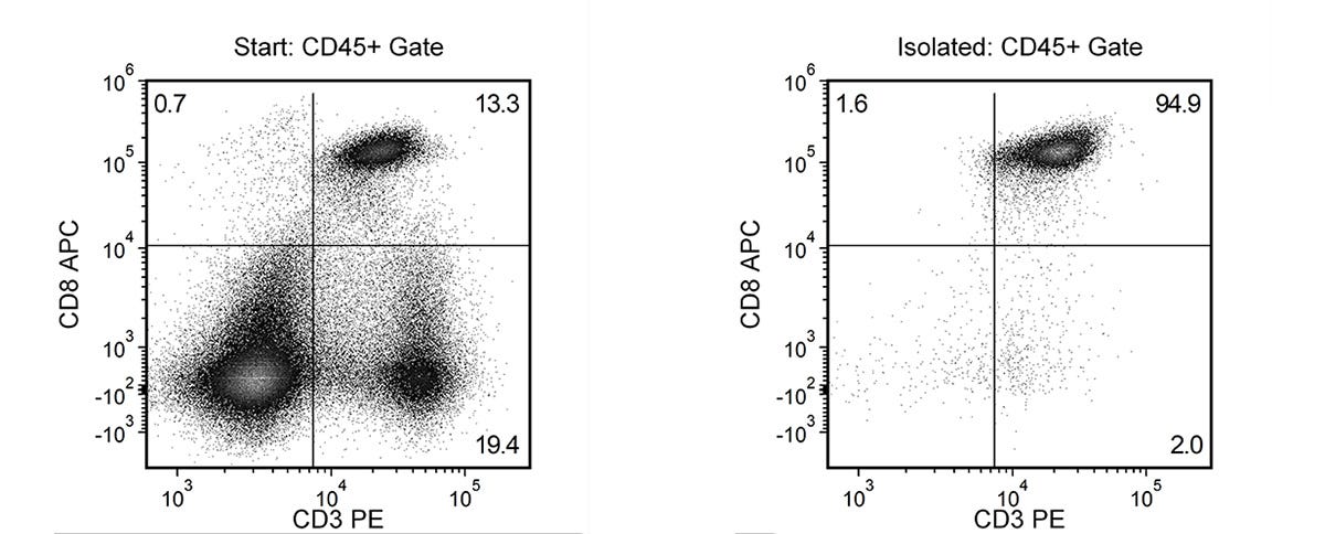
Typical EasySep™ Mouse CD8+ T Cell Isolation Profile

Figure 1. Typical EasySep™ Mouse CD8+ T Cell Isolation Profile

Starting with mouse splenocytes, the CD8+ T cell content (CD3+CD8+) of the isolated fraction is 94.4 ± 0.7% (mean ± SD), using the purple EasySep™ Magnet.

Protocols and Documentation

Find supporting information and directions for use in the Product Information Sheet or explore additional protocols below.

Document Type
Product Name
Catalog #
Lot #
Language
Catalog #
19853RF
Lot #
1000120417 or higher
Language
English
Catalog #
19853
Lot #
1000120417 or higher
Language
English
Document Type
Safety Data Sheet 1
Catalog #
19853RF
Lot #
All
Language
English
Document Type
Safety Data Sheet 2
Catalog #
19853RF
Lot #
All
Language
English
Document Type
Safety Data Sheet 3
Catalog #
19853RF
Lot #
All
Language
English
Document Type
Safety Data Sheet 4
Catalog #
19853RF
Lot #
All
Language
English
Document Type
Safety Data Sheet 5
Catalog #
19853RF
Lot #
All
Language
English
Document Type
Safety Data Sheet 1
Catalog #
19853
Lot #
All
Language
English
Document Type
Safety Data Sheet 2
Catalog #
19853
Lot #
All
Language
English
Document Type
Safety Data Sheet 3
Catalog #
19853
Lot #
All
Language
English
Document Type
Safety Data Sheet 4
Catalog #
19853
Lot #
All
Language
English

Applications

This product is designed for use in the following research area(s) as part of the highlighted workflow stage(s). Explore these workflows to learn more about the other products we offer to support each research area.

Resources and Publications

Frequently Asked Questions

Can EasySep™ Streptavidin RapidSpheres™ be used for either positive or negative selection?

Currently, EasySep™ Streptavidin RapidSphere™ kits are only available for negative selection and work by targeting and removing unwanted cells.

How does the separation work?

Streptavidin RapidSphere™ magnetic particles are crosslinked to unwanted cells using biotinylated antibodies. When placed in the EasySep™ Magnet, labeled cells migrate to the wall of the tube. The unlabeled cells are then poured off into a new tube.

Which columns do I use?

The EasySep™ procedure is column-free. That's right - no columns!

How can I analyze the purity of my enriched sample?

The Product Information Sheet provided with each EasySep™ kit contains detailed staining information.

Can EasySep™ Streptavidin RapidSphere™ separations be automated?

Yes. RoboSep™, the fully automated cell separator, automates all EasySep™ labeling and cell separation steps.

Are cells isolated using EasySep™ RapidSphere™ products FACS-compatible?

Yes. Desired cells are unlabeled and ready to use in downstream applications, such as FACS analysis.

Can I alter the separation time in the magnet?

Yes; however, this may impact the kit's performance. The provided EasySep™ protocols have already been optimized to balance purity, recovery and time spent on the isolation.

Publications (107)

PRECISE-seq reveals disease-relevant TCR repertoires with phenotypic plasticity S. Liu et al. The Journal of Experimental Medicine 2026 May

Abstract

Liu et al. develop PRECISE-seq, a single-cell platform linking TCR specificity and avidity to T cell phenotypes in vivo. They reveal that high-potency antiviral T cells become exhausted, while tumor-reactive CD8+ T cells acquire a regulatory Ly49+ state that is restrained by PD-1 blockade, resulting in effector revival within tumors. Linking T cell phenotypes with antigen specificity and functional avidity is critical for understanding in vivo immune responses in infection and cancer. Here, we develop PRECISE-seq, a method that integrates multi-omics T cell analysis with contact-dependent proximity labeling for rapid screening of disease-relevant T cell repertoires, and for linking the relative TCR avidity with T cell phenotypes at single-cell resolution. PRECISE-seq accurately retrieves CMV-specific clonotypes from human peripheral blood and quantitatively measures functional avidity in physiological contexts. We find that high-potency CMV-specific T cells preferentially acquire an exhausted phenotype. In tumors, polyclonal tumor-reactive CD8+ T cells predominantly differentiate into a protumor Ly49+ regulatory state (TLy49), characterized by inhibitory killer cell lectin-like receptor expression and originating from effector memory T cells along a trajectory distinct from exhaustion. Notably, PD-1 blockade reduces TLy49 formation and promotes effector revival, which correlates with responsiveness to immunotherapy. Together, PRECISE-seq enables high-resolution mapping of TCR potency and T cell phenotype, revealing a regulatory axis shaping T cell fate in tumors. Graphical Abstract A multi-part diagram shows workflow linking T-cell antigen specificity, phenotype, PD-1 response mechanisms, and prediction of clinical outcomes.Panel A: An illustration shows the identification of antigen specificity. It includes a depiction of CD 8 cells interacting with antigens and labeled with biotin. Single-cell RNA sequencing (scRNA-seq) and single-cell TCR sequencing (scTCR-seq) are illustrated with colored bars representing different gene segments. Panel B: A UMAP plot shows the phenotype and TCR potency. The plot is labeled with UMAP_1 and UMAP_2 axes and shows clusters of cells with different TCR potency and exhaustion scores. Panel C: A diagram illustrates the Ly49 plus regulatory state. It shows immune checkpoints, NK receptors, SPP1, granzyme C, and Ly49 genes interacting with a cell. Panel D: A diagram depicting the mechanism of discovery of alpha PD-1 therapy. It shows different types of T cells (T subscript EM, T subscript EFF, T subscript EX, T subscript Ly49) and their responses to alpha PD-1 therapy. Panel E: A diagram predicts clinical response. It shows two figures representing responders and non-responders based on the levels of T subscript EM, T subscript EFF, and T subscript Ly49/KIR cells.
Nanobioconjugate Trispecific Antibody Augments Antitumor Immunity of Triple Negative Breast Cancer. Q. Chang and L. Liu International journal of nanomedicine 2026 Jun

Abstract

INTRODUCTION: Triple-negative breast cancer (TNBC) is characterized as the most unfavorable prognosis of the breast cancer subtype. Chemotherapy is currently the primary treatment owing to a persistent lack of effective alternative medicine. To fulfill this unaddressed clinical requirement, we utilized the PGLU-Fc-III-4C MsAb platform to develop a nanobioconjugate trispecific antibody (TsAb; CD3×CD137×CD276) that targeted CD3, CD137, and CD276, aiming to restrict the growth of TNBC tumors (4T1 model) and provide a novel therapeutic strategy. METHODS: The capacity for binding to target cells and anti-tumor effects of TsAb in vitro were evaluated. In vivo antitumor efficacy and biosafety were further assessed in the 4T1 subcutaneous tumor model. Anti-tumor immune responses induced by TsAb on tumor-infiltrating CD8+ T cells were monitored. RESULTS: The TsAb effectively bound and facilitated interactions between 4T1 tumor cells and T cells, significantly boosting the anti-tumor effect. TsAb promoted the expansion of tumor and splenic T lymphocytes and facilitated the recruitment of splenic and blood T lymphocytes to tumor tissues. Compared with the PGLU-Fc-III-4C-IgG treatment group, the TsAb treatment group had varying degrees of increase in CD8+ tissue-resident memory T cells (TRM), central memory T cells (TCM), and terminal effector memory T cells (TEM) in tumor tissues. The TsAb treatment group exhibited a significant increase of PD-1+ CD8+ T cells and TCF1-Tim-3+ CD8+ terminally exhausted T cells in tumor tissues. The safety profile demonstrated no obvious systemic toxicity. DISCUSSION: Briefly, TsAb mediated CD8+ T cell activation, proliferation, and terminal differentiation, accompanied by increasing cytokine production to eliminate the tumor. Meanwhile, no obvious systemic toxicity was observed. In general, the CD3×CD137×CD276 nanobioconjugate trispecific antibody provides a promising immunotherapeutic approach via regulating CD8+ T cell immune response for the treatment of triple-negative breast cancer.
A Circulating GPNMB-Based Multimodal Model Integrates Tumor-Immune Crosstalk to Predict Immunotherapy Response in Esophageal Cancer. L. Zhu et al. Cancer discovery 2026 Jul

Abstract

UNLABELLED: Neoadjuvant immunotherapy improves outcomes in esophageal squamous cell carcinoma (ESCC), yet ∼70% of patients fail to respond. Pretreatment biopsies and plasma provide critical opportunities for biomarker discovery. In this study, we performed plasma proteomic profiling and identified soluble glycoprotein nonmetastatic melanoma protein B (sGPNMB) as the most elevated circulating protein in nonresponders. Mechanistically, tumor cell-derived sGPNMB suppressed CD8+ T-cell receptor signaling via the SDC4-CD148 axis to induce functional exhaustion, with secretion being required for its immunosuppressive activity. Cancer-associated fibroblast-epithelial (CAF-Epi) niches promoted SOX2 upregulation in tumor cells, transcriptionally activating GPNMB expression. In humanized patient-derived xenograft models, circulating GPNMB levels predicted response to PD-1 blockade, and GPNMB inhibition synergized with therapy. Across retrospective cohorts and a prospective clinical trial, a multimodal model combining plasma GPNMB levels, CAF-Epi niche detection, and clinical-pathologic features achieved robust predictive accuracy for immunotherapy response and survival. These findings establish a spatial-circulating biomarker framework for precision ESCC immunotherapy. SIGNIFICANCE: Tumor-derived soluble GPNMB, transcriptionally activated by SOX2 within CAF-Epi niches, drives CD8+ T-cell exhaustion and resistance to PD-1 blockade in ESCC. Integrating circulating GPNMB levels with CAF-Epi niche features and clinical-pathologic factors, we develop and validate a clinically scalable multimodal model for predicting immunotherapy response.
Before performing cell isolation using EasySep™, consult the product information sheet (PIS) to determine whether red blood cell (RBC) lysis is required for your sample type. RBC lysis should only be performed if indicated in the PIS. It is often recommended for blood samples; however, RBC lysis is not recommended for mouse splenocytes as it may reduce cell recovery. For the most accurate cell recovery calculation, we recommend performing total nucleated cell (TNC) count.

See How to Calculate Cell Recovery & Measure Purity After Cell Isolation >