MesenCult™-ACF Chondrogenic Differentiation Kit

Animal component-free medium for the differentiation of MSCs into chondrocytes
MesenCult™-ACF Chondrogenic Differentiation Kit

Animal component-free medium for the differentiation of MSCs into chondrocytes

From: 360 USD
Catalog #
(Select a product)
Animal component-free medium for the differentiation of MSCs into chondrocytes
Add to Wish List

Product Advantages


  • Animal component-free (ACF) formulation

  • Robust chondrogenic differentiation with as few as 3 x 10^5 MSCs and as early as day 14

  • Strong expression of chondrogenic transcripts - Acan, Col2a, Sox9 and Col10a; low expression of hypertrophic transcript Mmp13

  • Completes optimized ACF workflow for MSC isolation, expansion, cryopreservation and chondrogenic differentiation

What's Included

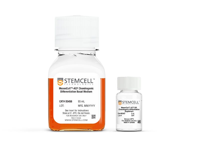
  • MesenCult™-ACF Chondrogenic Differentiation Basal Medium, 95 mL
  • MesenCult™-ACF 20X Chondrogenic Differentiation Supplement, 5 mL

Overview

Generate chondrogenic lineage cells, including chondrocytes from human mesenchymal stromal cells (MSCs) and hPSC-derived mesenchymal progenitors, using the MesenCult™-ACF Chondrogenic Differentiation Kit. This fully animal component-free (ACF) formulation enables efficient chondrogenic differentiation and matrix deposition starting with as few as 3 × 10⁵ cells. Generated chondrocytes will have detectable chondrogenesis as early as Day 14. The medium is compatible with MSCs derived from bone marrow, adipose tissue, or synovium and is suitable for cells expanded in serum-containing or ACF conditions.

Use this kit as part of a complete MSC functional assessment workflow. Pair with the MesenCult™ Adipogenic Differentiation Kit (Human) and MesenCult™ Osteogenic Differentiation Kit (Human) to evaluate trilineage differentiation potential and verify MSC identity and potency. These ready-to-use reagents enable reliable assessment of MSC identity and potency while ensuring reproducible lineage-specific outcomes across the complete MesenCult™ workflow.
Subtype
Specialized Media
Cell Type
Chondrocytes, Mesenchymal Stem and Progenitor Cells
Species
Human
Application
Cell Culture, Differentiation
Brand
MesenCult
Area of Interest
Drug Discovery and Toxicity Testing, Stem Cell Biology
Formulation Category
Animal Component-Free, Serum-Free

Data Figures

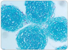
MesenCult™-ACF Chondrogenic Differentiation Medium Induces Robust Chondrogenic Differentiation of Human MSCs

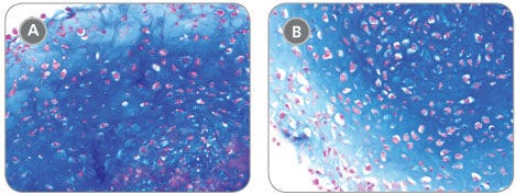
Figure 1. MesenCult™-ACF Chondrogenic Differentiation Medium Induces Robust Chondrogenic Differentiation of Human MSCs

Human BM-derived MSCs were cultured in MesenCult™-ACF Medium then differentiated to the chondrogenic lineage using MesenCult™-ACF Chondrogenic Differentiation Medium. Robust chondrogenic differentiation was observed (A) starting with as few as 3 x 10⁵ MSCs, or (B) when differentiating for just 14 days starting with 5 x 10⁵ MSCs.

Chondrogenic Differentiation of Human MSCs Is More Robust With Fewer Hypertrophic Chondrocytes Using MesenCult™-ACF Chondrogenic Differentiation Medium Compared to Competitor Media

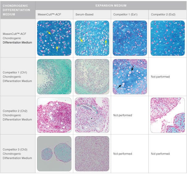
Figure 2. Chondrogenic Differentiation of Human MSCs Is More Robust With Fewer Hypertrophic Chondrocytes Using MesenCult™-ACF Chondrogenic Differentiation Medium Compared to Competitor Media

Human BM-derived MSCs culture-expanded for up to two passages in MesenCult™-ACF Medium, serum-based medium, or one of two commercially available media (Competitor 1 (Ex1) and Competitor 2 (Ex2)), were then differentiated to the chondrogenic lineage starting with 5 x 10⁵ MSCs and either using MesenCult™-ACF Chondrogenic Differentiation Medium or one of several commercially available chondrogenic differentiation media (Ch1, Ch2 or Ch3) for 21 days. More robust and uniform chondrogenic differentiation was observed when the MSCs were differentiated in MesenCult™-ACF Chondrogenic Differentiation Medium compared to the other commercially available chondrogenic differentiation media (Ch1, Ch2 and Ch3), irrespective of the expansion medium used to culture the MSCs prior to differentiation. Cultures differentiated using MesenCult™-ACF Chondrogenic Differentiation Medium displayed an abundance of isogenous groups (yellow arrows), suggesting there is proliferation of differentiating chondrocyte progenitors. Few hypertrophic chondrocytes (black arrows) are seen in cultures differentiated with MesenCult™-ACF Chondrogenic Differentiation Medium, suggesting the maintenance of chondrogenic activity throughout the culturing period.

MesenCult™-ACF Chondrogenic Differentiation Medium Induces Stronger and More Sustained Chondrogenic Transcript Expression Compared to Competitor Media

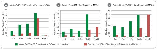
Figure 3. MesenCult™-ACF Chondrogenic Differentiation Medium Induces Stronger and More Sustained Chondrogenic Transcript Expression Compared to Competitor Media

Human BM-derived MSCs expanded in (A) MesenCult™-ACF Medium, (B) a serum-based medium or (C) Competitor 2 (Ex2) medium, were differentiated for 21 days with MesenCult™-ACF Chondrogenic Differentiation Medium and Competitor 2 (Ch2) chondrogenic differentiation medium. Regardless of the expansion medium initially used to culture the MSCs, differentiation using MesenCult™-ACF Chondrogenic Differentiation Medium led to a substantial up-regulation of the chondrogenic transcripts compared to Ch2. In addition, expression of the terminally-differentiated hypertrophic transcript Mmp13 was higher for Ch2 differentiated cultures compared to cultures differentiated with MesenCult™-ACF Chondrogenic Differentiation Medium.

Mouse MSCs Cultured in MesenCult™-ACF Chondrogenic Differentiation Medium Differentiate to Chondrocytes

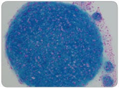
Figure 4. Mouse MSCs Cultured in MesenCult™-ACF Chondrogenic Differentiation Medium Differentiate to Chondrocytes

Mouse compact bone-derived MSCs were cultured using the MesenCult™ Proliferation Kit with MesenPure™ (Mouse, Catalog #05512) for 2 passages then differentiated by pellet culture with MesenCult™-ACF Chondrogenic Differentiation Medium for 21 days under normoxic (20% O2) conditions. Strong chondrogenic differentiation is indicated by dark-blue staining of the cartilage extracellular matrix and an abundance of isogenous chondrocyte groups.

Human MSC Chondrogenic Differentiation With AggreWell™800 Plates

Figure 5. Human MSC Chondrogenic Differentiation With AggreWell™800 Plates

Using centrifugation, 1 x 10^6 human MSCs were distributed evenly among 800 µm microwells in one well of an AggreWell™800 plate. Small aggregates of only ~3,300 cells per pellet were then differentiated to chondrocytes using MesenCult™-ACF Chondrogenic Differentiation Medium for 21 days under normoxic (20% O2) conditions.

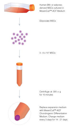
Procedure Overview: Human MSC Chondrogenic Differentiation Using MesenCult™-ACF Chondrogenic Differentiation Medium

Figure 6. Procedure Overview: Human MSC Chondrogenic Differentiation Using MesenCult™-ACF Chondrogenic Differentiation Medium

Aggregate culture is a useful method for inducing chondrogenic differentiation of human BM- and adipose-derived MSCs in a three-dimensional in vitro culture environment. MSCs are efficiently differentiated to the chondrogenic lineage using MesenCult™-ACF Chondrogenic Differentiation Medium in 14 - 21 days with 3 - 5 x 10⁵ cells.

Protocols and Documentation

Find supporting information and directions for use in the Product Information Sheet or explore additional protocols below.

Document Type
Product Name
Catalog #
Lot #
Language
Catalog #
05455
Lot #
All
Language
English
Document Type
Safety Data Sheet 1
Catalog #
05455
Lot #
All
Language
English
Document Type
Safety Data Sheet 2
Catalog #
05455
Lot #
All
Language
English

Applications

This product is designed for use in the following research area(s) as part of the highlighted workflow stage(s). Explore these workflows to learn more about the other products we offer to support each research area.

Resources and Publications

Publications (18)

Single-cell transcriptomics reveals critical role of CD74 in bone marrow-derived mesenchymal stem cells. Q. Xu et al. Stem cells translational medicine 2026 Sep

Abstract

BACKGROUND: Human bone marrow aspirates have been widely used for skeletal tissue regeneration. However, clinical outcomes of the tissue repair after transplantation are variable. METHODS: In this study, we analyzed the molecular signatures and osteogenic differentiation potential of bone marrow-derived mesenchymal stromal cells (BMSCs) from different donors. RESULTS: Our results clearly showed that BMSCs isolated from different patients exhibited different osteogenic capacities and growth rates. Moreover, single-cell RNA sequencing revealed that these BMSCs expressed elevated levels of genes associated with early osteogenesis, chondrogenesis, and adipogenesis. Differentially expressed genes related to cell cycles were enriched in clusters from all 5 different samples, suggesting these genes play an important role in BMSC maintenance and differentiation. More importantly, the BMSCs with the lowest osteogenic differentiation potential showed a dramatic decrease in the expression of cell surface protein CD74 when compared to other osteogenic genes. Knockdown of CD74 in BMSCs with high osteogenic potential significantly reduced their osteogenic capacity. CONCLUSIONS: These findings suggest that CD74 is a novel predictive marker of outcomes following BMSC transplantation. Furthermore, CD74-high BMSCs may represent a subpopulation of bone marrow cells responsible for bone marrow aspirate-mediated tissue regeneration.
Prednisolone attenuates the immunomodulatory capacity and reshapes the functional phenotype of canine adipose-derived mesenchymal stromal cells. Y. Yasumura et al. Regenerative therapy 2026 Dec

Abstract

INTRODUCTION: Mesenchymal stromal cells (MSCs) are a promising therapeutic option for refractory canine chronic inflammatory enteropathy (CIE), primarily because of their immunomodulatory properties. However, MSC function may be influenced by concomitant medications, including prednisolone. Therefore, we investigated the effects of prednisolone on canine adipose-derived MSCs (cADSCs), with particular focus on immunomodulatory capacity and migratory behavior. METHODS: Effects on the biological and functional properties of cADSCs were evaluated following treatment with prednisolone ranging from 0.01 to 100 μM. Morphology, senescence, apoptosis, proliferation, cell-cycle distribution, mitochondrial activity, migration, and invasion were assessed. Immunomodulatory function was assessed by co-culture assays using DH82 macrophages and canine peripheral blood mononuclear cells (PBMCs), including analyses of macrophage polarization, regulatory T-cell induction, T-cell subset composition, activation, and proliferation. RNA sequencing was performed to characterize transcriptomic changes in cADSCs treated with or without prednisolone and cultured with or without activated PBMCs. RESULTS: Prednisolone did not induce apparent morphological changes, senescence, or apoptosis in cADSCs, but altered proliferation, mitochondrial activity, and migratory behavior and attenuated key immunomodulatory functions. RNA sequencing revealed that prednisolone suppressed inflammatory signaling in cADSCs under basal conditions, whereas these cells retained robust inflammatory transcriptional responsiveness to signals derived from activated PBMC. CONCLUSIONS: Prednisolone reshapes cADSC function in a context-dependent manner, reducing immunomodulatory capacity without substantially impairing viability. These findings suggest that glucocorticoid exposure and inflammatory activation interact to modulate cADSC function and should be considered when optimizing MSC-based therapy for canine CIE.
Increased BMP/SMAD Signaling by PD-MSCs Promotes Bone Formation in an Ovariectomized Mouse Model of Osteoporosis D. Lee et al. International Journal of Molecular Sciences 2025 Oct

Abstract

Mesenchymal stem cells (MSCs) have emerged as a promising therapeutic approach for degenerative diseases due to their ability to modulate disease progression through paracrine mechanisms. Among various MSC sources, placenta-derived MSCs (PD-MSCs) offer significant advantages, including high proliferation capacity, reduced senescence, and low immunogenicity, making them ideal for allogeneic applications. In this study, we investigated the therapeutic effects of PD-MSC transplantation in an estrogen-deficiency-induced osteoporosis mouse model. Mice were divided into three groups: a normal control group, a non-transplanted osteoporosis group, and a PD-MSC-transplanted group. Our findings demonstrated that PD-MSC transplantation significantly improved osteoporosis-related parameters, including increased femur weight, bone volume, bone mineral density, and calcium deposition. Additionally, estrogen levels were elevated, bone formation markers were upregulated, and bone resorption markers were downregulated. PD-MSCs also reduced inflammatory cytokine levels while enhancing anti-inflammatory factors. Notably, the BMP/SMAD signaling pathway, crucial for bone formation, was significantly upregulated. These results suggest that PD-MSC transplantation effectively restores bone homeostasis by inhibiting osteoclast activity, promoting osteogenesis, and modulating inflammation. This study provides strong evidence supporting the potential of PD-MSCs as a novel therapeutic strategy for osteoporosis, offering a regenerative and anti-inflammatory approach to bone disease management.