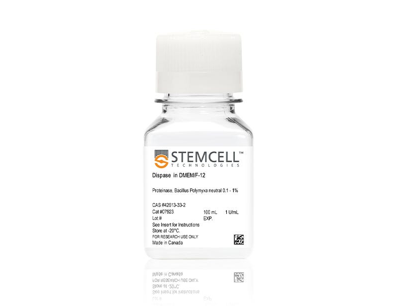
Dispase (1 U/mL)

1 U/mL dispase in DMEM/F-12

Dispase (1 U/mL)

1 U/mL dispase in DMEM/F-12

From: 74 USD
Catalog #
(Select a product)
1 U/mL dispase in DMEM/F-12
Add to Wish List

Product Advantages


  • Achieve gentle dissociation in a wide variety of tissues

  • Optimized for enzymatic passaging of human ES and iPS cells

Overview

Use Dispase (1 U/mL) for gentle dissociation of a wide variety of tissues. This proteolytic dissociation reagent has been optimized for use in enzymatic passaging of human embryonic stem (ES) cells and human induced pluripotent stem (iPS) cells. Incubation of minced tissue with pre-warmed dispase and gentle agitation will liberate cells with minimal cell damage. Pre-warmed dispase can also be used to harvest cells from tissue culture plastic.

This product contains 1 U/mL Dispase II (neutral protease from Bacillus polymyxa) dissolved in DMEM/F-12. Unlike trypsin, Dispase is not inhibited by serum. Dispase activity is inhibited by EDTA and EGTA. Dispase should be removed from cell suspensions by centrifuging the cells followed by washing the cells with buffer or culture medium.
Subtype
Enzymatic
Alternative Names
Neutral protease; Proteinase
Cell Type
Intestinal Cells, Mammary Cells, Other, Pluripotent Stem Cells, Prostate Cells
Species
Human, Mouse, Non-Human Primate, Other, Rat
Application
Cell Culture
Area of Interest
Epithelial Cell Biology, Stem Cell Biology

Protocols and Documentation

Find supporting information and directions for use in the Product Information Sheet or explore additional protocols below.

Document Type
Product Name
Catalog #
Lot #
Language
Product Name
Dispase (1 U/mL)
Catalog #
07923
Lot #
All
Language
English
Document Type
Safety Data Sheet
Product Name
Dispase (1 U/mL)
Catalog #
07923
Lot #
All
Language
English

Applications

This product is designed for use in the following research area(s) as part of the highlighted workflow stage(s). Explore these workflows to learn more about the other products we offer to support each research area.

Resources and Publications

Publications (72)

Directed differentiation of human embryonic stem cells into keratinocyte progenitors in vitro: an attempt with promise of clinical use. Li H et al. In vitro cellular & developmental biology. Animal 2016 SEP

Abstract

Human embryonic stem cells (hESCs) can differentiate into all somatic lineages including stratified squamous epithelia. Thus, efficient methods are required to direct hESC differentiation to obtain a pure subpopulation for tissue engineering. The study aimed to assess the effects of retinoic acid (RA), bone morphogenetic protein-4 (BMP4), and ascorbic acid (AA) on the differentiation of hESCs into keratinocyte progenitors in vitro. The first media contained AA and BMP4; the second contained RA, AA, and BMP4; the third was commercial-defined keratinocyte serum-free medium, which was used to differentiate H9 hESCs (direct approach) or embryoid bodies (EBs) (indirect approach) into keratinocyte progenitors. Real-time RT-PCR, immunofluorescence, and flow-cytometry were used to characterize the differentiated cells. Cells induced by AA + BMP4 + RA showed the typical epithelial morphology, while cells induced by AA + BMP4 showed multiple appearances. CK14 and p63 messenger RNA (mRNA) expressions in the AA + BMP4 + RA-treated cells were higher than those of the AA + BMP4-treated cells (CK14: 22.4-fold; p63: 84.7-fold). Epithelial marker CK18 mRNA expressions at 14 d of differentiation and keratinocyte marker CK14 and transcription factor p63 mRNA expressions at 35 d of differentiation were higher in cells differentiated from hESCs compared with those differentiated from EBs (CK18 10.51 ± 3.26 vs. 6.67 ± 1.28; CK14 9.27 ± 3.61 vs. 5.32 ± 1.86; p63 0.73 ± 0.06 vs. 0.44 ± 0.12, all P textless 0.05) After hESC induction by AA+BMP4+RA, CK14 mRNA expression was upregulated after day 21, peaking by 35 d of differentiation. Combined RA, BMP4, and AA could effectively induce differentiation of hESCs into keratinocyte progenitors in vitro. These keratinocytes could be used for oral mucosa and skin tissue engineering.
Efficient generation of endothelial cells from human pluripotent stem cells and characterization of their functional properties Song W et al. Journal of Biomedical Materials Research - Part A 2016 OCT

Abstract

Although endothelial cells (ECs) have been derived from human pluripotent stem cells (hPSCs), large-scale generation of hPSC-ECs remains challenging and their functions are not well characterized. Here we report a simple and efficient three-stage method that allows generation of approximately 98 and 9500 ECs on day 16 and day 34, respectively, from each human embryonic stem cell (hESC) input. The functional properties of hESC-ECs derived in the presence and absence of a TGF$$-inhibitory molecule SB431542 were characterized and compared with those of human umbilical vein endothelial cells (HUVECs). Confluent monolayers formed by SB431542(+) hESC-ECs, SB431542(-) hESC-ECs, and HUVECs showed similar permeability to 10,000 Da dextran, but these cells exhibited striking differences in forming tube-like structures in 3D fibrin gels. The SB431542(+) hESC-ECs were most potent in forming tube-like structures regardless of whether VEGF and bFGF were present in the medium; less potent SB431542(-) hESC-ECs and HUVECs responded differently to VEGF and bFGF, which significantly enhanced the ability of HUVECs to form tube-like structures but had little impact on SB431542(-) hESC-ECs. This study offers an efficient approach to large-scale hPSC-EC production and suggests that the phenotypes and functions of hPSC-ECs derived under different conditions need to be thoroughly examined before their use in technology development. This article is protected by copyright. All rights reserved.
Generation of induced pluripotent stem cells as a potential source of hematopoietic stem cells for transplant in PNH patients. Phondeechareon T et al. Annals of hematology 2016 OCT

Abstract

Paroxysmal nocturnal hemoglobinuria (PNH) is an acquired hemolytic anemia caused by lack of CD55 and CD59 on blood cell membrane leading to increased sensitivity of blood cells to complement. Hematopoietic stem cell transplantation (HSCT) is the only curative therapy for PNH, however, lack of HLA-matched donors and post-transplant complications are major concerns. Induced pluripotent stem cells (iPSCs) derived from patients are an attractive source for generating autologous HSCs to avoid adverse effects resulting from allogeneic HSCT. The disease involves only HSCs and their progeny; therefore, other tissues are not affected by the mutation and may be used to produce disease-free autologous HSCs. This study aimed to derive PNH patient-specific iPSCs from human dermal fibroblasts (HDFs), characterize and differentiate to hematopoietic cells using a feeder-free protocol. Analysis of CD55 and CD59 expression was performed before and after reprogramming, and hematopoietic differentiation. Patients' dermal fibroblasts expressed CD55 and CD59 at normal levels and the normal expression remained after reprogramming. The iPSCs derived from PNH patients had typical pluripotent properties and differentiation capacities with normal karyotype. After hematopoietic differentiation, the differentiated cells expressed early hematopoietic markers (CD34 and CD43) with normal CD59 expression. The iPSCs derived from HDFs of PNH patients have normal levels of CD55 and CD59 expression and hold promise as a potential source of HSCs for autologous transplantation to cure PNH patients.