How to Culture Human Pluripotent Stem Cell (hPSC)-Derived Forebrain Neurons for MEA Analysis Using the Maestro MEA™ System
Micro-electrode array (MEA) systems are powerful tools to measure activity from a population of cells simultaneously. They detect activity-associated voltage changes in the extracellular space around electrically active cells, such as neurons, that are grown on specialized cell culture plates containing a grid of electrodes in each well. By measuring activity from several electrodes simultaneously, researchers can characterize the activity of neuronal networks. Using MEAs to measure cellular activity is non-invasive and does not require cell labeling, allowing for long-term monitoring of electrophysiological maturation. This makes MEA systems ideal for both disease modeling and drug studies.
Because cells must cover the electrodes uniformly and consistently to produce high-fidelity data, culture substrates, and seeding densities must be optimized for each cell type. Read on for a step-by-step guide for culturing human pluripotent stem cell (hPSC)-derived forebrain neurons on CytoView MEA™ plates to produce mature, consistent, and stable neuronal activity using Maestro MEA™ systems.
Workflow Specifications
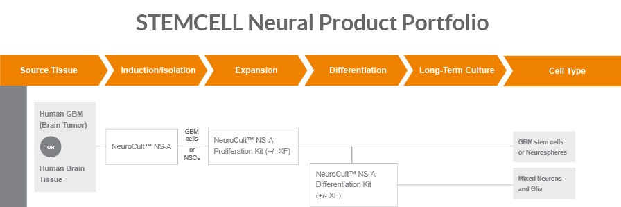
Obtaining hPSC-Derived Forebrain Neuron Precursor Cells
Important Note: For this protocol, cells must be seeded on the MEA plate at the “forebrain neuron precursor” stage. This stage corresponds to the time point after the differentiation stage and before the maturation stage of the STEMdiff™ Forebrain Neuron workflow (see below). Forebrain neuron precursor cells cannot be further dissociated or replated once seeded on MEA plates in STEMdiff™ Forebrain Neuron Maturation Medium.
There are multiple options for generating or sourcing forebrain neuron precursor cells:
- Start with a high-quality hPSC line such as Healthy Control Human iPSC Line, Female, SCTi003-A (Catalog #200-0511) or Healthy Control Human iPSC Line, Male, SCTi004-A (Catalog #200-0769).
- Use STEMdiff™ SMADi Neural Induction Kit (Catalog #08581) followed by STEMdiff™ Forebrain Neuron Differentiation Kit (Catalog #08600) to differentiate hPSCs to forebrain neuron precursor cells.
- Refer to the Technical Manual for STEMdiff™ Neural Induction System and the Product Information Sheet for STEMdiff™ Forebrain Neuron Kits for detailed instructions and additional required materials.
- Start with a high-quality neural progenitor intermediate such as Human iPSC-Derived Neural Progenitor Cells (Catalog #200-0620).
- Use STEMdiff™ Forebrain Neuron Differentiation Kit (Catalog #08600) to differentiate NPCs to forebrain neuron precursor cells.
- Refer to the Product Information Sheet for Human iPSC-Derived Neural Progenitor Cells and the Product Information Sheet for STEMdiff™ Forebrain Neuron Kits for detailed instructions and additional required materials.
- Start with high-quality, ready-to-use forebrain neuron precursor cells such as Human iPSC-Derived Forebrain Neuron Precursor Cells (Catalog #200-0770).
- Refer to the Product Information Sheet for the Human iPSC-Derived Forebrain Neuron Precursor Cells for detailed thawing instructions and additional required materials.
This protocol can also be used with hPSC-derived midbrain neurons instead of forebrain neurons. If working with midbrain neurons, cells should be seeded onto MEA plates at the “midbrain neuron precursor” stage using STEMdiff™ Midbrain Neuron Maturation Kit (Catalog #100-0041). Refer to the Product Information Sheet for STEMdiff™ Midbrain Neuron Kits for detailed instructions and additional materials required to generate midbrain neuron precursor cells. All other protocol details outlined below remain unchanged (e.g. coating procedures, recommended seeding density ranges, etc).
Choosing an MEA Plate Format
This protocol is optimized for culturing cells on polyethyleneimine (PEI)-coated 24-, 48-, and 96-well CytoView MEA™ plates. Refer to Figure 1 and Table 1 below for a schematic diagram showing the bottom surfaces of the wells for each of these plate formats, as well as plate specifications.
We recommended seeding at least 3 - 8 replicate wells per condition to calculate sample-to-sample variability alongside well-to-well variability.

Figure 1. CytoView MEA™ Plate Electrode Layouts.
Schematic diagrams of the bottom surfaces of different CytoView MEA™ Plate formats depicting recording electrodes (blue), grounds (orange), and on-plate spotting guides (gray), and where present, a large dedicated stimulation electrode (blue; 24-well format).
Table 1. CytoView MEA™ Plate Specifications and Compatibility
Selecting a Coating/Plating Method
Two different coating/plating methods are outlined throughout this protocol. Refer to the guidance below to determine which method is best suited for your application and experimental design.
Whole-Well Method
- This approach involves coating and seeding cells over the entire growth area of each well.
- This option is well-suited for those requiring higher throughput, as it tends to be easier from a technical standpoint and is more amenable to the use of multichannel pipettes or automated platforms.
Spot Method
- This approach involves coating and seeding cells using a small droplet placed only over the recording electrodes within each well.
- This option is advantageous for researchers using high plating densities, as it concentrates the cells over the recording area and, therefore, reduces the total cell number required for an assay compared to that for the whole-well method. However, the spot method tends to be more technically difficult and less amenable to the use of multichannel pipettes or automated platforms.
Materials and Reagents
- STEMdiff™ Forebrain Neuron Maturation Kit (Catalog #08605)
- BrainPhys™ Neuronal Medium (Catalog #05797)
- STEMdiff™ Forebrain Neuron Maturation Supplement (Catalog #08602)
- D-PBS (Without Ca++ and Mg++) (Catalog #37350)
- 50% (w/v) Poly(ethyleneimine) solution (Sigma P3143)
- Boric acid (Sigma B6768)
- Sodium tetraborate (Sigma 221732)
- D-(+)-glucose (Sigma G8270 or G8644)
- Trypan Blue (Catalog #07050)
- Hemocytometer and cover slip (e.g. Hausser Scientific™ Bright-Line Hemocytometer; Catalog #100-1181)
Protocol
Part I. Reagent Preparation
Borate Buffer, pH 8.4
- Add 100 mL of distilled water to a glass beaker and heat to 50°C.
- Stir at 300 rpm and add the following to the beaker of water:
- 310 mg of boric acid
- 475 mg of sodium tetraborate
- Continue to stir at 300 rpm at 50°C for 30 minutes.
- Adjust pH to pH 8.4 with 12 N HCl.
Note: Adjust pH while the chemicals are dissolving. pH adjustment can accelerate dissolution.
- Stir until all chemicals are dissolved and the solution becomes clear.
- Measure pH and adjust to pH 8.4 with HCl or NaOH accordingly.
- Remove the beaker from the hot plate and allow the solution to equilibrate to room temperature (15 - 25°C).
- Filter sterilize and store at room temperature until use.
5% Polyethyleneimine (PEI) Intermediate Stock Solution in Borate Buffer
- Pour approximately 2 mL of 50% (w/v) PEI stock solution into a tared 50 mL tube.
- Centrifuge at 1000 rpm for 5 minutes.
- Re-weigh the tube to determine the weight (in grams) of 50% PEI solution added to the tared tube.
- Add borate buffer (pH 8.4) to dilute the 50% PEI solution to a final concentration of 5% (w/v). Vortex to mix.
- E.g. 2 g of 50% (w/v) PEI solution is diluted with 18 mL of borate buffer to reach a final volume of 20 mL, resulting in a 5% PEI solution.
- Store the 5% PEI solution at 2 - 8°C for up to 6 months.
1 M Glucose Stock Solution
- The following instructions are for preparing 10 mL of 1 M D-(+)-glucose stock solution from D-(+)-glucose powder. If preparing other volumes, adjust accordingly. Alternatively, a sterile 10% w/v D-(+)-glucose solution can be used by diluting to a 1 M solution with distilled water.
- Use sterile technique to prepare 1 M glucose stock solution.
- Dispense 7 mL distilled water into a 50 mL tube.
- Weigh out 1.8 g of D-(+)-glucose powder and add to the tube containing 7 mL water.
- Mix to dissolve.
- Check the volume of the solution and add distilled water to bring the total volume up to 10 mL.
- Filter sterilize the glucose solution and store at 4°C.
Glucose-Supplemented STEMdiff™ Forebrain Neuron Maturation Medium
- BrainPhys™ contains glucose at a physiological level of 2.5 mM. However, in applications with high cellular metabolic demands, this glucose level may deplete rapidly. To ensure cell survival and support neuronal activity, supplementing the cultures with additional glucose is recommended. For MEA cultures of hPSC-derived neurons, add 7.5 mM glucose to achieve a final concentration of 10 mM. This process is described below.
- Use sterile technique to prepare STEMdiff™ Forebrain Neuron Maturation Medium. The following example is for preparing 125 mL of medium. If preparing other volumes, adjust accordingly.
- Thaw STEMdiff™ Forebrain Neuron Maturation Supplement at room temperature (15 - 25°C) or at 2 - 8°C overnight. Mix thoroughly.
Note: If not used immediately, aliquot supplement and store at -20°C. Do not exceed the shelf life of the supplement. After thawing aliquots, use immediately. Do not refreeze.
- Add 25 mL of STEMdiff™ Forebrain Neuron Maturation Supplement and 0.938 mL of 1 M glucose stock solution to 100 mL of BrainPhys™ Neuronal Medium. Mix thoroughly.
Note: If not used immediately, store STEMdiff™ Forebrain Neuron Maturation Medium at 2 - 8°C for up to 4 weeks. Warm medium to 37°C before using and protect from light.
Part II. Coating MEA Plates
- Prepare a suitable volume of 0.1% PEI solution in borate buffer (pH 8.4). Filter sterilize and use immediately.
- E.g. To prepare 10 mL of 0.1% PEI solution, add 200 μL of 5% PEI solution to 9.8 mL of borate buffer (pH 8.4).
- Add 0.1% PEI solution to each well:
- For the whole-well method: Pipette 200 μL of solution into each well to cover the entire growth area.
- For the spot method: Pipette a 5 µL droplet of solution just over the electrode array at the center of each well.
- Incubate the MEA plate for at least 2 hours at room temperature.
- Gently tilt the PEI-coated cultureware onto one side. Aspirate the excess solution, ensuring that the coating is not scratched.
Note: This step is optional for the spot method, in which case it is not strictly necessary to aspirate the 5 µL PEI droplet before Step 5.
- Wash the PEI-coated cultureware 3 times (refer to Table 2 for recommended volumes) by pipetting sterile distilled water toward the edge of each well to avoid scratching the PEI substrate. To remove each wash volume, tilt the plate and aspirate carefully from the bottom edge of the well.
- Replace the plate lid and allow the plate to air dry overnight at room temperature in a biosafety cabinet.
Note: This step may be shortened to ~ 2 hours at room temperature if desired, provided the plate completely dries out during this time.
Table 2. Recommended Volumes for CytoView MEA™ Plates
Part III. Plating and Culturing Forebrain Neuron Precursors on MEA Plates
- There is normal biological variation between cell lines in terms of proliferative capacity at the neuronal precursor stage and during early maturation. Continued proliferation during the first few days of maturation on CytoView MEA™ plates may result in cell clumping. If this issue is encountered, it is advised to supplement the STEMdiff™ Forebrain Neuron Maturation Medium with 5 μM of DAPT for the first 7 days of maturation to promote more rapid and coordinated cell cycle exit.
- Use sterile technique for this part of the protocol.
Whole-Well Method
- Obtain and resuspend a single-cell suspension of forebrain neuron precursor cells in STEMdiff™ Forebrain Neuron Maturation Medium supplemented with 10 μM Y-27632. Refer to Section ‘Workflow Specifications’ for an overview of options for generating or sourcing forebrain neuron precursor cells.
- Perform a viable cell count using trypan blue and a hemocytometer.
- Adjust the cell suspension using additional STEMdiff™ Forebrain Neuron Maturation Medium + 10 μM Y-27632 to an appropriate concentration to allow for the desired cell number to be seeded in 200 μL of suspension per well. Suggested cell numbers range from 50,000 to 150,000 per well, which corresponds to an adjusted cell suspension concentration of 0.25 - 0.75 million live cells/mL.
Note: Optimal plating density is cell line dependent. We recommend testing multiple seeding densities within this range for each new cell line to determine the ideal conditions for the application of interest.
- Add 200 μL of the cell suspension onto the entire growth area of each coated MEA plate well.
- Add sterile distilled water to reservoirs around the edge of the plate to prevent evaporation.
- Incubate the MEA plate with seeded cells at 37°C, 5% CO2 for 1 hour.
- If required (i.e. for 24- and 48-well plate formats), top up the volume of each well with warm (37°C) STEMdiff™ Forebrain Neuron Maturation Medium + 10 μM Y-27632 to the recommended final culture volume (refer to Table 2). Expel medium slowly down the wall of the well to avoid cell detachment.
- Perform a half-medium change every 2 days for the remainder of the culture period with warm (37°C) STEMdiff™ Forebrain Neuron Maturation Medium.
Notes:
- Supplementation of the medium with Y-27632 is no longer required after the initial cell plating step.
- Ensure that the MEA culture is incubated in a humidified environment to avoid medium evaporation. Evaporation may increase the osmolality of the culture medium and can negatively affect cell survival and neuronal activity. Placing the MEA plate into a larger covered tray containing additional water dishes can further help to maintain humidity and avoid evaporation of the culture medium.
- To avoid weekend feedings, add twice the required volume of medium on Friday. Remove the same volume of spent medium on Monday and resume half medium changes as described above (e.g. for a 96-well plate, remove 100 μL per well and add 200 μL of fresh medium on Friday. On Monday, remove 200 μL per well and replace with 100 μL of fresh medium).
Spot Method
- Obtain a single-cell suspension of forebrain neuron precursor cells in STEMdiff™ Forebrain Neuron Maturation Medium supplemented with 10 μM Y-27632. Refer to Section “Workflow Specifications" for an overview of options for generating or sourcing forebrain neuron precursor cells.
- Perform a viable cell count using trypan blue and a hemocytometer.
- Adjust the cell suspension using additional STEMdiff™ Forebrain Neuron Maturation Medium + 10 μM Y-27632 to an appropriate concentration to allow for the desired cell number to be seeded in one 5 μL droplet of cell suspension per well. This may require concentration of the cell suspension by centrifugation at 400 x g for 5 minutes, followed by resuspension in the appropriate final volume. Suggested cell numbers range from 15,000 to 40,000 cells per well (i.e. per 5 μL droplet), which corresponds to an adjusted cell suspension concentration of 3 million - 8 million live cells/mL).
Note: Optimal plating density is cell-line dependent. We recommended testing multiple seeding densities within this range for each new cell line to determine the ideal conditions for the application of interest.
- Pipette a 5 µL droplet of the cell suspension over the recording electrode area of each MEA plate well.
- Add sterile distilled water to reservoirs around the edge of the plate to prevent evaporation.
- Incubate the MEA plate with seeded cells at 37°C, 5% CO2 for 1 hour.
- Gently add half of the final culture volume of warm (37°C) STEMdiff™ Forebrain Neuron Maturation Medium + 10 μM Y-27632 to each well (refer to Table 2 for recommended final culture volumes). Expel medium slowly down the wall of the well to avoid cell detachment.
- Immediately repeat step 7.
- Perform a half-medium change every 2 days for the remainder of the culture period with warm (37°C) STEMdiff™ Forebrain Neuron Maturation Medium.
Notes:
- Supplementation of the medium with Y-27632 is no longer required after the initial cell plating step.
- Ensure that the MEA culture is incubated in a humidified environment to avoid medium evaporation. Evaporation may increase the osmolality of the culture medium, and can negatively affect cell survival and neuronal activity. Placing the MEA plate into a larger covered tray containing additional water dishes can further help to maintain humidity and avoid evaporation of the culture medium.
- To avoid weekend feedings, add twice the required volume of medium on Friday. Remove the same volume of spent medium on Monday and resume half medium changes as described above (e.g. for a 96-well plate, remove 100 μL per well and add 200 μL of fresh medium on Friday. On Monday, remove 200 μL per well and replace with 100 μL of fresh medium).
Part IV. MEA Data Acquisition and Analysis
- Neurons are sensitive cell types. Medium changes and fluctuations in environmental temperature can introduce stress to the neurons that may negatively impact cell survival, overall culture health, and measured activity.
- It is advisable to standardize the length of time between medium changes and subsequent recordings. E.g. if a recording is planned for the afternoon, perform a medium change approximately 4-6 hours before the recording, keeping this time frame as consistent as possible between different recording days.
- Transfer the plate from the incubator to a Maestro MEA™ system pre-equilibrated to 37°C and 5% CO2.
- Place the MEA plate into the instrument and allow an additional 5-10 minutes before beginning data acquisition to let the system and culture equilibrate.
- Refer to the AxIS Navigator™ Software Manual for the Maestro MEA™ systems for detailed guidance on data acquisition.
- Return the plate to the incubator to maintain the culture until the next recording day.
- Analyze recorded data by using either commercially available software (e.g. Neural Module for Maestro Pro™ or Maestro Edge™) or customized algorithms (e.g. MATLAB).
Part V. Example Results

Figure 2. Human iPSC-Derived Forebrain Neuron Precursor Cells Display Increasing Neuronal Activity and Synchrony Over 42 Days of Maturation
Human iPSC-Derived Forebrain Neuron Precursor Cells (Catalog #200-0770) were generated from the hiPSC line SCTi003-A (Catalog #200-0511). The neuron precursors were then matured on a 48-well CytoView MEA™ plate (Catalog #200-0872 or #200-0873) with STEMdiff™ Forebrain Neuron Maturation Kit (Catalog #08605). Electrical activity from 16 electrodes was measured over time using the Maestro Pro™ MEA system (Catalog #200-0887). (A - D) Detected spikes (black lines), single channel bursts (blue lines; a collection of at least 5 spikes, each separated by an ISI of no more than 100 ms), and network bursts (orange boxes; a collection of at least 50 spikes from a minimum of 35% of participating electrodes, each separated by an ISI of no more than 100 ms) were recorded for each time point. Neuronal activity can be detected by Day 14 and increases over time throughout the 42-day culture period. (E) Mean firing rate, (F) number of bursts, and (G) synchrony index were all shown to increase over the 42-day culture period. hiPSC = human induced pluripotent stem cell; MEA = micro-electrode array; ISI = inter-spike interval
Request Pricing
Thank you for your interest in this product. Please provide us with your contact information and your local representative will contact you with a customized quote. Where appropriate, they can also assist you with a(n):
Estimated delivery time for your area
Product sample or exclusive offer
In-lab demonstration

