EasySep™ Mouse Mesenchymal Stem/Progenitor Cell Enrichment Kit

Immunomagnetic negative selection cell isolation kit
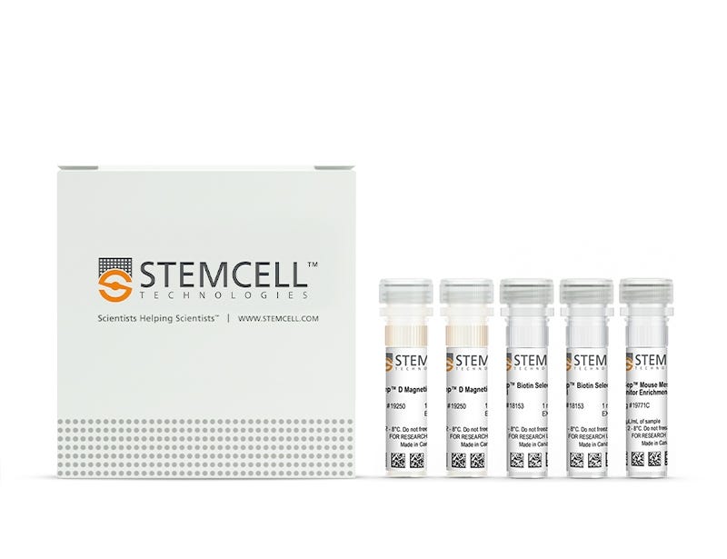
New look, same high quality and support! You may notice that your instrument or reagent packaging looks slightly different from images displayed on the website, or from previous orders. We are updating our look but rest assured, the products themselves and how you should use them have not changed. Learn more

EasySep™ Mouse Mesenchymal Stem/Progenitor Cell Enrichment Kit

Immunomagnetic negative selection cell isolation kit

From: 845 USD
Catalog #
(Select a product)
Immunomagnetic negative selection cell isolation kit
Add to Wish List

Product Advantages


  • Fast, easy-to-use and column-free

  • Up to 99% purity

  • Isolated cells are untouched

What's Included

  • EasySep™ Mouse Mesenchymal Stem/Progenitor Cell Enrichment Kit (Catalog #19771)
    • EasySep™ Mouse Mesenchymal Stem/Progenitor Cell Enrichment Cocktail, 0.4 mL
    • EasySep™ Biotin Selection Cocktail, 2 x 1 mL
    • EasySep™ Magnetic Particles, 2 x 1 mL

Overview

The EasySep™ Mouse Mesenchymal Stem/Progenitor Cell Enrichment Kit is designed to isolate mesenchymal stem/progenitor cells from mouse compact bone by negative selection. Unwanted cells are targeted for removal with biotinylated antibodies that are directed against non-mesenchymal stem/progenitor cells (CD45, TER119). Labeled cells are then recognized by Tetrameric Antibody Complexes that are directed against biotin and dextran. These cells are bound to magnetic particles and separated using an EasySep™ magnet without the use of columns. Desired cells are poured off into a new tube.
Magnet Compatibility
• EasySep™ Magnet (Catalog #18000)
Subtype
Cell Isolation Kits
Cell Type
Mesenchymal Stem and Progenitor Cells
Species
Mouse
Sample Source
Compact Bone
Selection Method
Negative
Application
Cell Isolation
Brand
EasySep
Area of Interest
Stem Cell Biology

Data Figures

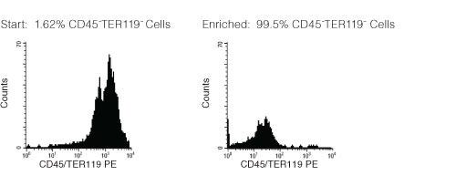
FACS Histogram Results with EasySep™ Mouse Mesenchymal Progenitor Cell Enrichment Kit

Figure 1. FACS Histogram Results with EasySep™ Mouse Mesenchymal Progenitor CellEnrichment Kit

Starting with compact bone, the CD45-TER119- cell content of the enriched cells typically ranges from 50 - 99%. CFU-F enrichment: 50 - 200 fold.

Protocols and Documentation

Find supporting information and directions for use in the Product Information Sheet or explore additional protocols below.

Document Type
Product Name
Catalog #
Lot #
Language
Catalog #
19771
Lot #
18L96978 or higher
Language
English
Document Type
Safety Data Sheet 1
Catalog #
19771
Lot #
All
Language
English
Document Type
Safety Data Sheet 2
Catalog #
19771
Lot #
All
Language
English
Document Type
Safety Data Sheet 3
Catalog #
19771
Lot #
All
Language
English

Applications

This product is designed for use in the following research area(s) as part of the highlighted workflow stage(s). Explore these workflows to learn more about the other products we offer to support each research area.

Resources and Publications

Frequently Asked Questions

Can EasySep™ be used for either positive or negative selection?

Yes. The EasySep™ kits use either a negative selection approach by targeting and removing unwanted cells or a positive selection approach targeting desired cells. Depletion kits are also available for the removal of cells with a specific undesired marker (e.g. GlyA).

How does the separation work?

Magnetic particles are crosslinked to cells using Tetrameric Antibody Complexes (TAC). When placed in the EasySep™ Magnet, labeled cells migrate to the wall of the tube. The unlabeled cells are then poured off into a separate fraction.

Which columns do I use?

The EasySep™ procedure is column-free. That's right - no columns!

How can I analyze the purity of my enriched sample?

The Product Information Sheet provided with each EasySep™ kit contains detailed staining information.

Can EasySep™ separations be automated?

Yes. RoboSep™, the fully automated cell separator, automates all EasySep™ labeling and cell separation steps.

Can EasySep™ be used to isolate rare cells?

Yes. We recommend a cell concentration of 2x10⁸ cells/mL and a minimum working volume of 100 µL. Samples containing 2x10⁷ cells or fewer should be suspended in 100 µL of buffer.

Are the EasySep™ magnetic particles FACS-compatible?

Yes, the EasySep™ particles are flow cytometry-compatible, as they are very uniform in size and about 5000X smaller than other commercially available magnetic beads used with column-free systems.

Can the EasySep™ magnetic particles be removed after enrichment?

No, but due to the small size of these particles, they will not interfere with downstream applications.

Can I alter the separation time in the magnet?

Yes; however, this may impact the kit's performance. The provided EasySep™ protocols have already been optimized to balance purity, recovery and time spent on the isolation.

For positive selection, can I perform more than 3 separations to increase purity?

Yes, the purity of targeted cells will increase with additional rounds of separations; however, cell recovery will decrease.

How does the binding of the EasySep™ magnetic particle affect the cells? is the function of positively selected cells altered by the bound particles?

Hundreds of publications have used cells selected with EasySep™ positive selection kits for functional studies. Our in-house experiments also confirm that selected cells are not functionally altered by the EasySep™ magnetic particles.

If particle binding is a key concern, we offer two options for negative selection. The EasySep™ negative selection kits can isolate untouched cells with comparable purities, while RosetteSep™ can isolate untouched cells directly from whole blood without using particles or magnets.

Publications (4)

Chemokine CXCL12 drives pericyte accumulation and airway remodeling in allergic airway disease. R. Bignold et al. Respiratory research 2022 jul

Abstract

BACKGROUND Airway remodeling is a significant contributor to impaired lung function in chronic allergic airway disease. Currently, no therapy exists that is capable of targeting these structural changes and the consequent loss of function. In the context of chronic allergic inflammation, pericytes have been shown to uncouple from the pulmonary microvasculature, migrate to areas of inflammation, and significantly contribute to airway wall remodeling and lung dysfunction. This study aimed to elucidate the mechanism by which pulmonary pericytes accumulate in the airway wall in a model of chronic allergic airway inflammation. METHODS Mice were subjected to a protocol of chronic airway inflammation driven by the common environmental aeroallergen house dust mite. Phenotypic changes to lung pericytes were assessed by flow cytometry and immunostaining, and the functional capacity of these cells was evaluated using in vitro migration assays. The molecular mechanisms driving these processes were targeted pharmacologically in vivo and in vitro. RESULTS Pericytes demonstrated increased CXCR4 expression in response to chronic allergic inflammation and migrated more readily to its cognate chemokine, CXCL12. This increase in migratory capacity was accompanied by pericyte accumulation in the airway wall, increased smooth muscle thickness, and symptoms of respiratory distress. Pericyte uncoupling from pulmonary vessels and subsequent migration to the airway wall were abrogated following topical treatment with the CXCL12 neutraligand LIT-927. CONCLUSION These results provide new insight into the role of the CXCL12/CXCR4 signaling axis in promoting pulmonary pericyte accumulation and airway remodeling and validate a novel target to address tissue remodeling associated with chronic inflammation.
HS1 deficiency protects against sepsis by attenuating neutrophil-inflicted lung damage. I. M. Guerrero-Fonseca et al. European journal of cell biology 2022 apr

Abstract

Sepsis remains an important health problem worldwide due to inefficient treatments often resulting in multi-organ failure. Neutrophil recruitment is critical during sepsis. While neutrophils are required to combat invading bacteria, excessive neutrophil recruitment contributes to tissue damage due to their arsenal of molecular weapons that do not distinguish between host and pathogen. Thus, neutrophil recruitment needs to be fine-tuned to ensure bacterial killing, while avoiding neutrophil-inflicted tissue damage. We recently showed that the actin-binding protein HS1 promotes neutrophil extravasation; and hypothesized that HS1 is also a critical regulator of sepsis progression. We evaluated the role of HS1 in a model of lethal sepsis induced by cecal-ligation and puncture. We found that septic HS1-deficient mice had a better survival rate compared to WT mice due to absence of lung damage. Lungs of septic HS1-deficient mice showed less inflammation, fibrosis, and vascular congestion. Importantly, systemic CLP-induced neutrophil recruitment was attenuated in the lungs, the peritoneum and the cremaster in the absence of HS1. Lungs of HS1-deficient mice produced significantly more interleukin-10. Compared to WT neutrophils, those HS1-deficient neutrophils that reached the lungs had increased surface levels of Gr-1, ICAM-1, and L-selectin. Interestingly, HS1-deficient neutrophils had similar F-actin content and phagocytic activity, but they failed to polymerize actin and deform in response to CXCL-1 likely explaining the reduced systemic neutrophil recruitment in HS1-deficient mice. Our data show that HS1 deficiency protects against sepsis by attenuating neutrophil recruitment to amounts sufficient to combat bacterial infection, but insufficient to induce tissue damage.
The effect of mesenchymal stem cell sheets on structural allograft healing of critical sized femoral defects in mice. Long T et al. Biomaterials 2014 MAR

Abstract

Structural bone allografts are widely used in the clinic to treat critical sized bone defects, despite lacking the osteoinductive characteristics of live autografts. To address this, we generated revitalized structural allografts wrapped with mesenchymal stem/progenitor cell (MSC) sheets, which were produced by expanding primary syngenic bone marrow derived cells on temperature-responsive plates, as a tissue-engineered periosteum. In vitro assays demonstrated maintenance of the MSC phenotype in the sheets, suggesting that short-term culturing of MSC sheets is not detrimental. To test their efficacy in vivo, allografts wrapped with MSC sheets were transplanted into 4-mm murine femoral defects and compared to allografts with direct seeding of MSCs and allografts without cells. Evaluations consisted of X-ray plain radiography, 3D microCT, histology, and biomechanical testing at 4- and 6-weeks post-surgery. Our findings demonstrate that MSC sheets induce prolonged cartilage formation at the graft-host junction and enhanced bone callus formation, as well as graft-host osteointegration. Moreover, a large periosteal callus was observed spanning the allografts with MSC sheets, which partially mimics live autograft healing. Finally, biomechanical testing showed a significant increase in the structural and functional properties of MSC sheet grafted femurs. Taken together, MSC sheets exhibit enhanced osteogenicity during critical sized bone defect repair, demonstrating the feasibility of this tissue engineering solution for massive allograft healing.
New look, same high quality and support! You may notice that your instrument or reagent packaging looks slightly different from images displayed on the website, or from previous orders. We are updating our look but rest assured, the products themselves and how you should use them have not changed. Learn more