Make more informed purchasing decisions with our new product availability and delivery estimate feature, now available on all product pages, in your cart, and during checkout.
Sign In
New to STEMCELL?
Register for an account to quickly and easily purchase products online and for one-click access to all educational content.
New look, same high quality and support! You may notice that your instrument or reagent packaging looks slightly different from images displayed on the website, or from previous orders. We are updating our look but rest assured, the products themselves and how you should use them have not changed. Learn more
Request Pricing
Thank you for your interest in this product.
Please provide us with your contact information and your local representative
will contact you with a customized quote. Where appropriate, they can also assist you with a(n):
Estimated delivery time for your area
Product sample or exclusive offer
In-lab demonstration
By submitting this form, you are providing your consent to STEMCELL Technologies Canada Inc. and its subsidiaries and affiliates (“STEMCELL”) to collect and use your information, and send you newsletters and emails in accordance with our privacy policy. Please contact us with any questions that you may have. You can unsubscribe or change your email preferences at any time.
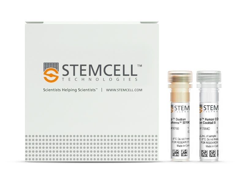
Isolate highly purified human CD19+ cells from fresh or previously frozen human peripheral blood mononuclear cells (PBMCs) or washed leukapheresis samples by immunomagnetic positive selection, with the EasySep™ Human CD19 Positive Selection Kit II. Widely used in published research for more than 20 years, EasySep™ combines the specificity of monoclonal antibodies with the simplicity of a column-free magnetic system.
In this EasySep™ positive selection procedure, desired cells are labeled with antibody complexes recognizing CD19 and magnetic particles. The cocktail in this kit also contains an antibody to human Fc receptor to prevent non-specific binding. Labeled cells are separated using an EasySep™ magnet and by simply pouring or pipetting off the unwanted cells. The cells of interest remain in the tube. Following magnetic cell isolation in as little as 18 minutes, the desired CD19+ cells are ready for downstream applications such as flow cytometry, culture, or DNA/RNA extraction.
This product replaces the EasySep™ Human CD19 Positive Selection Kit (Catalog #18054) for even faster cell isolations.
Learn more about how immunomagnetic EasySep™ technology works or how to fully automate immunomagnetic cell isolation with RoboSep™. Alternatively, choose ready-to-use, ethically sourced, primary Human Peripheral Blood CD19+ B Cells, Frozen isolated with EasySep™ Human CD19 Positive Selection Kit II. Explore additional products optimized for your workflow, including culture media, supplements, antibodies, and more.
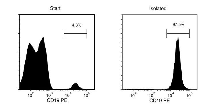
Figure 1. Typical EasySep™ Human CD19 Positive Selection Profile
Starting with a single cell suspension of human PBMCs, the CD19+ cell content of the isolated fraction is typically 98 ± 1% (mean ± SD) using the purple EasySep™ Magnet.
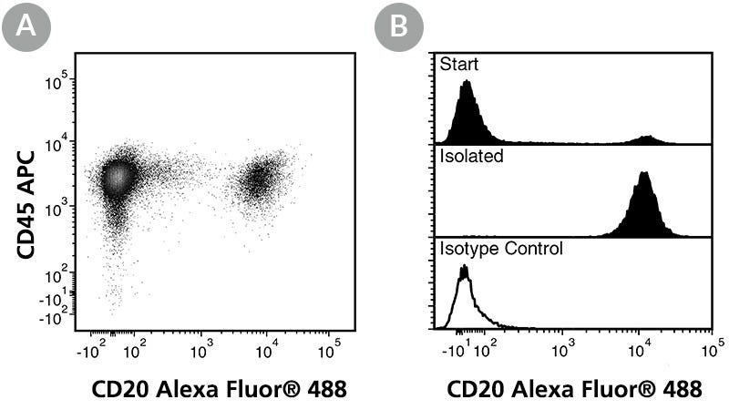
Figure 2. FACS Data for Anti-Human CD20 Antibody, Clone 2H7, Alexa Fluor® 488-Conjugated
(B) Flow cytometry analysis of human PBMCs processed with the EasySep™ Human CD19 Positive Selection Kit (Catalog #17854) and labeled with Anti-Human CD20 Antibody, Clone 2H7, Alexa Fluor® 488. Histograms show labeling of the PBMCs (Start) and isolated cells (Isolated). Labeling of start cells with a mouse IgG2b, kappa Alexa Fluor® 488 isotype control antibody is shown in the bottom panel (open histogram).
This product is designed for use in the following research area(s) as part
of the highlighted workflow stage(s). Explore these workflows to learn more about the other products we
offer to support each research area.
Can EasySep™ be used for either positive or negative selection?
Yes. The EasySep™ kits use either a negative selection approach by targeting and removing unwanted cells or a positive selection approach targeting desired cells. Depletion kits are also available for the removal of cells with a specific undesired marker (e.g. GlyA).
How does the separation work?
Magnetic particles are crosslinked to cells using Tetrameric Antibody Complexes (TAC). When placed in the EasySep™ Magnet, labeled cells migrate to the wall of the tube. The unlabeled cells are then poured off into a separate fraction.
Which columns do I use?
The EasySep™ procedure is column-free. That's right - no columns!
How can I analyze the purity of my enriched sample?
The Product Information Sheet provided with each EasySep™ kit contains detailed staining information.
Can EasySep™ separations be automated?
Yes. RoboSep™, the fully automated cell separator, automates all EasySep™ labeling and cell separation steps.
Can EasySep™ be used to isolate rare cells?
Yes. We recommend a cell concentration of 2x10⁸ cells/mL and a minimum working volume of 100 µL. Samples containing 2x10⁷ cells or fewer should be suspended in 100 µL of buffer.
Are the EasySep™ magnetic particles FACS-compatible?
Yes, the EasySep™ particles are flow cytometry-compatible, as they are very uniform in size and about 5000X smaller than other commercially available magnetic beads used with column-free systems.
Can the EasySep™ magnetic particles be removed after enrichment?
No, but due to the small size of these particles, they will not interfere with downstream applications.
Can I alter the separation time in the magnet?
Yes; however, this may impact the kit's performance. The provided EasySep™ protocols have already been optimized to balance purity, recovery and time spent on the isolation.
For positive selection, can I perform more than 3 separations to increase purity?
Yes, the purity of targeted cells will increase with additional rounds of separations; however, cell recovery will decrease.
How does the binding of the EasySep™ magnetic particle affect the cells? is the function of positively selected cells altered by the bound particles?
Hundreds of publications have used cells selected with EasySep™ positive selection kits for functional studies. Our in-house experiments also confirm that selected cells are not functionally altered by the EasySep™ magnetic particles.
If particle binding is a key concern, we offer two options for negative selection. The EasySep™ negative selection kits can isolate untouched cells with comparable purities, while RosetteSep™ can isolate untouched cells directly from whole blood without using particles or magnets.
BA.2.12.1, BA.4 and BA.5 escape antibodies elicited by Omicron infection.
Y. Cao et al.
Nature 2022 aug
Abstract
Severe acute respiratory syndrome coronavirus 2 (SARS-CoV-2) Omicron sublineages BA.2.12.1, BA.4 and BA.5 exhibit higher transmissibility than the BA.2 lineage1. The receptor binding and immune-evasion capability of these recently emerged variants require immediate investigation. Here, coupled with structural comparisons of the spike proteins, we show that BA.2.12.1, BA.4 and BA.5 (BA.4 and BA.5 are hereafter referred collectively to as BA.4/BA.5) exhibit similar binding affinities to BA.2 for the angiotensin-converting enzyme 2 (ACE2) receptor. Of note, BA.2.12.1 and BA.4/BA.5 display increased evasion of neutralizing antibodies compared with BA.2 against plasma from triple-vaccinated individuals or from individuals who developed a BA.1 infection after vaccination. To delineate the underlying antibody-evasion mechanism, we determined the escape mutation profiles2, epitope distribution3 and Omicron-neutralization efficiency of 1,640 neutralizing antibodies directed against the receptor-binding domain of the viral spike protein, including 614 antibodies isolated from people who had recovered from BA.1 infection. BA.1 infection after vaccination predominantly recalls humoral immune memory directed against ancestral (hereafter referred to as wild-type (WT)) SARS-CoV-2 spike protein. The resulting elicited antibodies could neutralize both WT SARS-CoV-2 and BA.1 and are enriched on epitopes on spike that do not bind ACE2. However, most of these cross-reactive neutralizing antibodies are evaded by spike mutants L452Q, L452R and F486V. BA.1 infection can also induce new clones of BA.1-specific antibodies that potently neutralize BA.1. Nevertheless, these neutralizing antibodies are largely evaded by BA.2 and BA.4/BA.5 owing to D405N and F486V mutations, and react weakly to pre-Omicron variants, exhibiting narrow neutralization breadths. The therapeutic neutralizing antibodies bebtelovimab4 and cilgavimab5 can effectively neutralize BA.2.12.1 and BA.4/BA.5, whereas the S371F, D405N and R408S mutations undermine most broadly sarbecovirus-neutralizing antibodies. Together, our results indicate that Omicron may evolve mutations to evade the humoral immunity elicited by BA.1 infection, suggesting that BA.1-derived vaccine boosters may not achieve broad-spectrum protection against new Omicron variants.
Novel genes exhibiting DNA methylation alterations in Korean patients with chronic lymphocytic leukaemia: a methyl-CpG-binding domain sequencing study.
M. Kim et al.
Scientific reports 2020 jan
Abstract
Chronic lymphocytic leukaemia (CLL) exhibits differences between Asians and Caucasians in terms of incidence rate, age at onset, immunophenotype, and genetic profile. We performed genome-wide methylation profiling of CLL in an Asian cohort for the first time. Eight Korean patients without somatic immunoglobulin heavy chain gene hypermutations underwent methyl-CpG-binding domain sequencing (MBD-seq), as did five control subjects. Gene Ontology, pathway analysis, and network-based prioritization of differentially methylated genes were also performed. More regions were hypomethylated (2,062 windows) than were hypermethylated (777 windows). Promoters contained the highest proportion of differentially methylated regions (0.08{\%}), while distal intergenic and intron regions contained the largest number of differentially methylated regions. Protein-coding genes were the most abundant, followed by long noncoding and short noncoding genes. The most significantly over-represented signalling pathways in the differentially methylated gene list included immune/cancer-related pathways and B-cell receptor signalling. Among the top 10 hub genes identified via network-based prioritization, four (UBC, GRB2, CREBBP, and GAB2) had no known relevance to CLL, while the other six (STAT3, PTPN6, SYK, STAT5B, XPO1, and ABL1) have previously been linked to CLL in Caucasians. As such, our analysis identified four novel candidate genes of potential significance to Asian patients with CLL.
Specific memory B cell response in humans upon infection with highly pathogenic H7N7 avian influenza virus.
B. Westerhuis et al.
Scientific reports 2020 feb
Abstract
H7 avian influenza viruses represent a major public health concern, and worldwide outbreaks raise the risk of a potential pandemic. Understanding the memory B cell response to avian (H7) influenza virus infection in humans could provide insights in the potential key to human infection risks. We investigated an epizootic of the highly pathogenic A(H7N7) in the Netherlands, which in 2003 led to infection of 89 persons and one fatal case. Subtype-specificity of antibodies were determined for confirmed H7N7 infected individuals (cases) (n = 19), contacts of these cases (n = 21) and a comparison group controls (n = 16), by microarray, using recombinant hemagglutinin (HA)1 proteins. The frequency and specificity of memory B cells was determined by detecting subtype-specific antibodies in the culture supernatants from in vitro stimulated oligoclonal B cell cultures, from peripheral blood of cases and controls. All cases (100{\%}) had high antibody titers specific for A(H7N7)2003 (GMT {\textgreater} 100), whereas H7-HA1 antigen binding was detected in 29{\%} of contacts and 31{\%} of controls, suggesting that some of the H7 reactivity stems from cross reactive antibodies. To unravel homotypic and heterotypic responses, the frequency and specificity of memory B cells were determined in 2 cases. Ten of 123 HA1 reactive clones isolated from the cases bound to only H7- HA1, whereas 5 bound both H7 and other HA1 antigens. We recovered at least four different epitopal reactivities, though none of the H7 reactive antibodies were able to neutralize H7 infections in vitro. Our study serologically confirms the infection with H7 avian influenza viruses, and shows that H7 infection triggers a mixture of strain -specific and cross-reactive antibodies.
New look, same high quality and support! You may notice that your instrument or reagent packaging looks slightly different from images displayed on the website, or from previous orders. We are updating our look but rest assured, the products themselves and how you should use them have not changed. Learn more
Quality Statement:
PRODUCTS ARE FOR RESEARCH USE ONLY AND NOT INTENDED FOR HUMAN OR ANIMAL DIAGNOSTIC OR THERAPEUTIC USES UNLESS OTHERWISE STATED. FOR ADDITIONAL INFORMATION ON QUALITY AT STEMCELL, REFER TO WWW.STEMCELL.COM/COMPLIANCE.
 EasySep™ Magnet
 EasySep™ Buffer