Make more informed purchasing decisions with our new product availability and delivery estimate feature, now available on all product pages, in your cart, and during checkout.
Sign In
New to STEMCELL?
Register for an account to quickly and easily purchase products online and for one-click access to all educational content.
Thank you for your interest in this product.
Please provide us with your contact information and your local representative
will contact you with a customized quote. Where appropriate, they can also assist you with a(n):
Estimated delivery time for your area
Product sample or exclusive offer
In-lab demonstration
By submitting this form, you are providing your consent to STEMCELL Technologies Canada Inc. and its subsidiaries and affiliates (“STEMCELL”) to collect and use your information, and send you newsletters and emails in accordance with our privacy policy. Please contact us with any questions that you may have. You can unsubscribe or change your email preferences at any time.
The TC15-12F12.2 antibody reacts with CD150 (signaling lymphocyte activation molecule or SLAM), an ~75 kDa type I transmembrane glycoprotein that plays multiple roles in the immune response by serving as a cell adhesion molecule and/or coreceptor. It is differentially expressed by T cells, immature thymocytes, B cells, dendritic cells, macrophages, and endothelial cells. The expression pattern differs according to cell type and activation status. Expression is rapidly induced upon activation of T cells, B cells and dendritic cells, with synthesis by T cells being maintained on Th1 but not Th2 clones. CD150-mediated co-stimulation of TCR-activated T cells enhances the production of IFN-γ by Th1 cells, a response that can be augmented by binding of the TC15-12F12.2 antibody. CD150 is thought to mediate signal transduction by associating with the intracellular protein tyrosine phosphatase, SHP-2. CD150 also has functions in hematopoietic cell development and is a useful marker for detection of multipotent hematopoietic stem cells (in concert with other markers such as CD48 and CD41). CD150 is not expressed on non-multipotent hematopoietic progenitor cells.
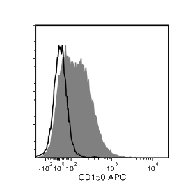
Flow cytometry analysis of C57BL/6 mouse splenocytes labeled with Anti-Mouse CD150 Antibody, Clone TC15-12F12.2, Alexa Fluor® 488 (filled histogram) or a rat IgG2a, lambda Alexa Fluor® 488 isotype control antibody (solid line histogram).
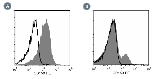
Figure 2. Data for PE-Conjugated
(A) Flow cytometry analysis of C57BL/6 mouse splenocytes labeled with Anti-Mouse CD150 Antibody, Clone TC15-12F12.2, PE (filled histogram) or a rat IgG2a, lambda PE isotype control antibody (solid line histogram).
(B) Flow cytometry analysis of C57BL/6 mouse bone marrow cells labeled with Anti-Mouse CD150 Antibody, Clone TC15-12F12.2, PE (filled histogram) or a rat IgG2a, lambda PE isotype control antibody (solid line histogram).
Figure 3. Data for Unconjugated
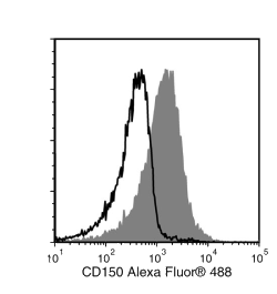
Flow cytometry analysis of C57BL/6 mouse splenocytes labeled with Anti-Mouse CD150 Antibody, Clone TC15-12F12.2, followed by a mouse anti-rat IgG2a antibody, FITC (filled histogram), or a rat IgG2a isotype control antibody followed by a mouse anti-rat IgG2a antibody, FITC (solid line histogram).
Figure 4. Data for APC-Conjugated
Flow cytometry analysis of C57BL/6 mouse splenocytes labeled with Anti-Mouse CD150 Antibody, Clone TC15-12F12.2, APC (filled histogram) or a rat IgG2a isotype control antibody, APC (solid line histogram).
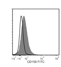
Figure 5. Data for Biotin-Conjugated
Flow cytometry analysis of C57BL/6 mouse splenocytes labeled with Anti-Mouse CD150 Antibody, Clone TC15-12F12.2, Biotin followed by streptavidin (SAV) APC (filled histogram), or a biotinylated rat IgG2a isotype control antibody followed by SAV APC (solid line histogram).
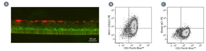
(A) Primary human airway epithelial cells were cultured in PneumaCult™-ALI Medium (Catalog #05001) at the air-liquid interface, then cryo-sectioned and
labeled with Anti-Human MUC1 (CD227) Antibody, Clone 16A, followed by a goat anti-rabbit IgG antibody, Alexa Fluor® 594 (red), and an anti-human
NGF Receptor/p75NTR (CD271) antibody, followed by a donkey anti-mouse IgG antibody, Alexa Fluor® 488 (green).
(B) Flow cytometry analysis of human peripheral blood lymphocytes following stimulation with PHA for 3 days. Cells were labeled with Anti-Human MUC1
(CD227) Antibody, Clone 16A, followed by an anti-mouse IgG1 antibody, PE and anti-human CD3 antibody, Clone HIT3a, Pacific Blue™.
(C) Flow cytometry analysis of human peripheral blood lymphocytes following stimulation with PHA for 3 days. Cells were labeled with Mouse IgG1, kappa
Isotype Control Antibody, Clone MOPC-21 (Catalog #60070), followed by an anti-mouse IgG1 antibody, PE, and anti-human CD3 antibody, clone HIT3a,
Pacific Blue™.
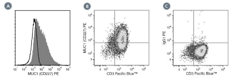
(A) Flow cytometry analysis of human airway epithelial cells cultured in PneumaCult™-ALI Medium (Catalog #05001) at the air-liquid interface. Cells were
enzymatically dissociated and labeled with Anti-Human MUC1 (CD227) Antibody, Clone 16A, PE (filled histogram) or Mouse IgG1, kappa Isotype Control
Antibody, Clone MOPC-21, PE (Catalog #60070PE, solid line histogram).
(C) Flow cytometry analysis human peripheral blood lymphocytes following stimulation with PHA for 3 days. Cells were labeled with Anti-Human MUC1
(CD227) Antibody, Clone 16A, PE and anti-human CD3 antibody, clone HIT3a, Pacific Blue™.
(D) Flow cytometry analysis of PHA-activated human peripheral blood lymphocytes following stimulation with PHA for 3 days. Cells were labeled with
Mouse IgG1, kappa Isotype Control Antibody, Clone MOPC-21, PE, and anti-human CD3 antibody, clone HIT3a, Pacific Blue™.
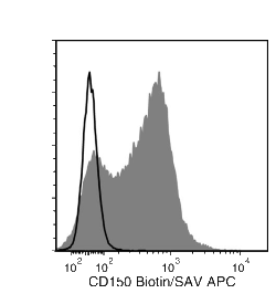
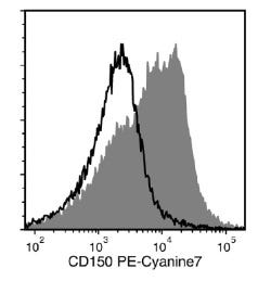
Flow cytometry analysis of C57BL/6 mouse splenocytes labeled with Anti-Mouse CD150 Antibody, Clone TC15-12F12.2, PE-Cyanine7 (filled histogram) or
a rat IgG2a isotype control antibody, PE-Cyanine7 (solid line histogram).
This product is designed for use in the following research area(s) as part
of the highlighted workflow stage(s). Explore these workflows to learn more about the other products we
offer to support each research area.
Rat monoclonal IgG2a, kappa isotype control antibody
Item added to your cart
Anti-Mouse CD150 Antibody, Clone TC15-12F12.2
Legal Statement:
Alexa Fluor is a registered trademark of Life Technologies Corporation. Antibodies conjugated to Alexa Fluor® are licensed for internal research use only and sale is expressly conditioned on the buyer not using the antibody for manufacturing, performing a service or medical test, or otherwise generating revenue. For use other than research, contact Life Technologies Corporation, 5791 Van Allen Way, Carlsbad, CA 92008 USA or outlicensing@lifetech.com.
Quality Statement:
PRODUCTS ARE FOR RESEARCH USE ONLY AND NOT INTENDED FOR HUMAN OR ANIMAL DIAGNOSTIC OR THERAPEUTIC USES UNLESS OTHERWISE STATED. FOR ADDITIONAL INFORMATION ON QUALITY AT STEMCELL, REFER TO WWW.STEMCELL.COM/COMPLIANCE.