ImmunoCult™-XF

cGMP, serum- and xeno-free medium for the expansion of human immune cells

ImmunoCult™-XF

cGMP, serum- and xeno-free medium for the expansion of human immune cells

From: 233 USD
Catalog #
(Select a product)
cGMP, serum- and xeno-free medium for the expansion of human immune cells
Add to Wish List

Product Advantages


  • Confidently expand immune cells for use in clinical applications by using medium produced under relevant cGMPs

  • Reduce variability by expanding cells in serum- and xeno-free culture conditions

  • Achieve robust immune cell expansion with high viability

  • Produce cytokines from functional T cells after restimulation

  • Activate T cells with an easy-to-use, bead-free protocol by combining with ImmunoCult™ Human T Cell Activators

Overview

Support the culture of human immune cells and enhance your cell therapy development by using high-performance, cGMP-manufactured ImmunoCult™-XF medium. Designed for robust immune cell expansion for cell and gene therapy applications, ImmunoCult™-XF does not require serum or serum derivatives. It is also phenol red-free and xeno-free, and has no added cytokines, providing complete flexibility for your workflows. T cells can be harvested after just 10 - 12 days of culture and are able to produce cytokines, including interferon-gamma (IFN-γ) and interleukin 4 (IL4), upon restimulation. This ready-to-use medium is produced according to current GMP guidelines, ensuring the highest quality and consistency for reproducible results. ImmunoCult™-XF is available in a 500 mL bottle or a 1 L bag equipped with a needleless injection port and weldable non-DEHP PVC tubing to support closed, aseptic workflows.

ImmunoCult™-XF can be used individually or in combination with ImmunoCult™ Human CD3/CD28 T Cell Activator or ImmunoCult™ Human CD3/CD28/CD2 T Cell Activator for gentle, bead-free activation of T cells. For more information on the expansion of T cells with ImmunoCult™, please explore the Product Information Sheet. In addition to T cells, ImmunoCult™-XF can be used for high-viability expansion of natural killer (NK) cells, extending its utility across cell therapy workflows.

To request a Letter of Authorization (LOA) for the FDA Master File for ImmunoCult™-XF, click here.
Subtype
Specialized Media
Cell Type
NK Cells, T Cells, T Cells, CD4+, T Cells, CD8+, T Cells, Other Subsets, T Cells, Regulatory
Species
Human
Application
Expansion
Brand
ImmunoCult
Area of Interest
Cancer, Immunology, Infectious Diseases, Cell Therapy Development
Formulation Category
Serum-Free, Xeno-Free

Data Figures

Culture Protocol for T Cell Activation And Expansion with ImmunoCult™-XF

Figure 1. Culture Protocol for T Cell Activation and Expansion

(A) T cells were isolated from fresh leukopaks (Catalog #70500) from healthy donors using EasySep™ Human T Cell Isolation Kit (Catalog #17951) and cryopreserved in CryoStor® CS10 (Catalog #07930). On Day 0, T cells were thawed, washed, and resuspended at 1 x 10⁶ cells/mL in ImmunoCult™-XF (Catalog #100-0956) supplemented with 180 IU/mL Human Recombinant IL-2, ACF (rhIL-2; Catalog #78145). Cells were seeded into 24-well tissue culture plates (1 mL/well) and stimulated with 25 µL/mL ImmunoCult™ Human CD3/CD28/CD2 T Cell Activator (Catalog #100-0785)*. On Days 3, 5, 7, and 10, cells were re-seeded in fresh ImmunoCult™-XF + rhIL-2 at low or high cell densities as outlined in the table (B). Cells were harvested on Day 12.

*A similar protocol was performed using ImmunoCult™ Human CD3/CD28 T Cell Activator (Catalog #100-0784). Please reach out to our Product and Scientific Support team for additional information.

ImmunoCult™ Reagents Result in Robust T Cell Expansion Under Low Cell Density Culture Conditions

Figure 2. ImmunoCult™ Reagents Result in Robust T Cell Expansion Under Low Cell Density Culture Conditions

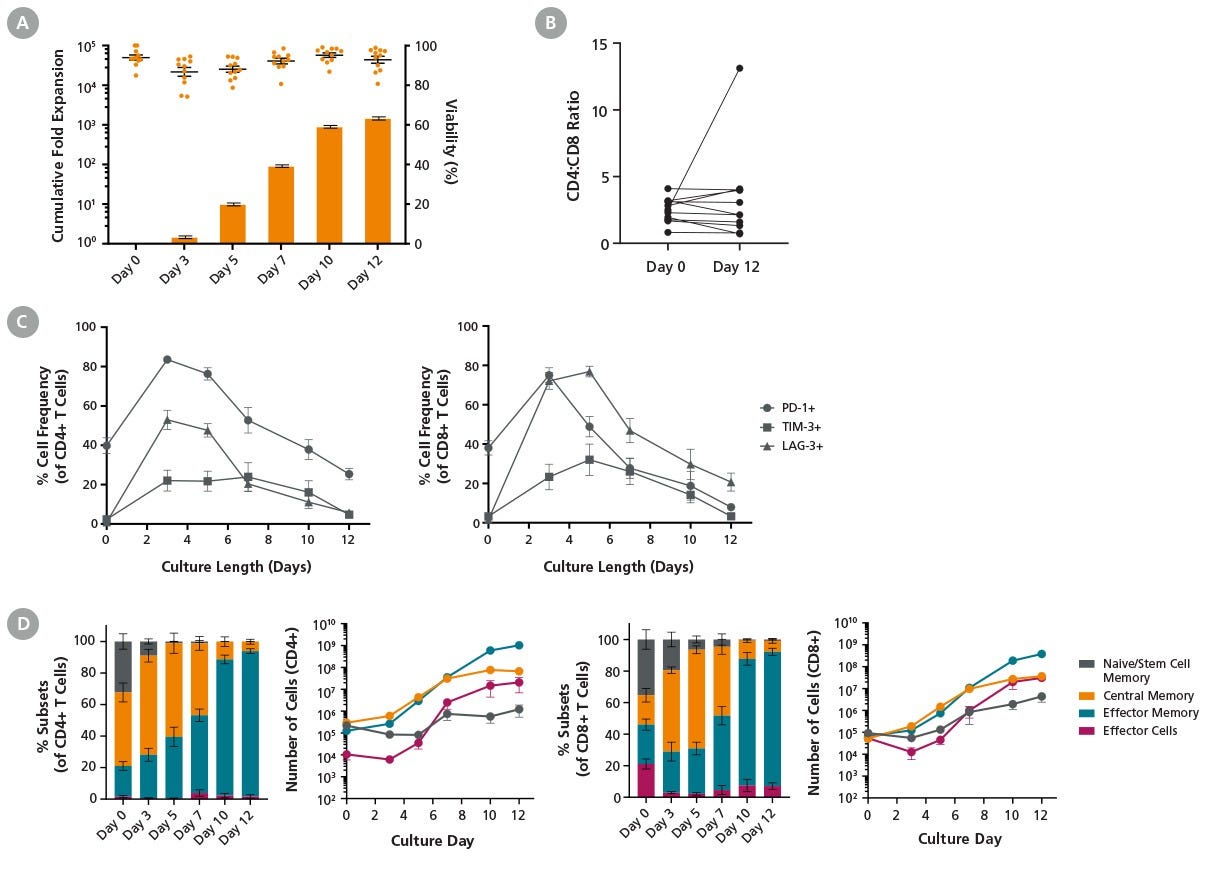
T cells were cultured with ImmunoCult™-XF (Catalog #100-0956), Human Recombinant IL-2, ACF (Catalog #78145), and ImmunoCult™ Human CD3/CD28/CD2 T Cell Activator (Catalog #100-0785) for 12 days following the low cell density protocol described in Figure 1. Cells were harvested and analyzed at different time points. Cell count and viability were assessed using the NucleoCounter® NC-250™ automated cell analyzer, and cell surface marker expression was analyzed by flow cytometry. (A) Points and bars in the graph represent T cell viability and average cumulative fold expansion, respectively, for each assessed culture time point. An average viability of 92.8 ± 1.8% and a total fold expansion of 1497 ± 143 were achieved by Day 12 in the low cell density cultures. Data represent mean ± SEM (n = 11). (B) The ratio of CD4+ T cells to CD8+ T cells (CD4:CD8) was comparable between Day 0 and Day 12 in 10 out of 11 donors. (C) The frequency of T cells expressing PD-1, TIM-3, and LAG-3 was upregulated upon stimulation and subsequently decreased to basal levels (n = 4). (D) The frequency and number of T cell subtypes in CD4+ and CD8+ populations during the 12-day culture period are shown. Cell numbers were calculated based on 10⁶ starting T cells. The number of central memory T cells remained constant from day 7 to 12. T cell subsets: naive and T memory stem cell (TN/SCM) CCR7+CD45RO-; central memory (TCM) CCR7+CD45RO+; effector memory (TEM) CCR7-CD45RO+; effector cells (TEFF) CCR7-CD45RO- (n = 4 - 11).

ImmunoCult™ Reagents Support T Cell Activation and Expansion Following High Cell Density Protocol

Figure 3. ImmunoCult™ Reagents Support T Cell Activation and Expansion Following High Cell Density Protocol

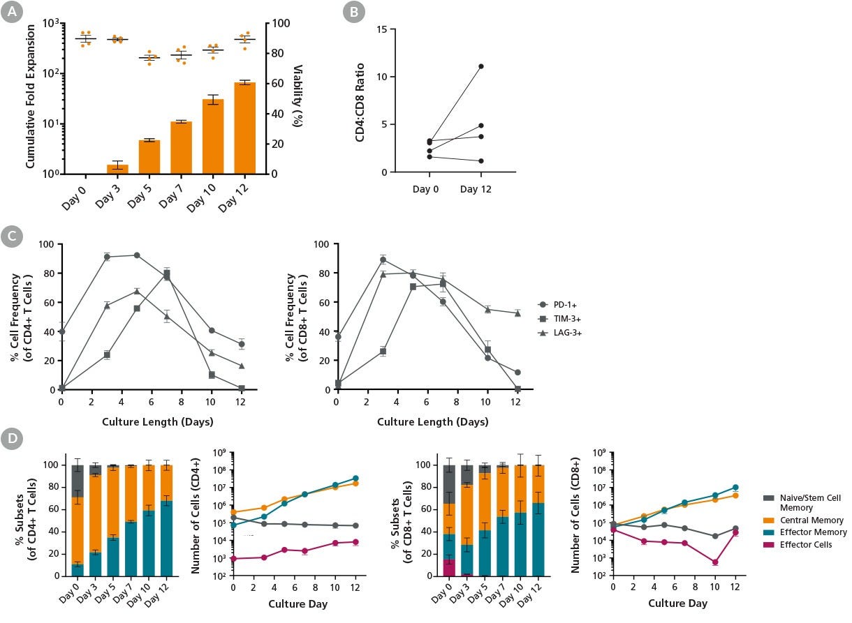
T cells were cultured with ImmunoCult™-XF (Catalog #100-0956), Human Recombinant IL-2, ACF (Catalog #78145), and ImmunoCult™ Human CD3/CD28/CD2 T Cell Activator (Catalog #100-0785) for 12 days following the high cell density protocol described in Figure 1. Cells were harvested at different time points and analyzed. Cell count and viability were assessed using the NucleoCounter® NC-250™ automated cell analyzer, and cell surface marker expression was analyzed by flow cytometry. (A) Points and bars in the graph represent T cell viability and average cumulative fold expansion, respectively, for each assessed culture time point. An average viability of 89.3 ± 2.3% and a total fold expansion of 67 ± 7 were achieved by Day 12 in the high cell density cultures. Data represent mean ± SEM (n = 4). (B) The ratio of CD4+ T cells to CD8+ T cells (CD4:CD8) was measured at the beginning and end of culture. The CD4:CD8 ratios increased in two out of four donors tested. (C) The frequency of T cells expressing PD-1, TIM-3, and LAG-3 increased upon stimulation and subsequently decreased to basal levels by Day 12 of culture (n = 4), with the exception of LAG-3 expression in the CD8⁺ population. (D) Data shows the frequency and number of T cell subtypes in CD4+ and CD8+ populations at the indicated time points. Cell numbers were calculated based on 10⁶ starting T cells. The number of central memory T cells continued to increase from Day 7 to Day 12 of culture. T cell subsets: naive and T memory stem cells (TN/SCM) CCR7+CD45RO-; central memory (TCM) CCR7+CD45RO+; effector memory (TEM) CCR7-CD45RO+; effector cells (TEFF) CCR7-CD45RO- (n = 4).

Culture Protocol for Expansion of T Cells with ImmunoCult™ Reagents in the Xuri™ Cell Expansion System W25

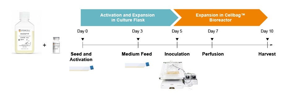
Figure 4. Culture Protocol for Expansion of T Cells with ImmunoCult™ Reagents in the Xuri™ Cell Expansion System W2

T cell expansion workflow in Xuri™ Cell Expansion System W25: T cells were isolated from fresh leukopaks (Catalog #70500) using the EasySep™ Human T Cell Isolation Kit (Catalog #17951) and cryopreserved in Cryostor® CS10 (Catalog #07930). On Day 0, cryopreserved cells were thawed and resuspended at 1 x 10⁶ cells/mL in ImmunoCult™-XF (Catalog #100-0956) supplemented with 180 IU/mL Human Recombinant IL-2, ACF (Catalog #78145). 5 x 10⁷ T cells were seeded in a T175 tissue culture flask and activated with ImmunoCult™ Human CD3/CD28/CD2 T Cell Activator (Catalog #100-0785) at 25 μL/mL. On Day 3, cell density was adjusted to 1 - 2.5 x 10⁵ cells/mL by adding fresh ImmunoCult™-XF with IL-2. On Day 5, the cell suspension containing 3 x 10⁸ cells/L of culture volume was inoculated into a Xuri™ 2L Cellbag™ Bioreactor and cultured until Day 10. Once the cell density reached 1.5 x 10⁶ cells/mL, perfusion was initiated at 0.5 L/day on Day 7 and increased to 1 L/day on Day 9. The culture was harvested on Day 10.

Expansion of T Cells with ImmunoCult™ Reagents in the Xuri™ Cell Expansion System W25

Figure 5. Expansion of T Cells with ImmunoCult™ Reagents in the Xuri™ Cell Expansion System W25

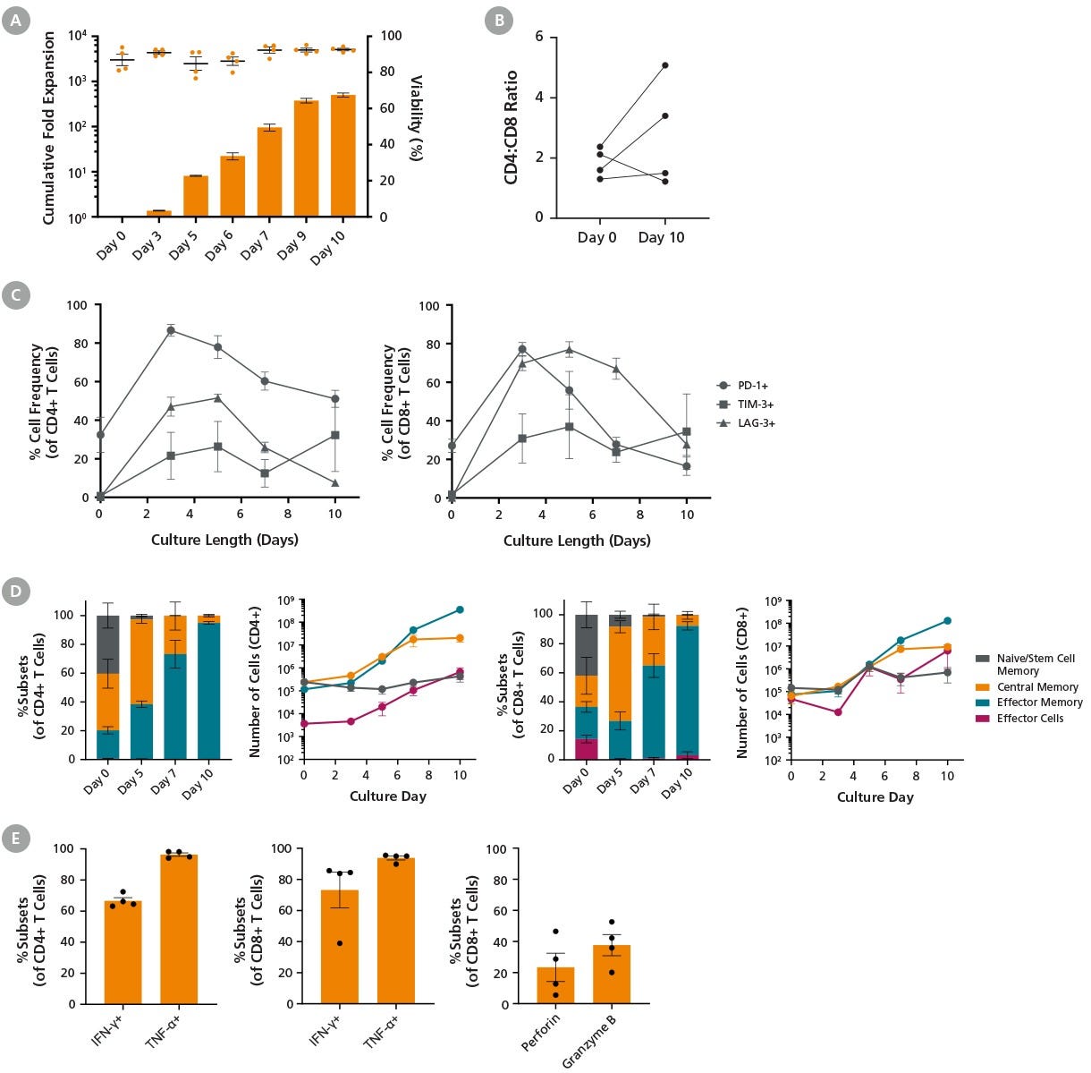
T cells were cultured with ImmunoCult™-XF (Catalog #100-0956), Human Recombinant IL-2, ACF (Catalog #78145), and ImmunoCult™ Human CD3/CD28/CD2 T Cell Activator (Catalog #100-0785) for 10 days following the protocol described in Figure 4. (A) Points and bars in the graph represent T cell viability and average cumulative fold expansion, respectively, for each assessed culture time point. On average, 92.7 ± 0.7% viability and a total fold expansion of 503 ± 51 were achieved by Day 10. Data represent mean ± SEM (n = 4). (B) Changes in the ratio of CD4+ T cells to CD8+ T cells (CD4:CD8) were measured at the beginning and end of culture. Some variability among donors was observed (n = 4). (C) The frequency of T cells expressing PD-1, TIM-3, and LAG-3 increased by Day 3 and subsequently decreased from Day 3 to Day 10 (n = 4). (D) The frequency and number of T cell subtypes in CD4+ and CD8+ populations are shown. Cell numbers were calculated based on 10⁶ starting T cells. The number of central memory T cells peaked on Day 7 of the culture. T cell subsets: naive and T memory stem cells (TN/SCM) CCR7+CD45RO-; central memory (TCM) CCR7+CD45RO+; effector memory (TEM) CCR7-CD45RO+; effector cells (TEFF) CCR7-CD45RO- (n = 4). (E) The frequencies of expanded T cells expressing IFN-ɣ and TNF-ɑ upon stimulation with PMA and ionomycin were 66.6 ± 2.1% and 96.3 ± 1.1%, respectively, in the CD4+ population, and 73.2 ± 11.4% and 93.8 ± 1.3%, in the CD8⁺ population. Additionally, the frequencies of expanded CD8+ T cells expressing perforin and granzyme B without stimulation were 23.3 ± 9.1% and 37.7 ± 6.8%, respectively (n = 4).

High-Efficiency TRAC Knockout of T Cells

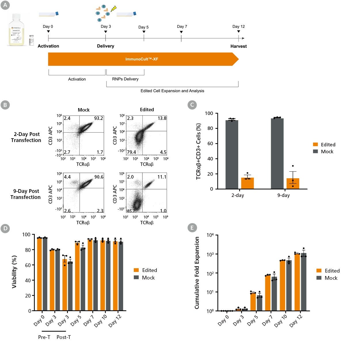
Figure 6. High-Efficiency TRAC Knockout of T Cells

(A) The protocol for non-viral genetic modification and expansion of T cells: On Day 0, T cells were isolated from fresh leukopaks using the EasySep™ Human T Cell Isolation Kit (Catalog #17951). Cells were resuspended at 1 x 10⁶ cells/mL in ImmunoCult™-XF (Catalog #100-0956) supplemented with 180 IU/mL Human Recombinant IL-2, ACF (rhIL-2; Catalog #78145). Cells were seeded into 24-well tissue culture plates at 1 mL/well and stimulated with ImmunoCult™ Human CD3/CD28/CD2 T Cell Activator (Catalog #100-0785) at 25 μL/mL. Following 3 days of activation, RNP complexes containing ArciTect™ Cas9 Nuclease (Catalog #76004) and sgRNA targeting the TRAC gene were delivered to T cells by electroporation (Neon® Transfection System). Cells were then returned to culture in ImmunoCult™-XF supplemented with rhIL-2 for another 9 days of expansion following the low cell density protocol. (B) Representative flow cytometry plots of TCRαβ and CD3 expression analysis on Days 2 and 9 post-T cell editing. (C) The gene editing efficiency was assessed by measuring the frequency of the remaining TCRαβ+CD3+ cells in the mock (91.8 ± 1.1%) and edited T cells (16.1 ± 2.6%) 2 days post-editing. The frequency of TRAC-edited cells was maintained throughout the culture duration. (D) The viability of T cells was measured using the NucleoCounter® NC-250™ automated cell analyzer across culture periods. On Day 3, the viability of T cells further decreased immediately following transfection. (E) Cumulative cell expansion post-electroporation was comparable between mock-treated and TRAC-edited T cells, suggesting that the edited T cells are functional. Data represent mean ± SEM (n = 3).

ImmunoCult™ NK Cell Base Medium and GMP ImmunoCult™-XF Support Natural Killer Cell Expansion

Figure 7. ImmunoCult™ NK Cell Base Medium and GMP ImmunoCult™-XF Support Natural Killer Cell Expansion

Natural killer (NK) cells were isolated from human peripheral blood samples using EasySep™ Human NK Cell Isolation Kit (Catalog #17955), then cultured in media supplemented with ImmunoCult™ NK Cell Expansion Supplement (Catalog #110-0715) for a total of 14 days. Cells were stimulated with ImmunoCult™ NK Cell Expansion Coating Material (Catalog #100-0714) on Days 0, 7, and 10 of culture. The medium used was either ImmunoCult™ NK Cell Base Medium (Catalog #100-0712) or GMP ImmunoCult™-XF (Catalog #100-0956). (A) NK cells undergo robust expansion over 14 days of expansion with ImmunoCult™ NK Cell Base Medium or GMP ImmunoCult™-XF. (B) NK cells remain highly viable after 14 days of expansion in ImmunoCult™ NK Cell Base Medium or GMP ImmunoCult™-XF. Each column with error bars represents the mean ± S.E.M. across three individual donors tested across three different manufactured lots of media.

Protocols and Documentation

Find supporting information and directions for use in the Product Information Sheet or explore additional protocols below.

Document Type
Product Name
Catalog #
Lot #
Language
Product Name
ImmunoCult™-XF
Catalog #
100-2243
Lot #
All
Language
English
Product Name
ImmunoCult™-XF
Catalog #
100-0956
Lot #
All
Language
English
Document Type
Safety Data Sheet
Product Name
ImmunoCult™-XF
Catalog #
100-2243
Lot #
All
Language
English
Document Type
Safety Data Sheet
Product Name
ImmunoCult™-XF
Catalog #
100-0956
Lot #
All
Language
English

Applications

This product is designed for use in the following research area(s) as part of the highlighted workflow stage(s). Explore these workflows to learn more about the other products we offer to support each research area.

Resources and Publications

Publications (51)

Phase II Metabolite-Inspired Tripeptide Editing Generates Anti-Inflammatory Candidates for Treatment-Refractory Ulcerative Colitis. X. Shi et al. International journal of molecular sciences 2026 Sep

Abstract

Ulcerative colitis remains a major clinical challenge due to limited availability of safe, orally active anti-inflammatory drugs. Although phase II metabolism is traditionally viewed as a detoxification process, certain metabolites retain or exhibit enhanced pharmacological activity, offering an underexplored drug discovery opportunity. Here, we investigated phase II metabolite-inspired tripeptide editing using M13, a glutathione-conjugated metabolite of the anti-inflammatory natural product 6-shogaol, as a lead scaffold. A focused library of 38 analogs was generated by systematic tripeptide editing while preserving the central cysteine-containing 6-shogaol-derived scaffold. Phenotypic nuclear factor-κB (NF-κB) reporter screening identified 29 of 38 analogs with greater inhibitory activity than M13 at the screening concentration. Two selected leads, MLY2 and MLY8, demonstrated enhanced anti-inflammatory activity in Dextran Sulfate Sodium-induced experimental colitis, including restoration of colon length and reductions in fecal lipocalin-2 and pro-inflammatory cytokines. Both compounds also suppressed inflammatory cytokine production more effectively than M13 in ex vivo colonic biopsies from patients with treatment-refractory ulcerative colitis. In parallel, MLY2 and MLY8 showed no detectable mutagenicity under the Ames assay conditions and were well tolerated in a preliminary single-dose maximum tolerated dose study. Collectively, these findings establish phase II metabolite-inspired tripeptide editing as a productive lead-discovery strategy and identify MLY2 and MLY8 as anti-inflammatory leads warranting further preclinical characterization.
Tregs transduced with a Ro60-reactive TCR have increased immunosuppressive functions in Sjögren's disease. Z. Sherman Lim et al. iScience 2026 Oct

Abstract

Sjögren's disease (SjD) is a T cell-driven autoimmune condition strongly associated with the HLA-DRB1∗03:01 allele and autoantibodies against the Ro60 protein. Previous studies have identified deficiencies in regulatory T cells (Treg) in patients, indicating a breakdown in immune tolerance. Current treatments remain limited to symptomatic relief or broad immunosuppression, without addressing the underlying immune dysregulation. In this study, we present the first preclinical evidence supporting a TCR-engineered Treg (TCR-Treg) therapy specifically targeting SjD. We designed a TCR-Treg that recognizes a dominant epitope of the Ro60 antigen (Ro60369-383) presented by HLA-DRB1∗03:01 and demonstrate that these cells can selectively suppress autoreactive T cells more effectively than polyclonal Treg. These findings underscore the pathogenic significance of the Ro-HLA-DRB1∗03:01 axis and provide proof-of-concept for a targeted, antigen-specific Treg therapy. This approach offers a promising strategy to restore immune tolerance and represents a potential disease-modifying treatment for genetically defined subsets of patients with SjD.
Single-allele nanoscale mapping of regulatory variants. J. Hamley et al. Nature genetics 2026 Oct

Abstract

Millions of genetic variants are linked to human disease but identifying underlying mechanisms is challenging because most variants are noncausal and lie within the noncoding genome. We developed a Micro Capture-C variant-to-function platform (MCCv) based on analysis of single-allele chromatin structure. This can identify changes in nanoscale chromatin architecture and link variants in cis-regulatory elements to target genes. Furthermore, MCCv can phase other heterozygous variants within a locus to link regulatory variants to allelically imbalanced gene expression and directly read out variant effects on chromatin interactions after genome editing. With this approach, we investigated 405 cis-regulatory elements linked to immune-mediated inflammatory disease in CD4+ T cells. We uncovered a previously undescribed gain-of-function mechanism, which increases risk of autoimmunity through creation of a neo-CTCF motif that blocks super-enhancer contacts with the SESN3 promoter. We showed that SESN3 regulates mammalian target of rapamycin by sensing tryptophan and demonstrated its role in autoimmunity using mouse models.