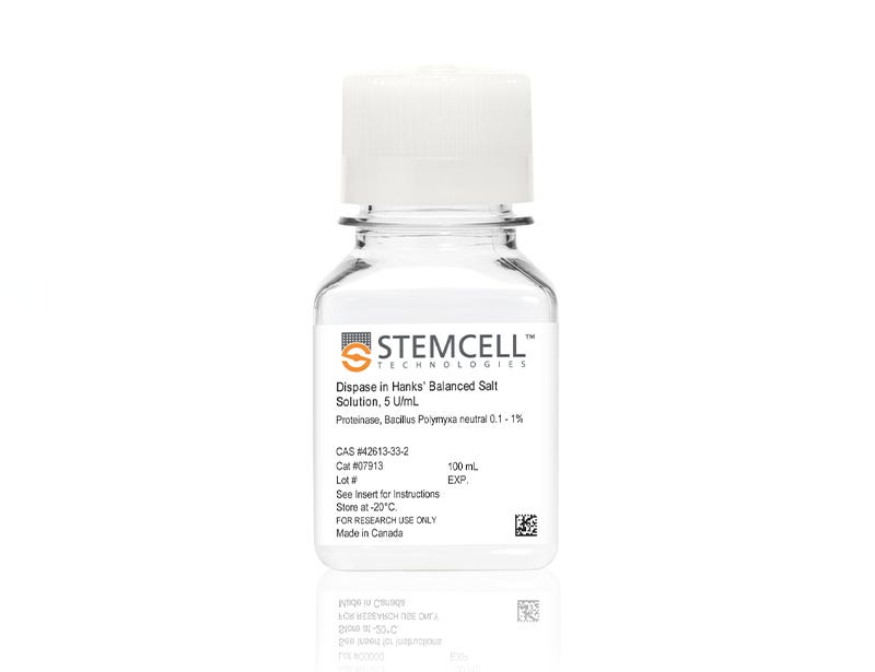
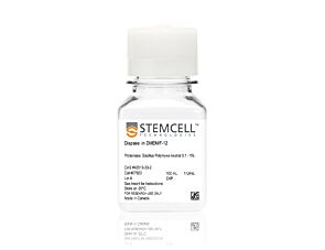
Dispase (5 U/mL)

5 U/mL dispase in Hanks' Balanced Salt Solution

Dispase (5 U/mL)

5 U/mL dispase in Hanks' Balanced Salt Solution

From: 288 USD
Catalog #
(Select a product)
5 U/mL dispase in Hanks' Balanced Salt Solution
Add to Wish List

Product Advantages


  • Achieve gentle dissociation in a wide variety of tissues

  • Optimized for for the generation of single-cell suspensions from dissociated human and mouse mammary tissue.

Overview

Use Dispase for gentle dissociation of a wide variety of tissues. Incubation of minced tissue with pre-warmed Dispase and gentle agitation will liberate cells with minimal cell damage. Pre-warmed Dispase can also be used to harvest cells from tissue culture plastic. This proteolytic dissociation reagent has been optimized for use in the generation of a single-cell suspension from dissociated human and mouse mammary tissue.

This product contains 5 U/mL Dispase II (neutral protease from Bacillus polymyxa) dissolved in Hanks’ Balanced Salt Solution. Unlike trypsin, Dispase is not inhibited by serum. Dispase activity is inhibited by EDTA and EGTA. Dispase should be removed from cell suspensions by centrifugation followed by washing the cells with
buffer or culture medium.
Subtype
Enzymatic
Alternative Names
Neutral protease; Proteinase
Cell Type
Intestinal Cells, Mammary Cells, Other, Pluripotent Stem Cells, Prostate Cells
Species
Human, Mouse, Non-Human Primate, Other, Rat
Application
Cell Culture
Area of Interest
Epithelial Cell Biology, Stem Cell Biology

Protocols and Documentation

Find supporting information and directions for use in the Product Information Sheet or explore additional protocols below.

Document Type
Product Name
Catalog #
Lot #
Language
Product Name
Dispase (5 U/mL)
Catalog #
07913
Lot #
All
Language
English
Document Type
Safety Data Sheet
Product Name
Dispase (5 U/mL)
Catalog #
07913
Lot #
All
Language
English

Applications

This product is designed for use in the following research area(s) as part of the highlighted workflow stage(s). Explore these workflows to learn more about the other products we offer to support each research area.

Resources and Publications

Publications (13)

Multisystemic Disease Modeling of Liver-Derived Protein Folding Disorders Using Induced Pluripotent Stem Cells (iPSCs). Leung A and Murphy GJ Methods in molecular biology (Clifton, N.J.) 2016 JAN

Abstract

Familial transthyretin amyloidosis (ATTR) is an autosomal dominant protein-folding disorder caused by over 100 distinct mutations in the transthyretin (TTR) gene. In ATTR, protein secreted from the liver aggregates and forms fibrils in target organs, chiefly the heart and peripheral nervous system, highlighting the need for a model capable of recapitulating the multisystem complexity of this clinically variable disease. Here, we describe detailed methodologies for the directed differentiation of protein folding disease-specific iPSCs into hepatocytes that produce mutant protein, and neural-lineage cells often targeted in disease. Methodologies are also described for the construction of multisystem models and drug screening using iPSCs.
Arterial specification of endothelial cells derived from human induced pluripotent stem cells in a biomimetic flow bioreactor. Sivarapatna A et al. Biomaterials 2015 JUN

Abstract

Endothelial cells (ECs) exist in different microenvironments in vivo, including under different levels of shear stress in arteries versus veins. Standard stem cell differentiation protocols to derive ECs and EC-subtypes from human induced pluripotent stem cells (hiPSCs) generally use growth factors or other soluble factors in an effort to specify cell fate. In this study, a biomimetic flow bioreactor was used to subject hiPSC-derived ECs (hiPSC-ECs) to shear stress to determine the impacts on phenotype and upregulation of markers associated with an anti-thrombotic, anti-inflammatory, arterial-like phenotype. The in vitro bioreactor system was able to efficiently mature hiPSC-ECs into arterial-like cells in 24 h, as demonstrated by qRT-PCR for arterial markers EphrinB2, CXCR4, Conexin40 and Notch1, as well protein-level expression of Notch1 intracellular domain (NICD). Furthermore, the exogenous addition of soluble factors was not able to fully recapitulate this phenotype that was imparted by shear stress exposure. The induction of these phenotypic changes was biomechanically mediated in the shear stress bioreactor. This biomimetic flow bioreactor is an effective means for the differentiation of hiPSC-ECs toward an arterial-like phenotype, and is amenable to scale-up for culturing large quantities of cells for tissue engineering applications.
Transcriptional profiling of ectoderm specification to keratinocyte fate in human embryonic stem cells Tadeu AMB et al. PLoS ONE 2015 APR

Abstract

In recent years, several studies have shed light into the processes that regulate epidermal specification and homeostasis. We previously showed that a broad-spectrum γ-secretase inhibitor DAPT promoted early keratinocyte specification in human embryonic stem cells triggered to undergo ectoderm specification. Here, we show that DAPT accelerates human embryonic stem cell differentiation and induces expression of the ectoderm protein AP2. Furthermore, we utilize RNA sequencing to identify several candidate regulators of ectoderm specification including those involved in epithelial and epidermal development in human embryonic stem cells. Genes associated with transcriptional regulation and growth factor activity are significantly enriched upon DAPT treatment during specification of human embryonic stem cells to the ectoderm lineage. The human ectoderm cell signature identified in this study contains several genes expressed in ectodermal and epithelial tissues. Importantly, these genes are also associated with skin disorders and ectodermal defects, providing a platform for understanding the biology of human epidermal keratinocyte development under diseased and homeostatic conditions.