Make more informed purchasing decisions with our new product availability and delivery estimate feature, now available on all product pages, in your cart, and during checkout.
Sign In
New to STEMCELL?
Register for an account to quickly and easily purchase products online and for one-click access to all educational content.
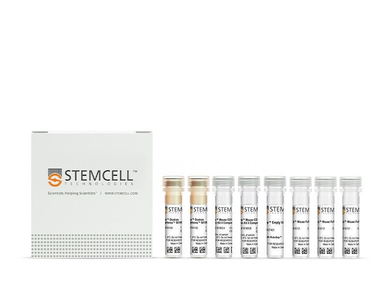
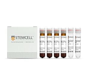
New format, same high quality! You may notice that your kit contents and packaging look slightly different from previous orders. We are currently updating the format of select EasySep™ Mouse kits to include a Mouse FcR blocker instead of Normal Rat Serum. With this change, all components will now be shipped in a single package, while providing the same cell isolation performance as before.
Request Pricing
Thank you for your interest in this product.
Please provide us with your contact information and your local representative
will contact you with a customized quote. Where appropriate, they can also assist you with a(n):
Estimated delivery time for your area
Product sample or exclusive offer
In-lab demonstration
By submitting this form, you are providing your consent to STEMCELL Technologies Canada Inc. and its subsidiaries and affiliates (“STEMCELL”) to collect and use your information, and send you newsletters and emails in accordance with our privacy policy. Please contact us with any questions that you may have. You can unsubscribe or change your email preferences at any time.
Easily isolate highly purified mouse CD11c+ cells from mouse splenocytes or cultured bone marrow cell samples, using immunomagnetic positive selection with the EasySep™ Mouse CD11c Positive Selection Kit II. Widely used in published research for more than 20 years, EasySep™ combines the specificity of monoclonal antibodies with the simplicity of a column-free magnetic system.
In this EasySep™ positive selection procedure, desired cells are labeled with antibody complexes recognizing CD11c and magnetic particles. Labeled cells are separated using an EasySep™ magnet and by simply pouring or pipetting off the unwanted cells. The cells of interest remain in the tube. Following magnetic cell isolation, the desired CD11c+ cells are ready for downstream applications such as flow cytometry, culture, and cell-based experiments.
Learn more about how immunomagnetic EasySep™ technology works or how to fully automate immunomagnetic cell isolation with RoboSep™. Explore additional products optimized for your workflow, including culture media, supplements, antibodies, and more.
Magnet Compatibility
• EasySep™ Magnet (Catalog #18000)
• “The Big Easy” EasySep™ Magnet (Catalog #18001)
• EasyEights™ EasySep™ Magnet (Catalog #18103)
• RoboSep™-S (Catalog #21000)
Subtype
Cell Isolation Kits
Cell Type
Dendritic Cells, Granulocytes and Subsets, Monocytes, NK Cells
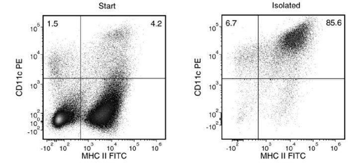
Starting with mouse splenocytes, the CD11c+ cell content of the enriched fraction is typically 86.8 ± 9.7% (gated on viable singlet cells, mean ± SD) using the purple EasySep™ Magnet. In the example above, the final purities of the start and isolated fraction are 5.7% and 92.3%, respectively.
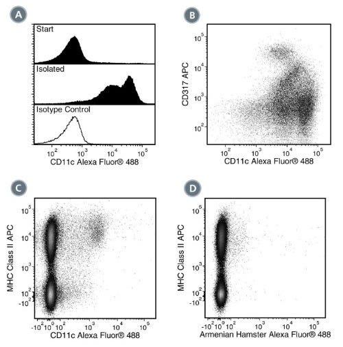
Figure 2. FACS Data for Anti-Mouse CD11c Antibody, Clone N418, Alexa Fluor® 488-Conjugated
(A) Flow cytometry analysis of C57BL/6 mouse splenocytes processed with EasySep™ Mouse CD11c Positive Selection Kit II (Catalog #18780) and labeled with Anti-Mouse CD11c Antibody, Clone N418, Alexa Fluor® 488 (Catalog # 60002AD). Histograms show labeling of splenocytes (Start) and isolated cells (Isolated). Labeling of the start cells with an Armenian hamster IgG Alexa Fluor® 488 isotype control antibody is shown in the bottom panel (solid line histogram).
(B) Flow cytometry analysis of C57BL/6 mouse splenocytes processed with EasySep™ Mouse CD11c Positive Selection Kit II and labeled with Anti-Mouse CD11c Antibody, Clone N418, Alexa Fluor® 488 and an anti-mouse CD317 antibody, APC.
(C) Flow cytometry analysis of C57BL/6 mouse splenocytes labeled with Anti-Mouse CD11c Antibody, Clone N418, Alexa Fluor® 488 and an anti-mouse MHC class II antibody, APC.
(D) Flow cytometry analysis of C57BL/6 mouse splenocytes labeled with an Armenian hamster IgG Alexa Fluor® 488 isotype control antibody and an anti-mouse MHC class II antibody, APC.
This product is designed for use in the following research area(s) as part
of the highlighted workflow stage(s). Explore these workflows to learn more about the other products we
offer to support each research area.
Can EasySep™ be used for either positive or negative selection?
Yes. The EasySep™ kits use either a negative selection approach by targeting and removing unwanted cells or a positive selection approach targeting desired cells. Depletion kits are also available for the removal of cells with a specific undesired marker (e.g. GlyA).
How does the separation work?
Magnetic particles are crosslinked to cells using Tetrameric Antibody Complexes (TAC). When placed in the EasySep™ Magnet, labeled cells migrate to the wall of the tube. The unlabeled cells are then poured off into a separate fraction.
Which columns do I use?
The EasySep™ procedure is column-free. That's right - no columns!
How can I analyze the purity of my enriched sample?
The Product Information Sheet provided with each EasySep™ kit contains detailed staining information.
Can EasySep™ separations be automated?
Yes. RoboSep™, the fully automated cell separator, automates all EasySep™ labeling and cell separation steps.
Can EasySep™ be used to isolate rare cells?
Yes. We recommend a cell concentration of 2x10⁸ cells/mL and a minimum working volume of 100 µL. Samples containing 2x10⁷ cells or fewer should be suspended in 100 µL of buffer.
Are the EasySep™ magnetic particles FACS-compatible?
Yes, the EasySep™ particles are flow cytometry-compatible, as they are very uniform in size and about 5000X smaller than other commercially available magnetic beads used with column-free systems.
Can the EasySep™ magnetic particles be removed after enrichment?
No, but due to the small size of these particles, they will not interfere with downstream applications.
Can I alter the separation time in the magnet?
Yes; however, this may impact the kit's performance. The provided EasySep™ protocols have already been optimized to balance purity, recovery and time spent on the isolation.
For positive selection, can I perform more than 3 separations to increase purity?
Yes, the purity of targeted cells will increase with additional rounds of separations; however, cell recovery will decrease.
How does the binding of the EasySep™ magnetic particle affect the cells? is the function of positively selected cells altered by the bound particles?
Hundreds of publications have used cells selected with EasySep™ positive selection kits for functional studies. Our in-house experiments also confirm that selected cells are not functionally altered by the EasySep™ magnetic particles.
If particle binding is a key concern, we offer two options for negative selection. The EasySep™ negative selection kits can isolate untouched cells with comparable purities, while RosetteSep™ can isolate untouched cells directly from whole blood without using particles or magnets.
Ethanol Intoxication and Burn Injury Increases Intestinal Regulatory T Cell Population and Regulatory T Cell Suppressive Capability.
M. E. Luck et al.
Shock (Augusta, Ga.) 2022 feb
Abstract
Traumatic injuries, such as burn, are often complicated by ethanol intoxication at the time of injury. This leads to a myriad of complications and post-burn pathologies exacerbated by aberrant immune responses. Recent findings suggest that immune cell dysfunction in the gastrointestinal system is particularly important in deleterious outcomes associated with burn injuries. In particular, intoxication at the time of burn injury leads to compromised intestinal T cell responses, which can diminish intestinal immunity and promote bacterial translocation, allowing for increased secondary infections in the injured host and associated sequelae, such as multiple organ failure and sepsis. Regulatory T cells (Treg) have been identified as important mediators of suppressing effector T cell function. Therefore, the goal of this study was to assess the effects of ethanol intoxication and burn injury on Treg populations in small intestinal immune organs. We also evaluated the suppressive capability of Tregs isolated from injured animals. Male C57BL/6 mice were gavaged with 2.9?Šg/kg ethanol before receiving a ˆ¼12.5% total body surface area scald burn. One day after injury, we identified a significant increase in Tregs number in small intestine Peyer's patches (ˆ¼?—1.5) and lamina propria (ˆ¼?—2). Tregs-producing cytokine IL-10 were also increased in both tissues. Finally, Tregs isolated from ethanol and burn-injured mice were able to suppress proliferation of effector T cells to a greater degree than sham vehicle Tregs. This was accompanied by increased levels of IL-10 and decreased levels of pro-proliferative cytokine IL-2 in cultures containing ethanol + burn Tregs compared with sham Tregs. These findings suggest that Treg populations are increased in intestinal tissues 1 day following ethanol intoxication and burn injury. Tregs isolated from ethanol and burn-injured animals also exhibit a greater suppression of effector T cell proliferation, which may contribute to altered T cell responses following injury.
Characterization of the Immunologic Phenotype of Dendritic Cells Infected With Herpes Simplex Virus 1.
J. Zhang et al.
Frontiers in immunology 2022
Abstract
Due to viral envelope glycoprotein D binding to cellular membrane HVEM receptor, HSV-1 can infect certain dendritic cells, which becomes an event in the viral strategy to interfere with the host's immune system. We previously generated the HSV-1 mutant strain M6, which produced an attenuated phenotype in mice and rhesus monkeys. The attenuated M6 strain was used to investigate how HSV-1 infection of dendritic cells interferes with both innate and adaptive immunity. Our study showed that dendritic cells membrane HVEM receptors could mediate infection of the wild-type strain and attenuated M6 strain and that dendritic cells infected by both viruses in local tissues of animals exhibited changes in transcriptional profiles associated with innate immune and inflammatory responses. The infection of pDCs and cDCs by the two strains promoted cell differentiation to the CD103+ phenotype, but varied transcriptional profiles were observed, implying a strategy that the HSV-1 wild-type strain interferes with antiviral immunity, probably due to viral modification of the immunological phenotype of dendritic cells during processing and presentation of antigen to T cells, leading to a series of deviations in immune responses, ultimately generating the deficient immune phenotype observed in infected individuals in the clinical.
PDE4B Is a Homeostatic Regulator of Cyclic AMP in Dendritic Cells.
A. M. Chinn et al.
Frontiers in pharmacology 2022
Abstract
Chronic decreases in the second messenger cyclic AMP (cAMP) occur in numerous settings, but how cells compensate for such decreases is unknown. We have used a unique system-murine dendritic cells (DCs) with a DC-selective depletion of the heterotrimeric GTP binding protein G$\alpha$s-to address this issue. These mice spontaneously develop Th2-allergic asthma and their DCs have persistently lower cAMP levels. We found that phosphodiesterase 4B (PDE4B) is the primary phosphodiesterase expressed in DCs and that its expression is preferentially decreased in G$\alpha$s-depleted DCs. PDE4B expression is dynamic, falling and rising in a protein kinase A-dependent manner with decreased and increased cAMP concentrations, respectively. Treatment of DCs that drive enhanced Th2 immunity with a PDE4B inhibitor ameliorated DC-induced helper T cell response. We conclude that PDE4B is a homeostatic regulator of cellular cAMP concentrations in DCs and may be a target for treating Th2-allergic asthma and other settings with low cellular cAMP concentrations.
New format, same high quality! You may notice that your kit contents and packaging look slightly different from previous orders. We are currently updating the format of select EasySep™ Mouse kits to include a Mouse FcR blocker instead of Normal Rat Serum. With this change, all components will now be shipped in a single package, while providing the same cell isolation performance as before.
Quality Statement:
PRODUCTS ARE FOR RESEARCH USE ONLY AND NOT INTENDED FOR HUMAN OR ANIMAL DIAGNOSTIC OR THERAPEUTIC USES UNLESS OTHERWISE STATED. FOR ADDITIONAL INFORMATION ON QUALITY AT STEMCELL, REFER TO WWW.STEMCELL.COM/COMPLIANCE.