Anti-Mouse CD11c Antibody, Clone N418

Hamster (Armenian) monoclonal IgG antibody against mouse CD11c

Anti-Mouse CD11c Antibody, Clone N418

Hamster (Armenian) monoclonal IgG antibody against mouse CD11c

From: 77 USD
Catalog #
(Select a product)
Hamster (Armenian) monoclonal IgG antibody against mouse CD11c
Add to Wish List

Overview

The N418 antibody reacts with CD11c (αX integrin), a 150 kDa type 1 transmembrane glycoprotein that associates non-covalently with CD18 (β2 integrin) to form a heterodimeric cell surface adhesion receptor. Through its interaction with ligands such as iC3b, fibrinogen, and CD54, the CD11c/CD18 receptor is involved in several immune response processes, including cell migration, stimulation of cytokine production by monocytes and macrophages, T cell proliferation, leukocyte recruitment, and phagocytosis. In mice, CD11c is expressed on dendritic cells, macrophages, monocytes, granulocytes, NK cells, and a subset of T cells.

This antibody clone has been verified for purity assessments of cells isolated with EasySep™ kits, including EasySep™ Mouse CD11c Positive Selection Kit II (Catalog #18780).
Subtype
Primary Antibodies
Target Antigen
CD11c
Alternative Names
alphaX integrin, CR4, integrin alphaX chain, p150
Reactive Species
Mouse
Conjugation
Biotin, FITC, PE, Unconjugated
Host Species
Hamster
Cell Type
Dendritic Cells
Species
Mouse
Application
CyTOF, Flow Cytometry, Immunocytochemistry, Immunofluorescence, Immunohistochemistry, Immunoprecipitation
Area of Interest
Immunology
Clone
N418
Gene ID
16411
Isotype
IgG

Data Figures

Data for Alexa Fluor® 488-Conjugated

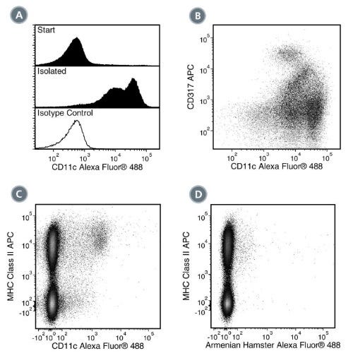
Figure 1. Data for Alexa Fluor® 488-Conjugated

(A) Flow cytometry analysis of C57BL/6 mouse splenocytes processed with EasySep™ Mouse CD11c Positive Selection Kit II and labeled with Anti-Mouse CD11c Antibody, Clone N418, Alexa Fluor® 488. Histograms show labeling of splenocytes (Start) and isolated cells (Isolated). Labeling of the start cells with an Armenian hamster IgG Alexa Fluor® 488 isotype control antibody is shown in the bottom panel (solid line histogram). (B) Flow cytometry analysis of C57BL/6 mouse splenocytes processed with EasySep™ Mouse CD11c Positive Selection Kit II and labeled with Anti-Mouse CD11c Antibody, Clone N418, Alexa Fluor® 488 and an anti-mouse CD317 antibody, APC. (C) Flow cytometry analysis of C57BL/6 mouse splenocytes labeled with Anti-Mouse CD11c Antibody, Clone N418, Alexa Fluor® 488 and an anti-mouse MHC class II antibody, APC. (D) Flow cytometry analysis of C57BL/6 mouse splenocytes labeled with an Armenian hamster IgG Alexa Fluor® 488 isotype control antibody and an anti-mouse MHC class II antibody, APC.

Data for PE-Conjugated

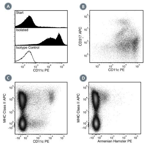
Figure 2. Data for PE-Conjugated

(A) Flow cytometry analysis of C57BL/6 mouse splenocytes processed with EasySep™ Mouse CD11c Positive Selection Kit II and labeled with Anti-Mouse CD11c Antibody, Clone N418, PE. Histograms show labeling of splenocytes (Start) and isolated cells (Isolated). Labeling of the start cells with an Armenian hamster IgG PE isotype control antibody is shown in the bottom panel (solid line histogram). (B) Flow cytometry analysis of C57BL/6 mouse splenocytes processed with EasySep™ Mouse CD11c Positive Selection Kit II and labeled with Anti-Mouse CD11c Antibody, Clone N418, PE and an anti-mouse CD317 antibody, APC. (C) Flow cytometry analysis of C57BL/6 mouse splenocytes labeled with Anti-Mouse CD11c Antibody, Clone N418, PE and an anti-mouse MHC class II antibody, APC. (D) Flow cytometry analysis of C57BL/6 mouse splenocytes labeled with an Armenian hamster IgG PE isotype control antibody and an anti-mouse MHC class II antibody, APC.

Data for Unconjugated

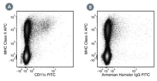
Figure 3. Data for Unconjugated

(A) Flow cytometry analysis of C57BL/6 mouse splenocytes labeled with Anti-Mouse CD11c Antibody, Clone N418, followed by anti-hamster (Armenian) IgG, FITC and anti-mouse MHC class II, APC. (B) Flow cytometry analysis of C57BL/6 mouse splenocytes labeled with a hamster (Armenian) IgG, isotype control antibody followed by anti-hamster (Armenian) IgG, FITC and anti-mouse MHC class II, APC.

Data for APC-Conjugated

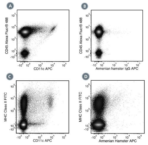
Figure 4. Data for APC-Conjugated

(A) Flow cytometry analysis of C57BL/6 mouse splenocytes labeled with Anti-Mouse CD11c Antibody, Clone N418, APC and Anti-Mouse CD45 Antibody, Clone 30-F11, Alexa Fluor® 488 (Catalog #60030AD). (B) Flow cytometry analysis of C57BL/6 mouse splenocytes labeled with an Armenian hamster IgG APC isotype control antibody and Anti-Mouse CD45 Antibody, Clone 30-F11, Alexa Fluor® 488. (C) Flow cytometry analysis of C57BL/6 mouse splenocytes labeled with Anti-Mouse CD11c Antibody, Clone N418, APC and an anti-mouse MHC class II antibody, FITC. (D) Flow cytometry analysis of C57BL/6 mouse splenocytes labeled with an Armenian hamster IgG APC isotype control antibody and an anti-mouse MHC class II antibody, FITC.

Data for Biotin-Conjugated

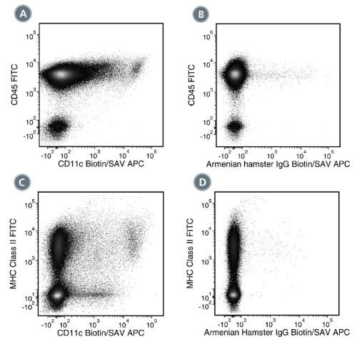
Figure 5. Data for Biotin-Conjugated

(A) Flow cytometry analysis of C57BL/6 mouse splenocytes labeled with Anti-Mouse CD11c Antibody, Clone N418, Biotin followed by streptavidin (SAV) APC and Anti-Mouse CD45 Antibody, Clone 30-F11, FITC (Catalog #60030FI). (B) Flow cytometry analysis of C57BL/6 mouse splenocytes labeled with a biotinylated Armenian hamster IgG, isotype control antibody followed by SAV APC and Anti-Mouse CD45 Antibody, Clone 30-F11, FITC. (C) Flow cytometry analysis of C57BL/6 mouse splenocytes labeled with Anti-Mouse CD11c Antibody, Clone N418, Biotin, followed by SAV APC and an anti-mouse MHC class II antibody, FITC. (D) Flow cytometry analysis of C57BL/6 mouse splenocytes labeled with a biotinylated Armenian hamster IgG, isotype control antibody followed by SAV APC and an anti-mouse MHC class II antibody, FITC.

Data for FITC-Conjugated

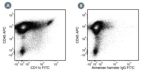
Figure 6. Data for FITC-Conjugated

(A) Flow cytometry analysis of C57BL/6 mouse splenocytes labeled with Anti-Mouse CD11c Antibody, Clone N418, FITC and Anti-Mouse CD45 Antibody, Clone 30-F11, APC (Catalog #60030AZ). (B) Flow cytometry analysis of C57BL/6 mouse splenocytes labeled with an Armenian hamster IgG FITC isotype control antibody and Anti-Mouse CD45 Antibody, Clone 30-F11, APC.

Data for PerCP-Cy55-Conjugated

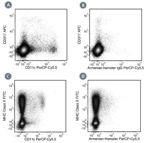
Figure 7. Data for PerCP-Cy55-Conjugated

(A) Flow cytometry analysis of C57BL/6 mouse splenocytes labeled with Anti-Mouse CD11c Antibody, Clone N418, PerCP-Cy5.5 and an anti-mouse CD317 antibody, APC. (B) Flow cytometry analysis of C57BL/6 mouse splenocytes labeled with an Armenian hamster IgG PerCP-Cy5.5 isotype control antibody and an anti-mouse CD317 antibody, APC. (C) Flow cytometry analysis of C57BL/6 mouse splenocytes labeled with Anti-Mouse CD11c Antibody, Clone N418, PerCP-Cy5.5 and an anti-mouse MHC class II antibody, FITC. (D) Flow cytometry analysis of C57BL/6 mouse splenocytes labeled with an Armenian hamster IgG PerCP-Cy5.5 isotype control antibody and an anti-mouse MHC class II antibody, FITC.

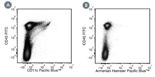
Figure 8. Data for PB-Conjugated

(A) Flow cytometry analysis of C57BL/6 mouse splenocytes labeled with Anti-Mouse CD11c Antibody, Clone N418, Pacific Blue™ and Anti-Mouse CD45 Antibody, Clone 30-F11, FITC (Catalog #60030FI). (B) Flow cytometry analysis of C57BL/6 mouse splenocytes labeled with an Armenian hamster IgG Pacific Blue™ isotype control antibody and Anti-Mouse CD45 Antibody, Clone 30-F11, FITC.

Protocols and Documentation

Find supporting information and directions for use in the Product Information Sheet or explore additional protocols below.

Document Type
Product Name
Catalog #
Lot #
Language
Catalog #
60002
Lot #
All
Language
English
Catalog #
100-0442, 60002BT, 100-0441
Lot #
All
Language
English
Catalog #
100-0444, 60002FI, 100-0443
Lot #
All
Language
English
Catalog #
100-0446, 100-0445
Lot #
All
Language
English
Catalog #
60002PB, 60002PB.1
Lot #
All
Language
English
Catalog #
60002AZ, 60002AZ.1
Lot #
All
Language
English
Catalog #
60002PS, 60002PS.1
Lot #
All
Language
English
Catalog #
100-0440
Lot #
All
Language
English
Catalog #
60002AD, 60002AD.1
Lot #
All
Language
English
Document Type
Safety Data Sheet
Catalog #
60002
Lot #
All
Language
English
Document Type
Safety Data Sheet
Catalog #
100-0442, 60002BT, 100-0441
Lot #
All
Language
English
Document Type
Safety Data Sheet
Catalog #
100-0444, 60002FI, 100-0443
Lot #
All
Language
English
Document Type
Safety Data Sheet
Catalog #
100-0446, 100-0445
Lot #
All
Language
English
Document Type
Safety Data Sheet
Catalog #
60002PB, 60002PB.1
Lot #
All
Language
English
Document Type
Safety Data Sheet
Catalog #
60002AZ, 60002AZ.1
Lot #
All
Language
English
Document Type
Safety Data Sheet
Catalog #
60002PS, 60002PS.1
Lot #
All
Language
English
Document Type
Safety Data Sheet
Catalog #
100-0440
Lot #
All
Language
English
Document Type
Safety Data Sheet
Catalog #
60002AD, 60002AD.1
Lot #
All
Language
English

Applications

This product is designed for use in the following research area(s) as part of the highlighted workflow stage(s). Explore these workflows to learn more about the other products we offer to support each research area.

Resources and Publications

Publications (1)

Simple suspension culture system of human iPS cells maintaining their pluripotency for cardiac cell sheet engineering. Haraguchi Y et al. Journal of Tissue Engineering and Regenerative Medicine 2015 DEC

Abstract

In this study, a simple three-dimensional (3D) suspension culture method for the expansion and cardiac differentiation of human induced pluripotent stem cells (hiPSCs) is reported. The culture methods were easily adapted from two-dimensional (2D) to 3D culture without any additional manipulations. When hiPSCs were directly applied to 3D culture from 2D in a single-cell suspension, only a few aggregated cells were observed. However, after 3 days, culture of the small hiPSC aggregates in a spinner flask at the optimal agitation rate created aggregates which were capable of cell passages from the single-cell suspension. Cell numbers increased to approximately 10-fold after 12 days of culture. The undifferentiated state of expanded hiPSCs was confirmed by flow cytometry, immunocytochemistry and quantitative RT-PCR, and the hiPSCs differentiated into three germ layers. When the hiPSCs were subsequently cultured in a flask using cardiac differentiation medium, expression of cardiac cell-specific genes and beating cardiomyocytes were observed. Furthermore, the culture of hiPSCs on Matrigel-coated dishes with serum-free medium containing activin A, BMP4 and FGF-2 enabled it to generate robust spontaneous beating cardiomyocytes and these cells expressed several cardiac cell-related genes, including HCN4, MLC-2a and MLC-2v. This suggests that the expanded hiPSCs might maintain the potential to differentiate into several types of cardiomyocytes, including pacemakers. Moreover, when cardiac cell sheets were fabricated using differentiated cardiomyocytes, they beat spontaneously and synchronously, indicating electrically communicative tissue. This simple culture system might enable the generation of sufficient amounts of beating cardiomyocytes for use in cardiac regenerative medicine and tissue engineering.