STEMdiff™ Cardiomyocyte Dissociation Kit

For dissociation of hPSC-derived cardiomyocytes

STEMdiff™ Cardiomyocyte Dissociation Kit

For dissociation of hPSC-derived cardiomyocytes
From: 228 USD
Catalog #
(Select a product)
For dissociation of hPSC-derived cardiomyocytes
Add to Wish List

Product Advantages


  • Maintains the integrity of hPSC-derived cardiomyocytes with greater than 80% cell viability.

  • Simple and robust method for harvesting and replating hPSC-derived cardiomyocytes

What's Included

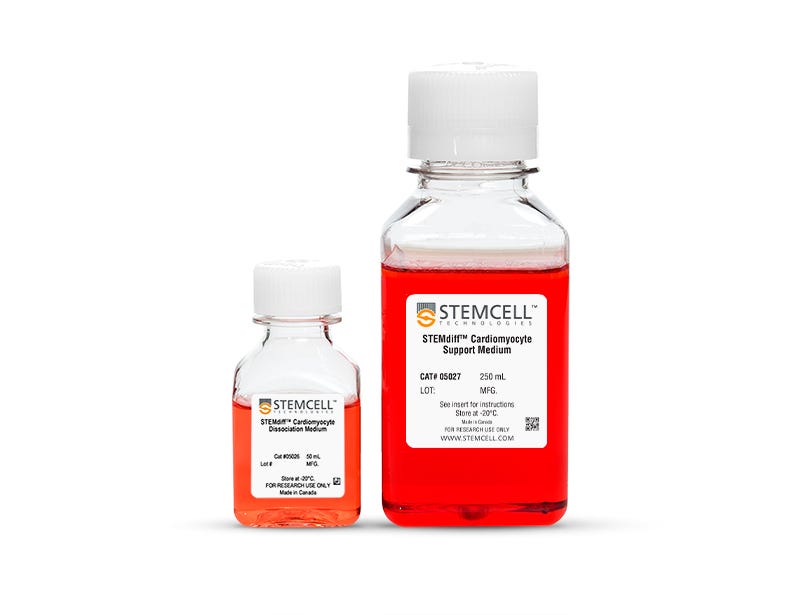
  • STEMdiff™ Cardiomyocyte Dissociation Medium, 50 mL
  • STEMdiff™ Cardiomyocyte Support Medium, 250 mL
Products for Your Protocol
To see all required products for your protocol, please consult the Protocols and Documentation.

Overview

STEMdiff™ Cardiomyocyte Dissociation Kit includes STEMdiff™ Cardiomyocyte Dissociation Medium and STEMdiff™ Cardiomyocyte Support Medium. STEMdiff™ Cardiomyocyte Dissociation Medium can be used to harvest cardiomyocytes that have been differentiated from human pluripotent stem cells (hPSCs) using STEMdiff™ Ventricular Cardiomyocyte Differentiation Kit (Catalog #05010) or STEMdiff™ Atrial Cardiomyocyte Differentiation Kit (Catalog #100-0215) and maintained in STEMdiff™ Cardiomyocyte Maintenance Medium (Catalog #05010/05020). STEMdiff™ Cardiomyocyte Support Medium reduces stress on these cardiomyocytes during harvesting and replating, maintaining their viability and functional capacity for downstream applications and analyses.
Subtype
Specialized Media
Cell Type
Cardiomyocytes, PSC-Derived
Species
Human
Application
Cell Culture
Brand
STEMdiff
Area of Interest
Stem Cell Biology

Protocols and Documentation

Find supporting information and directions for use in the Product Information Sheet or explore additional protocols below.

Document Type
Product Name
Catalog #
Lot #
Language
Catalog #
05025
Lot #
All
Language
English
Document Type
Safety Data Sheet 1
Catalog #
05025
Lot #
All
Language
English
Document Type
Safety Data Sheet 2
Catalog #
05025
Lot #
All
Language
English

Applications

This product is designed for use in the following research area(s) as part of the highlighted workflow stage(s). Explore these workflows to learn more about the other products we offer to support each research area.

Resources and Publications

Publications (14)

Dysregulation of sphingolipid-metabolizing enzymes in Friedreich's ataxia: In vitro and in vivo insights into therapeutic targeting. Z. Ramchunder et al. iScience 2026 Jul

Abstract

Friedreich's ataxia (FRDA) is an inherited neurodegenerative disorder caused by a GAA repeat expansion within the FXN gene, leading to reduced frataxin levels. This deficiency results in mitochondrial dysregulation, oxidative stress, and progressive cell death. Currently, only one approved treatment exists for FRDA in the United States, Canada, and the European Union, which improves neurological outcomes but has not been fully evaluated for broader disease symptoms. Therefore, identifying new therapeutic targets remains essential. Sphingolipids are increasingly recognized for their roles in neurodegeneration with emerging evidence indicating their dysregulation in FRDA. Here, we investigate whether sphingolipid-metabolizing enzymes are similarly affected and assess the therapeutic potential of targeting them. Our findings demonstrate that these enzymes are dysregulated across multiple FRDA models. Importantly, their modulation in vitro and in vivo significantly reduces mitochondrial dysfunction, enhances frataxin expression, and improves key pathological features of the disease, highlighting sphingolipid metabolism as a promising therapeutic target for FRDA.
Differentiation of human induced pluripotent stem cells into cardiac valve cells using 2D and 3D differentiation protocols. Z. Farzaneh et al. Stem cell research & therapy 2026 Jul

Abstract

BACKGROUND: Given the high relevance of human cardiac valve disease, recent research aims to differentiate human induced pluripotent stem cells (hiPSCs) into valve endothelial-like cells (VELCs) and, through endothelial-to-mesenchymal transition (EndMT), into valve interstitial-like cells (VILCs). METHODS: Here, we modified a 2D differentiation protocol demonstrating that VEGF can serve as the sole driver for differentiating cardiac progenitor cells (CPCs) into VELCs. Next, we utilized the so-called GiWi protocol (inhibition of glycogen synthase kinase, followed by inhibition of the Wnt pathway) to derive VELCs from 3D endocardial spheres. To this aim, hiPSCs were first differentiated into cardiac progenitor (CP) spheres using CHIR99021 (12 µM) and IWP2 (5 µM). Subsequent treatment with E8 medium containing high-dose FGF2 (100 ng/ml) resulted in endocardial spheres enriched for VELCs. For EndMT induction, endocardial spheres were MACS-sorted for PECAM1+ VELCs and transdifferentiated into ACTA2+/CDH5- VILCs using TGFβ1 (200 ng/ml). RESULTS: Using VEGF as main driver to differentiate CPCs into VELCs in 2D, the generated VELCs appeared stable over time, can be maintained in vitro and transdifferentiated into ACTA2+ VILCs using FGF2 (100 ng/ml) and TGFβ1 (50 ng/ml). Besides, our novel 3D differentiation protocol yielded endocardial spheres, which were highly enriched with VELCs, as shown by the expression of GATA4 (≈ 83%), PECAM1 (≈ 69%), and nuclear NFATC1 (≈ 76%), along with typical functional characteristics such as network formation, LDL uptake and the ability to undergo EndMT. CONCLUSIONS: Overall, we developed a 2D differentiation protocol that produces stable VELCs and a high percentage of VILCs upon EndMT induction. We also established a new, efficient, and cost-effective protocol for the 3D differentiation of endocardial spheres to better mimic a physiologically relevant environment, thereby enabling improved maturation.
Cryopreservation alters contractile function of human induced pluripotent stem cell-derived cardiomyocytes. K. Kowalski et al. Scientific reports 2026 Jul

Abstract

Advanced protocols are available for efficient generation of large quantities of human induced pluripotent stem cell-derived cardiomyocytes (hiPSC-CMs). Nevertheless, hiPSC-CMs show large batch to batch variations as well as fetal-like phenotype. Cryopreservation enables long-term storage of batches, leading to increased consistency and reproducibility of data while improving maturation. However, controversial data regarding comparability of fresh and cryopreserved hiPSC-CMs have been reported. Here, we compared fresh and cryopreserved hiPSC-CMs, demonstrating that both cryopreservation media (CryoStor®CS10 and KnockOut Serum Replacement) had a comparable recovery rate (CS10: 39%, KSR: 46%) and similar proportion of CMs in long-term culture. Cryopreservation altered cell morphology (increased cell area and shorter or longer sarcomere length) and contractile parameters (faster time to peak and half relaxation time and higher or shorter contraction amplitude) of recovered hiPSC-CMs with only slight changes in sarcomeric gene and protein expression. Some differential effects of both cryo-media on CM structure and function were observed. The data indicate an influence of cryopreservation on cell morphology as well as on contraction parameters that should be considered in downstream applications.