EasySep™ Mouse T Cell Isolation Kit

Immunomagnetic negative isolation of untouched mouse T cells

Before performing cell isolation using EasySep™, consult the product information sheet (PIS) to determine whether red blood cell (RBC) lysis is required for your sample type. RBC lysis should only be performed if indicated in the PIS. It is often recommended for blood samples; however, RBC lysis is not recommended for mouse splenocytes as it may reduce cell recovery. For the most accurate cell recovery calculation, we recommend performing total nucleated cell (TNC) count.

See How to Calculate Cell Recovery & Measure Purity After Cell Isolation >

EasySep™ Mouse T Cell Isolation Kit

Immunomagnetic negative isolation of untouched mouse T cells
From: 790 USD
Catalog #
(Select a product)
Immunomagnetic negative isolation of untouched mouse T cells
Add to Wish List

Product Advantages


  • Fast and easy-to-use

  • Up to 99% purity

  • No columns required

  • Untouched, viable cells

What's Included

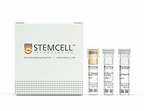
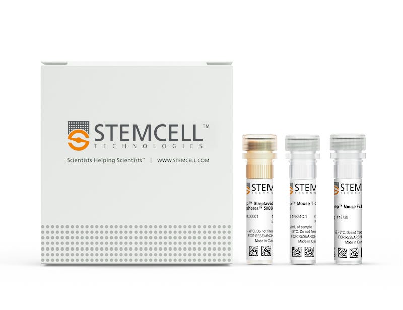
  • EasySep™ Mouse T Cell Isolation Kit (Catalog #19851)
    • EasySep™ Mouse T Cell Isolation Cocktail, 0.5 mL
    • EasySep™ Streptavidin RapidSpheres™ 50001, 1 mL
    • EasySep™ FcR Mouse Blocker, 0.2 mL
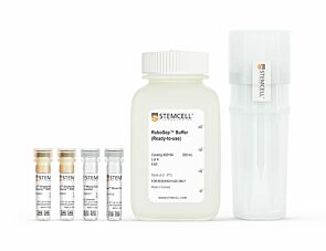
  • RoboSep™ Mouse T Cell Isolation Kit (Catalog #19851RF)
    • EasySep™ Mouse T Cell Isolation Cocktail, 0.5 mL
    • EasySep™ Streptavidin RapidSpheres™ 50001, 1.0 mL
    • EasySep™ FcR Mouse Blocker, 0.2 mL
    • RoboSep™ Buffer (Catalog #20104)
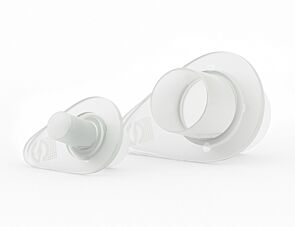
    • RoboSep™ Filter Tips (Catalog #20125)
Products for Your Protocol
To see all required products for your protocol, please consult the Protocols and Documentation.

Overview

Easily and efficiently isolate highly purified mouse T cells from single-cell suspensions of splenocytes or other tissues by immunomagnetic negative selection, with the EasySep™ Mouse T Cell Isolation Kit. Widely used in published research for more than 20 years, EasySep™ combines the specificity of monoclonal antibodies with the simplicity of a column-free magnetic system.

In this EasySep™ negative selection procedure, unwanted cells are labeled with antibody complexes and magnetic particles. Unwanted cells expressing the following markers are targeted for removal: CD11b, CD45R, Ter119, CD49b, CD19, and CD24. The magnetically labeled cells are then separated from the untouched desired mouse T cells by using an EasySep™ magnet and simply pouring or pipetting the desired cells into a new tube. Following magnetic cell isolation in as little as 15 minutes, the desired T cells are ready for downstream applications such as flow cytometry, culture, and cell-based experiments.

Learn more about how immunomagnetic EasySep™ technology works or how to fully automate immunomagnetic cell isolation with RoboSep™. Explore additional products optimized for your workflow, including culture media, supplements, antibodies, and more.
Magnet Compatibility
• EasySep™ Magnet (Catalog #18000)
• “The Big Easy” EasySep™ Magnet (Catalog #18001)
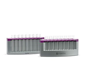
• EasyEights™ EasySep™ Magnet (Catalog #18103)
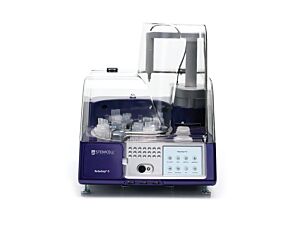
• RoboSep™-S (Catalog #21000)
Subtype
Cell Isolation Kits
Cell Type
T Cells
Species
Mouse
Sample Source
Other, Spleen
Selection Method
Negative
Application
Cell Isolation
Brand
EasySep, RoboSep
Area of Interest
Immunology

Data Figures

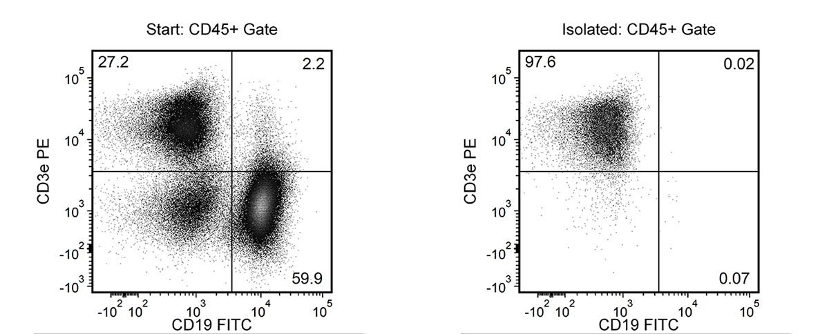
Typical EasySep™ Mouse T Cell Isolation Profile

Figure 1.Typical EasySep™ Mouse T Cell Isolation Profile

Starting with mouse splenocytes, the T cell content (CD3+CD19-) of the isolated fraction is 96.6 ± 2.0% (mean ± SD), using the purple EasySep™ magnet.

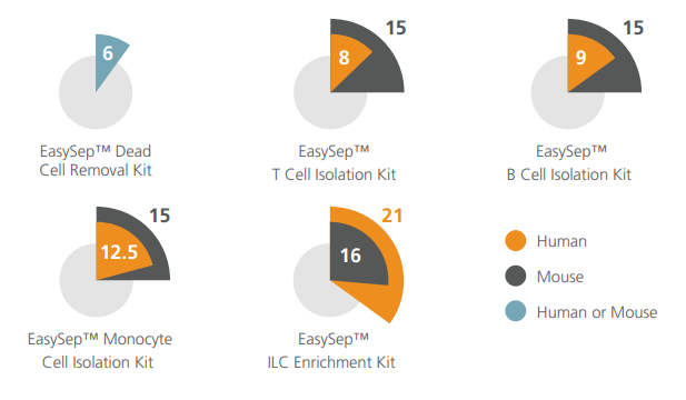
Cell Isolation Protocol Lengths

Figure 2.Cell Isolation Protocol Lengths

Typical time taken (in minutes) to isolate cells using select EasySep™ kits.

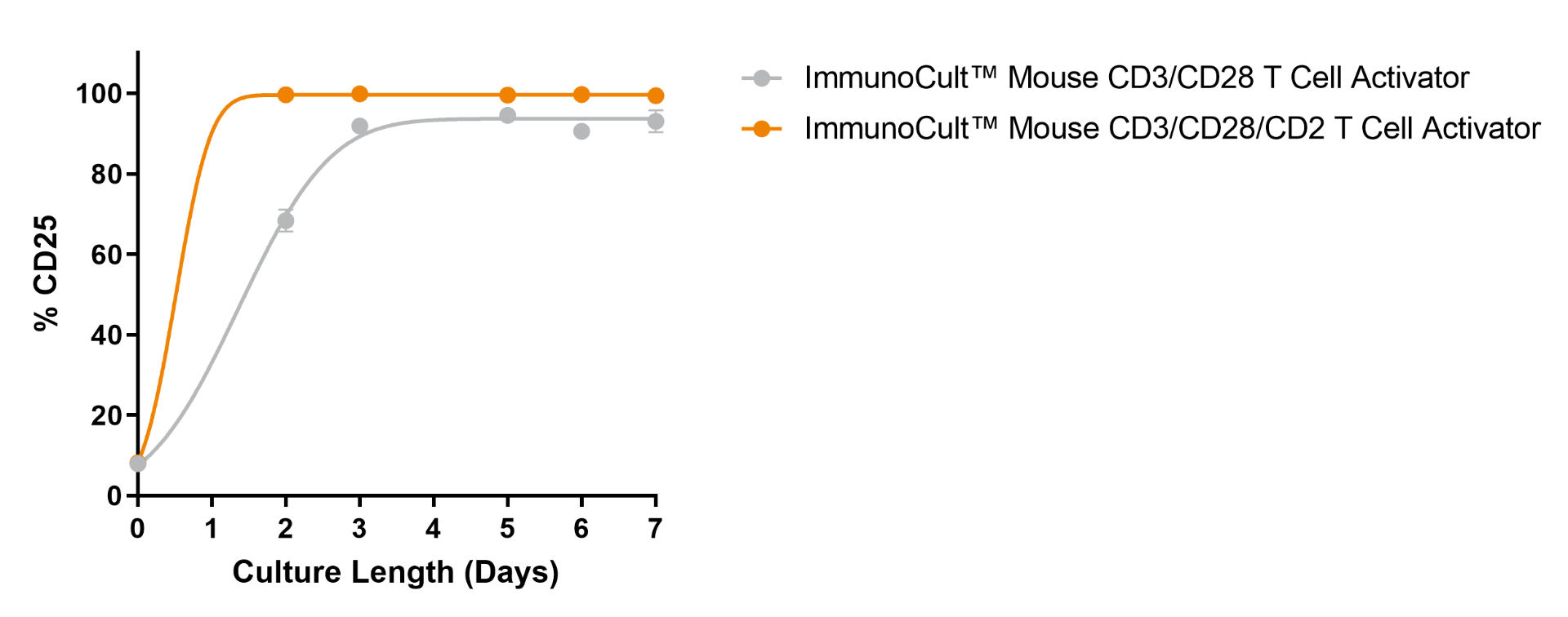
ImmunoCult™ Mouse T Cell Activator Kit Supports High Viability of Activated T Cells

Figure 3.ImmunoCult™ Mouse T Cell Activator Kit Supports High Viability of Activated T Cells

Mouse T cells were isolated using EasySep™ Mouse T Cell Isolation Kit (Catalog #19851), stimulated with ImmunoCult™ Mouse T Cell Activator Kit (Catalog #100-1572), and cultured in IMDM + FBS formulation. Following 3 days of culture, the mean ± SD frequency of CD25+ cells was 91.9 ± 5.1% (n = 11) or 99.9 ± 0.1% (n = 5), when stimulated with ImmunoCult™ Mouse CD3/CD28 T Cell Activator or ImmunoCult™ Mouse CD3/CD28/CD2 T Cell Activator, respectively. Stimulated mouse T cells maintained expression levels of CD25 throughout the 7-day culture period.

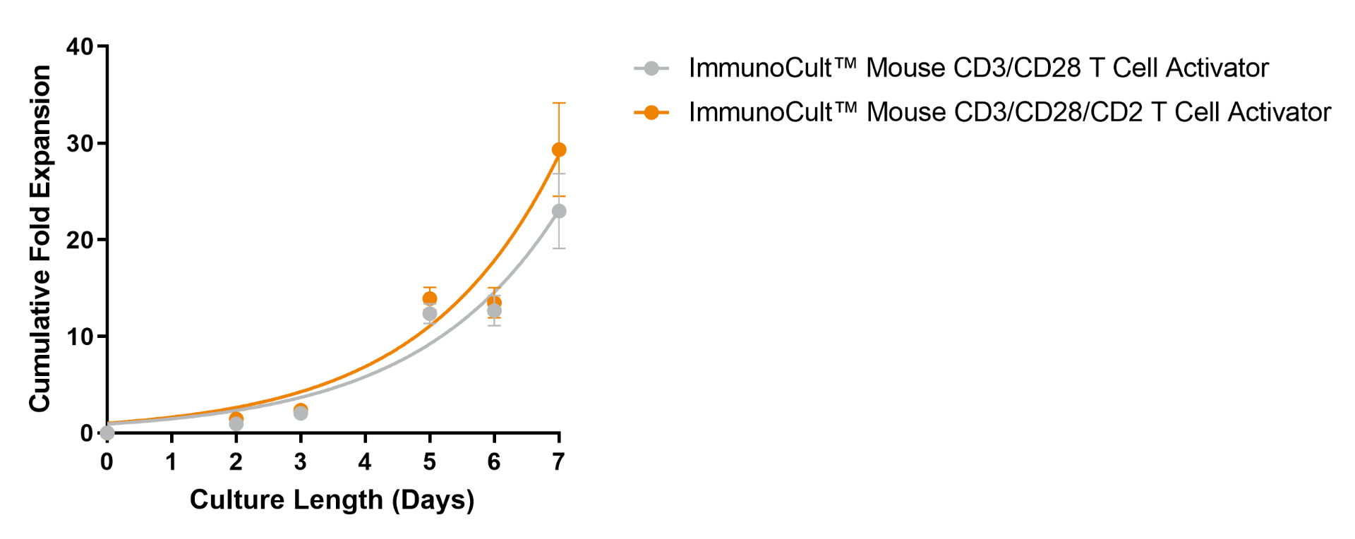
Robust Expansion of EasySep™-Isolated Mouse T Cells Can Be Achieved Following Stimulation with ImmunoCult™ Mouse T Cell Activator Kit

Figure 4.Robust Expansion of EasySep™-Isolated Mouse T Cells Can Be Achieved Following Stimulation with ImmunoCult™ Mouse T Cell Activator Kit

Mouse T cells isolated using EasySep™ Mouse T Cell Isolation Kit (Catalog #19851) were expanded with ImmunoCult™ Mouse T Cell Activator Kit (Catalog #100-1572) in IMDM + FBS formulation over 7 days. The number of viable cells was assessed every 2 - 3 days, and fresh medium supplemented with IL-2 was added. No additional ImmunoCult™ Mouse T Cell Activator was added during the 7-day culture period. After 7 days in culture with ImmunoCult™ Mouse CD3/CD28 T Cell Activator or ImmunoCult™ Mouse CD3/CD28/CD2 T Cell Activator, stimulation resulted in a fold expansion of 23 ± 3.4 or 29.3 ± 4.8 (mean ± SEM, n = 6), respectively.

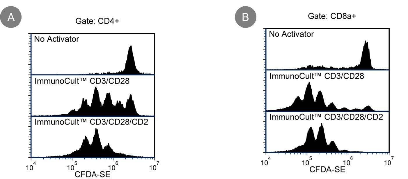
High Cell Proliferation is Observed in EasySep™-Isolated T cells After Stimulation with ImmunoCult™ Mouse T Cell Activator

Figure 5.High Cell Proliferation is Observed in EasySep™-Isolated T cells After Stimulation with ImmunoCult™ Mouse T Cell Activator

Mouse T cells isolated using EasySep™ Mouse T Cell Isolation Kit (Catalog #19851) were labeled with CFDA-SE (Catalog #75003), stimulated with ImmunoCult™ Mouse T Cell Activator Kit (Catalog #100-1572), and cultured in cultured in IMDM + FBS formulation. On Day 3, cells were harvested, stained with anti-mouse CD4 and CD8a antibodies, then measured by flow cytometry. Shown are CFDA-SE-labeled mouse T cells, gated on viable CD4+ (A) or CD8a+ (B) cells, cultured with no activator (top panel), with ImmunoCult™ Mouse CD3/CD28 T Cell Activator (middle panel), or with ImmunoCult™ Mouse CD3/CD28/CD2 T Cell Activator (bottom panel). Due to cell proliferation, the intensity of CFDA-SE signal is reduced by 50% for each cell division.

Protocols and Documentation

Find supporting information and directions for use in the Product Information Sheet or explore additional protocols below.

Document Type
Product Name
Catalog #
Lot #
Language
Catalog #
19851
Lot #
1000120298 or higher
Language
English
Catalog #
19851RF
Lot #
1000120298 or higher
Language
English
Document Type
Safety Data Sheet 1
Catalog #
19851
Lot #
All
Language
English
Document Type
Safety Data Sheet 2
Catalog #
19851
Lot #
All
Language
English
Document Type
Safety Data Sheet 3
Catalog #
19851
Lot #
All
Language
English
Document Type
Safety Data Sheet 4
Catalog #
19851
Lot #
All
Language
English
Document Type
Safety Data Sheet 1
Catalog #
19851RF
Lot #
All
Language
English
Document Type
Safety Data Sheet 2
Catalog #
19851RF
Lot #
All
Language
English
Document Type
Safety Data Sheet 3
Catalog #
19851RF
Lot #
All
Language
English
Document Type
Safety Data Sheet 4
Catalog #
19851RF
Lot #
All
Language
English
Document Type
Safety Data Sheet 5
Catalog #
19851RF
Lot #
All
Language
English

Applications

This product is designed for use in the following research area(s) as part of the highlighted workflow stage(s). Explore these workflows to learn more about the other products we offer to support each research area.

Resources and Publications

Frequently Asked Questions

Can EasySep™ Streptavidin RapidSpheres™ be used for either positive or negative selection?

Currently, EasySep™ Streptavidin RapidSphere™ kits are only available for negative selection and work by targeting and removing unwanted cells.

How does the separation work?

Streptavidin RapidSphere™ magnetic particles are crosslinked to unwanted cells using biotinylated antibodies. When placed in the EasySep™ Magnet, labeled cells migrate to the wall of the tube. The unlabeled cells are then poured off into a new tube.

Which columns do I use?

The EasySep™ procedure is column-free. That's right - no columns!

How can I analyze the purity of my enriched sample?

The Product Information Sheet provided with each EasySep™ kit contains detailed staining information.

Can EasySep™ Streptavidin RapidSphere™ separations be automated?

Yes. RoboSep™, the fully automated cell separator, automates all EasySep™ labeling and cell separation steps.

Are cells isolated using EasySep™ RapidSphere™ products FACS-compatible?

Yes. Desired cells are unlabeled and ready to use in downstream applications, such as FACS analysis.

Can I alter the separation time in the magnet?

Yes; however, this may impact the kit's performance. The provided EasySep™ protocols have already been optimized to balance purity, recovery and time spent on the isolation.

Publications (79)

Septin7 is essential in early hematopoiesis, but redundant at later stages. N. Ronkina et al. Life science alliance 2026 Oct

Abstract

The unique cytoskeletal protein Septin7 is generally considered to be required for cytokinesis in yeast and mammals. Whole-body genetic ablation of Septin7 in mice is embryonically lethal. Septin7-deficient fibroblasts and HeLa cells are defective in cytokinesis and undergo obligate multinucleation. Surprisingly, lymphocyte- and myeloid-specific deletion of Septin7 in mice caused no detectable defects in blood lineage development, indicating dispensability of Septin7 for hematopoiesis. However, Septin7-deficient hematopoietic stem cells failed to engraft and establish donor chimerism after transplantation. To reconcile these contradictory findings, we examined the consequences of Septin7 deletion at distinct stages of hematopoiesis. Our results demonstrate that Cre-induced deletion of the floxed Septin7 allele is inefficient during the early stages of hematopoiesis, suggesting strong selection pressure against Septin7 deficiency at this stage. In contrast, deletion of Septin7 at the common lymphoid progenitor stage, as well as in Hoxb8-immortalized hematopoietic progenitors, is efficient and does not result in any noticeable defects in cell division. Taken together, our findings indicate that Septin7 is essential during early hematopoiesis but becomes dispensable at later stages.
Protocol for immunomagnetic enrichment of T cells from complex murine tissues E. Trolio et al. STAR Protocols 2026 Mar

Abstract

SummaryT cells are the central effectors and regulators of the adaptive immune response. This protocol provides a step-by-step approach for isolating and enriching total T cells by negative selection from complex murine tissues, including bone marrow (BM), liver, heart, and kidneys. We describe steps for tissue harvesting, preparation of single-cell suspensions, and immunomagnetic enrichment. We then outline procedures for flow cytometric assessment of cell purity and viability. This protocol enables efficient recovery of high-quality T cells for reliable downstream analyses. Graphical abstract Highlights•Isolation of leukocytes from murine BM, liver, heart and kidneys•Non-enzymatic dissociation of kidney and heart tissue•Protocol for immunomagnetic enrichment of T cells•Flow cytometry analysis of T cell purity and viability Publisher’s note: Undertaking any experimental protocol requires adherence to local institutional guidelines for laboratory safety and ethics. T cells are the central effectors and regulators of the adaptive immune response. This protocol provides a step-by-step approach for isolating and enriching total T cells by negative selection from complex murine tissues, including bone marrow (BM), liver, heart, and kidneys. We describe steps for tissue harvesting, preparation of single-cell suspensions, and immunomagnetic enrichment. We then outline procedures for flow cytometric assessment of cell purity and viability. This protocol enables efficient recovery of high-quality T cells for reliable downstream analyses.
Inhibition of KDEL Receptors Remodels the Tumor Microenvironment for T Cell Independent Tumor Regression. S. Pattanayak et al. Advanced science (Weinheim, Baden-Wurttemberg, Germany) 2026 Jun

Abstract

Tumor immunotherapy is supported by low-grade inflammatory conditions in the microenvironment, triggered by immunogenic cell death (ICD). However, ICD is dampened when tumors acquire resistance, affecting immune recognition. KDEL receptors (KDELRs), through a retrograde Golgi-to-ER transport, prevent spontaneous secretion of KDEL proteins. We report that inhibition of a single KDELR in a minor fraction of tumor cells, primarily KDELR2, provokes robust infiltration of macrophages and neutrophils into the tumor microenvironment, resulting in regression of both immunogenic and non-immunogenic tumors initially independently of T cells. Importantly, in the course of regression, anti-tumor T cells are primed, conferring protection against a second challenge. Recapitulated by intratumoral delivery of siDKELR2 utilizing lipid nanoparticles, we implicate KDELR2 as a target to unleash an unusual robust innate immune response, which represents a tractable approach to initiate an adaptive response downstream, bypassing conventional ICD-inducing therapies. We propose KDELR targeting as a strategy to improve immunotherapy across tumor types, including "cold" tumors resistant to T cell-based immunotherapies.
Before performing cell isolation using EasySep™, consult the product information sheet (PIS) to determine whether red blood cell (RBC) lysis is required for your sample type. RBC lysis should only be performed if indicated in the PIS. It is often recommended for blood samples; however, RBC lysis is not recommended for mouse splenocytes as it may reduce cell recovery. For the most accurate cell recovery calculation, we recommend performing total nucleated cell (TNC) count.

See How to Calculate Cell Recovery & Measure Purity After Cell Isolation >