Human Peripheral Blood CD4+ T Cells, Frozen

Primary human cells, frozen
Save on high-quality cells! Take advantage of our bulk pricing discounts and price matching offer to get the best value for your purchase. Browse our Frequently Asked Questions for more information.

Human Peripheral Blood CD4+ T Cells, Frozen

Primary human cells, frozen

From: 295 USD
Catalog #
(Select a product)
Primary human cells, frozen
Add to Wish List

Overview

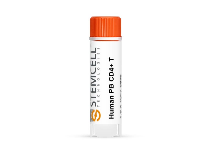
Primary human CD4+ T cells were isolated from peripheral blood (PB) mononuclear cells (MNCs) using negative immunomagnetic separation techniques. PB was collected using the anticoagulant acid-citrate-dextrose solution A (ACDA).

Cells were obtained using Institutional Review Board (IRB)-approved consent forms and protocols.

Certain products are only available in select territories. Please contact your local Sales representative or Product & Scientific Support at techsupport@stemcell.com for further information.

Browse our Frequently Asked Questions (FAQs) on Primary Cells.
Contains
• CryoStor® CS10
Subtype
Frozen
Cell Type
T Cells, T Cells, CD4+
Species
Human
Cell and Tissue Source
Peripheral Blood
Donor Status
Normal
Purity
≥ 85% CD4+ by flow cytometry

Protocols and Documentation

Find supporting information and directions for use in the Product Information Sheet or explore additional protocols below.

Document Type
Product Name
Catalog #
Lot #
Language
Catalog #
70026, 200-0165
Lot #
All
Language
English

Applications

This product is designed for use in the following research area(s) as part of the highlighted workflow stage(s). Explore these workflows to learn more about the other products we offer to support each research area.

Resources and Publications

Publications (2)

Isoform-selective activity-based profiling of ERK signaling. M. Shin et al. Chemical science 2018 MAR

Abstract

Extracellular signal-regulated kinases (ERKs) mediate downstream signaling of RAS-RAF-MEK as key regulators of the mitogen-activated protein kinase (MAPK) pathway. Activation of ERK signaling is a hallmark of cancer and upstream MAPK proteins have been extensively pursued as drug targets for cancer therapies. However, the rapid rise of resistance to clinical RAF and MEK inhibitors has prompted interest in targeting ERK (ERK1 and ERK2 isoforms) directly for cancer therapy. Current methods for evaluating activity of inhibitors against ERK isoforms are based primarily on analysis of recombinant proteins. Strategies to directly and independently profile native ERK1 and ERK2 activity would greatly complement current cell biological tools used to probe and target ERK function. Here, we present a quantitative chemoproteomic strategy that utilizes active-site directed probes to directly quantify native ERK activity in an isoform-specific fashion. We exploit a single isoleucine/leucine difference in ERK substrate binding sites to enable activity-based profiling of ERK1 versus ERK2 across a variety of cell types, tissues, and species. We used our chemoproteomic strategy to determine potency and selectivity of academic (VX-11e) and clinical (Ulixertinib) ERK inhibitors. Correlation of potency estimates by chemoproteomics with anti-proliferative activity of VX-11e and Ulixertinib revealed that {\textgreater}90{\%} inactivation of both native ERK1 and ERK2 is needed to mediate cellular activity of inhibitors. Our findings introduce one of the first assays capable of independent evaluation of native ERK1 and ERK2 activity to advance drug discovery of oncogenic MAPK pathways.
Evidence of an oncogenic role of aberrant TOX activation in cutaneous T-cell lymphoma. Huang Y et al. Blood 2015 FEB

Abstract

TOX is a nuclear factor essential for the development of CD4(+) T cells in the thymus. It is normally expressed in low amounts in mature CD4(+) T cells of the skin and the peripheral blood. We have recently discovered that the transcript levels of TOX were significantly increased in mycosis fungoides, the most common type of cutaneous T-cell lymphoma (CTCL), as compared to normal skin or benign inflammatory dermatoses. However, its involvement in advanced CTCL and its biological effects on CTCL pathogenesis have not been explored. In this study, we demonstrate that TOX expression is also enhanced significantly in primary CD4(+)CD7(-) cells from patients with Sézary syndrome, a leukemic variant of CTCL, and that high TOX transcript levels correlate with increased disease-specific mortality. Stable knockdown of TOX in CTCL cells promoted apoptosis and reduced cell cycle progression, leading to less cell viability and colony-forming ability in vitro and to reduced tumor growth in vivo. Furthermore, TOX knockdown significantly increased 2 cyclin-dependent kinase (CDK) inhibitors, CDKN1B and CDKN1C. Lastly, blocking CDKN1B and CDKN1C reversed growth inhibition of TOX knockdown. Collectively, these findings provide strong evidence that aberrant TOX activation is a critical oncogenic event for CTCL.
Save on high-quality cells! Take advantage of our bulk pricing discounts and price matching offer to get the best value for your purchase. Browse our Frequently Asked Questions for more information.