EasySep™ Mouse Monocyte Isolation Kit

15-Minute cell isolation kit using immunomagnetic negative selection
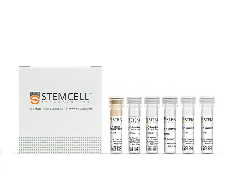
New format, same high quality! You may notice that your kit contents and packaging look slightly different from previous orders. We are currently updating the format of select EasySep™ Mouse kits to include a Mouse FcR blocker instead of Normal Rat Serum. With this change, all components will now be shipped in a single package, while providing the same cell isolation performance as before.

EasySep™ Mouse Monocyte Isolation Kit

15-Minute cell isolation kit using immunomagnetic negative selection

From: 847 USD
Catalog #
(Select a product)
15-Minute cell isolation kit using immunomagnetic negative selection
Add to Wish List

Product Advantages


  • Fast and easy-to-use

  • Up to 95% purity

  • No columns required

  • Untouched, viable cells

What's Included

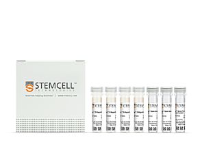
  • EasySep™ Mouse Monocyte Isolation Kit (Catalog #19861)
    • EasySep™ Mouse Monocyte Isolation Cocktail Component A, 0.5 mL
    • EasySep™ Mouse Monocyte Isolation Cocktail Component B, 0.5 mL
    • EasySep™ Dextran RapidSpheres™ 50103, 1 mL
    • EasySep™ Mouse FcR Blocker (Catalog #18731), 0.5 mL
  • RoboSep™ Mouse Monocyte Isolation Kit (Catalog #19861RF)
    • EasySep™ Mouse Monocyte Isolation Cocktail Component A, 0.5 mL
    • EasySep™ Mouse Monocyte Isolation Cocktail Component B, 0.5 mL
    • EasySep™ Dextran RapidSpheres™ 50103, 1 mL
    • EasySep™ Mouse FcR Blocker (Catalog #18731), 0.5 mL
    • RoboSep™ Empty Vial
    • RoboSep™ Buffer (Catalog #20104)
    • RoboSep™ Filter Tips (Catalog #20125)

Overview

The EasySep™ Mouse Monocyte Isolation Kit targets non-monocyte cells by labeling unwanted cells with antibodies and magnetic particles, and separates cells without columns using an EasySep™ magnet. Desired cells are simply poured off into a new tube. Isolated cells are immediately available for downstream applications such as flow cytometry, culture or cell-based assays.

This product replaces the EasySep™ Mouse Monocyte Enrichment Kit (Catalog #19761) for even faster cell isolations.
Magnet Compatibility
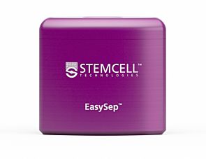
• EasySep™ Magnet (Catalog #18000)
• “The Big Easy” EasySep™ Magnet (Catalog #18001)
• EasyEights™ EasySep™ Magnet (Catalog #18103)
• EasyPlate™ EasySep™ Magnet (Catalog #18102)
• RoboSep™-S (Catalog #21000)
Subtype
Cell Isolation Kits
Cell Type
Monocytes
Species
Mouse
Sample Source
Bone Marrow, Spleen, Whole Blood
Selection Method
Negative
Application
Cell Isolation
Brand
EasySep, RoboSep
Area of Interest
Immunology

Data Figures

Typical EasySep™ Mouse Monocyte Isolation Profile

Figure 1. Typical FACS Profiles for EasySep™ Mouse Monocyte Isolation Kit

Starting with mouse bone marrow cells, the monocyte content (Lineage- (CD3, CD45R, CD117, CD49b, Siglec F) CD11b+Ly6G- Ly6Chi/lo) of the isolated fraction is 89.5 ± 4.8% (mean ± SD), using the purple EasySep™ Magnet. In the above example, monocyte purities in the start and final isolated fractions are 7.1% and 92.3%, respectively.

Data for Anti-Mouse CD11b Antibody, Clone M1/70, Alexa Fluor® 488-Conjugated

Figure 2. Data for Anti-Mouse CD11b Antibody, Clone M1/70, Alexa Fluor® 488-Conjugated

(A) Flow cytometry analysis of C57BL/6 mouse splenocytes labeled with Anti-Mouse CD11b Antibody, Clone M1/70, Alexa Fluor® 488 (Catalog #60001AD) and anti-mouse CD45 APC. (B) Flow cytometry analysis of C57BL/6 mouse splenocytes labeled with a rat IgG2b, kappa Alexa Fluor® 488 isotype control antibody and anti-mouse CD45 APC. (C) Flow cytometry analysis of C57BL/6 mouse splenocytes processed with the EasySep™ Mouse Monocyte Enrichment Kit (Catalog #19861) and labeled with Anti-Mouse CD11b Antibody, Clone M1/70, Alexa Fluor® 488 (Catalog #60001AD). Histograms show labeling of splenocytes (Start) and isolated cells (Isolated). Labeling of start cells with a rat IgG2b, kappa Alexa Fluor® 488 isotype control antibody is shown in the bottom panel (open histogram). (D) Flow cytometry analysis of C57BL/6 mouse bone marrow cells processed with the EasySep™ Mouse Monocyte Enrichment Kit (Catalog #19861) and labeled with Anti-Mouse CD11b Antibody, Clone M1/70, Alexa Fluor® 488 (Catalog #60001AD). Histograms show labeling of bone marrow cells (Start) and isolated cells (Isolated). Labeling of start cells with a rat IgG2b, kappa Alexa Fluor® 488 isotype control antibody is shown in the bottom panel (open histogram).

Cell Isolation Protocol Lengths

Figure 3. Cell Isolation Protocol Lengths

Typical time taken (in minutes) to isolate cells using select EasySep™ kits.

Protocols and Documentation

Find supporting information and directions for use in the Product Information Sheet or explore additional protocols below.

Document Type
Product Name
Catalog #
Lot #
Language
Catalog #
19861
Lot #
1000141231 or lower
Language
English
Catalog #
19861
Lot #
1000141232 or higher
Language
English
Catalog #
19861RF
Lot #
1000141231 or lower
Language
English
Catalog #
19861RF
Lot #
1000141232 or higher
Language
English
Document Type
Safety Data Sheet 1
Catalog #
19861
Lot #
All
Language
English
Document Type
Safety Data Sheet 2
Catalog #
19861
Lot #
All
Language
English
Document Type
Safety Data Sheet 3
Catalog #
19861
Lot #
All
Language
English
Document Type
Safety Data Sheet 4
Catalog #
19861
Lot #
All
Language
English
Document Type
Safety Data Sheet 1
Catalog #
19861RF
Lot #
All
Language
English
Document Type
Safety Data Sheet 2
Catalog #
19861RF
Lot #
All
Language
English
Document Type
Safety Data Sheet 3
Catalog #
19861RF
Lot #
All
Language
English
Document Type
Safety Data Sheet 4
Catalog #
19861RF
Lot #
All
Language
English
Document Type
Safety Data Sheet 5
Catalog #
19861RF
Lot #
All
Language
English

Applications

This product is designed for use in the following research area(s) as part of the highlighted workflow stage(s). Explore these workflows to learn more about the other products we offer to support each research area.

Resources and Publications

Publications (11)

Targeting Lymph Node Niches Enhances Type 1 Immune Responses to Immunization. J. Lian et al. Cell reports 2020 may

Abstract

Generating robust CD4+ T-helper cell type 1 (Th1) responses is essential for protective vaccine-induced type 1 immunity. Here, we examine whether immunization formulation associated with enhanced vaccine efficacy promotes antigen targeting and cell recruitment into lymph node (LN) niches associated with optimal type 1 responses. Immunization with antigen and Toll-like receptor agonist emulsified in oil leads to an increased differentiation of IFN$\gamma$/TNF-$\alpha$+ polyfunctional Th1 cells compared to an identical immunization in saline. Oil immunization results in a rapid delivery and persistence of antigen in interfollicular regions (IFRs) of the LN, whereas without oil, antigen is distributed in the medullary region. Following oil immunization, CXCL10-producing inflammatory monocytes accumulate in the IFR, which mobilizes antigen-specific CD4+ T cells into this niche. In this microenvironment, CD4+ T cells are advantageously positioned to encounter arriving IL-12-producing inflammatory dendritic cells (DCs). These data suggest that formulations delivering antigen to the LN IFR create an inflammatory niche that can improve vaccine efficacy.
The 10q26 Risk Haplotype of Age-Related Macular Degeneration Aggravates Subretinal Inflammation by Impairing Monocyte Elimination. F. Beguier et al. Immunity 2020 aug

Abstract

A minor haplotype of the 10q26 locus conveys the strongest genetic risk for age-related macular degeneration (AMD). Here, we examined the mechanisms underlying this susceptibility. We found that monocytes from homozygous carriers of the 10q26 AMD-risk haplotype expressed high amounts of the serine peptidase HTRA1, and HTRA1 located to mononuclear phagocytes (MPs) in eyes of non-carriers with AMD. HTRA1 induced the persistence of monocytes in the subretinal space and exacerbated pathogenic inflammation by hydrolyzing thrombospondin 1 (TSP1), which separated the two CD47-binding sites within TSP1 that are necessary for efficient CD47 activation. This HTRA1-induced inhibition of CD47 signaling induced the expression of pro-inflammatory osteopontin (OPN). OPN expression increased in early monocyte-derived macrophages in 10q26 risk carriers. In models of subretinal inflammation and AMD, OPN deletion or pharmacological inhibition reversed HTRA1-induced pathogenic MP persistence. Our findings argue for the therapeutic potential of CD47 agonists and OPN inhibitors for the treatment of AMD.
Distinct inactivated bacterial-based immune modulators vary in their therapeutic efficacies for treating disease based on the organ site of pathology. S. Kalyan et al. Scientific reports 2020 apr

Abstract

Recent developments in understanding how the functional phenotype of the innate immune system is programmed has led to paradigm-shifting views on immunomodulation. These advances have overturned two long-held dogmas: (1) only adaptive immunity confers immunological memory; and, (2) innate immunity lacks specificity. This work describes the observation that innate immune effector cells appear to be differentially recruited to specific pathological sites when mobilized by distinct inactivated bacterial-based stimuli administered subcutaneously. The studies presented suggest that the immune system, upon detecting the first signs of a potential infection by a specific pathogen, tends to direct its resources to the compartment from which that pathogen is most likely originating. The findings from this work puts forth the novel hypothesis that the immunotherapeutic efficacy of a microbial-based stimulus for innate immune mobilization depends on the correct selection of the microbial species used as the stimulant and its relationship to the organ in which the pathology is present.
New format, same high quality! You may notice that your kit contents and packaging look slightly different from previous orders. We are currently updating the format of select EasySep™ Mouse kits to include a Mouse FcR blocker instead of Normal Rat Serum. With this change, all components will now be shipped in a single package, while providing the same cell isolation performance as before.