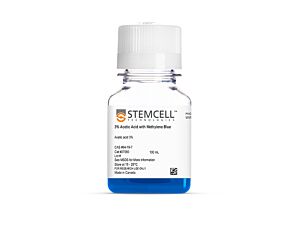
Before performing cell isolation using EasySep™, consult the product information sheet (PIS) to determine whether red blood cell (RBC) lysis is required for your sample type. RBC lysis should only be performed if indicated in the PIS. It is often recommended for blood samples; however, RBC lysis is not recommended for mouse splenocytes as it may reduce cell recovery. For the most accurate cell recovery calculation, we recommend performing total nucleated cell (TNC) count.
Thank you for your interest in this product.
Please provide us with your contact information and your local representative
will contact you with a customized quote. Where appropriate, they can also assist you with a(n):
Estimated delivery time for your area
Product sample or exclusive offer
In-lab demonstration
By submitting this form, you are providing your consent to STEMCELL Technologies Canada Inc. and its subsidiaries and affiliates (“STEMCELL”) to collect and use your information, and send you newsletters and emails in accordance with our privacy policy. Please contact us with any questions that you may have. You can unsubscribe or change your email preferences at any time.
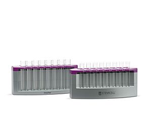
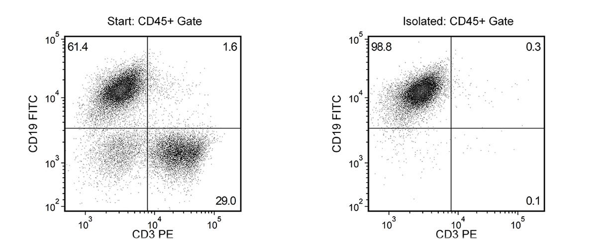
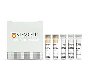
Easily and efficiently isolate highly purified mouse B cells from single-cell suspensions of splenocytes or other tissue samples by immunomagnetic negative selection, with the EasySep™ Mouse B Cell Isolation Kit. Widely used in published research for more than 20 years, EasySep™ combines the specificity of monoclonal antibodies with the simplicity of a column-free magnetic system.
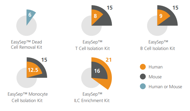
In this EasySep™ negative selection procedure, unwanted cells are labeled with antibody complexes and magnetic particles. Unwanted cells expressing the following markers are targeted for removal: CD11b, CD4, CD8a, Ly6G/C, Ter119, CD43, CD49b, and CD90.2. The magnetically labeled cells are then separated from the untouched desired B cells by using an EasySep™ magnet and simply pouring or pipetting the desired cells into a new tube. Following magnetic cell isolation in as little as 15 minutes, the desired B cells are ready for downstream applications such as flow cytometry, culture, and cell-based experiments.
Learn more about how immunomagnetic EasySep™ technology works or how to fully automate immunomagnetic cell isolation with RoboSep™. Explore additional products optimized for your workflow, including culture media, supplements, antibodies, and more.
This product is designed for use in the following research area(s) as part
of the highlighted workflow stage(s). Explore these workflows to learn more about the other products we
offer to support each research area.
Can EasySep™ Streptavidin RapidSpheres™ be used for either positive or negative selection?
Currently, EasySep™ Streptavidin RapidSphere™ kits are only available for negative selection and work by targeting and removing unwanted cells.
How does the separation work?
Streptavidin RapidSphere™ magnetic particles are crosslinked to unwanted cells using biotinylated antibodies. When placed in the EasySep™ Magnet, labeled cells migrate to the wall of the tube. The unlabeled cells are then poured off into a new tube.
Which columns do I use?
The EasySep™ procedure is column-free. That's right - no columns!
How can I analyze the purity of my enriched sample?
The Product Information Sheet provided with each EasySep™ kit contains detailed staining information.
Can EasySep™ Streptavidin RapidSphere™ separations be automated?
Yes. RoboSep™, the fully automated cell separator, automates all EasySep™ labeling and cell separation steps.
Are cells isolated using EasySep™ RapidSphere™ products FACS-compatible?
Yes. Desired cells are unlabeled and ready to use in downstream applications, such as FACS analysis.
Can I alter the separation time in the magnet?
Yes; however, this may impact the kit's performance. The provided EasySep™ protocols have already been optimized to balance purity, recovery and time spent on the isolation.
Bi-specific T cell-engaging antibody triggers protective immune memory and glioma microenvironment remodeling in immune-competent preclinical models
M. Zannikou et al.
Journal for Immunotherapy of Cancer 2025 Oct
Abstract
Bispecific T cell-engagers (BTEs) are engineered antibodies that redirect T cells to target antigen-expressing tumors. BTEs targeting tumor-specific antigens such as interleukin 13 receptor alpha 2 (IL13RA2) and epidermal growth factor receptor variant III (EGFRvIII) have been developed for glioblastoma (GBM). However, there is limited mechanistic understanding of the action of BTE since prior studies were mostly conducted in immunocompromised animal models. To close this gap, the function of BTEs was assessed in the immunosuppressive tumor microenvironment (TME) of orthotopic and genetically engineered mouse models (GEMM) with intact immune systems. Method: sA BTE that bridges CD3 epsilon on murine T cells to IL13RA2-positive GBM cells was developed, and the therapeutic mechanism was investigated in immunocompetent mouse models of GBM. Multicolor flow cytometry, single-cell RNA sequencing (scRNA-seq), multiplex immunofluorescence, and multiparametric MRI across multiple preclinical models of GBM were used to evaluate the mechanism of action and response. Results: BTE-mediated interactions between murine T cells and GBM cells triggered T cell activation and antigen-dependent killing of GBM cells. BTE treatment significantly extended the survival of mice bearing IL13RA2-expressing orthotopic glioma and de novo forming GBM in the GEMM. Quantified parametric MRI validated the survival data, showing a reduction in glioma volume and decreased glioma viability. Flow cytometric and scRNA-seq analyses of the TME revealed robust increases in activated and memory T cells and decreases in immunosuppressive myeloid cells within the brains of mice following BTE treatment. Conclusions: Our data demonstrate that the survival benefits of BTEs in preclinical models of glioma are due to the ability to engage the host immune system in direct killing, induction of immunological memory, and modulation of the TME. These findings provide a deeper insight into the mechanism of BTE actions in GBM.
PI3Kdelta-driven expansion of regulatory B cells impairs protective immune responses to Trypanosoma congolense parasite infection
F. Olayinka-Adefemi et al.
PLOS Pathogens 2025 Nov
Abstract
Phosphatidylinositol 3-kinase delta (PI3KCD) is a critical signaling enzyme for B cell development, activation, function and immune regulation. Gain-of-function mutations in PI3KCD result in the congenital immunodeficiency known as Activated PI3KCD Syndrome (APDS). APDS patients are prone to repeated infections and other serious clinical manifestations. Here, we determine how B cell-intrinsic expression of the APDS-associated PI3KCDE1021K mutation impacts immune responses to the protozoan parasite Trypanosoma congolense. PI3KCDE1021K/B mice exhibit a significant expansion of IL10-expressing B cells within the spleen and peritoneal cavity, which was associated with impaired control of T. congolense infection. Despite the generation of robust germinal center, plasma cell and antibody responses, PI3KCDE1021K/B mice show elevation in the first wave of parasitemia and increased mortality. We further characterize the phenotype of the expanded IL10-producing B cell population in PI3KCDE1021K/B mice, which show hallmarks of innate-like regulatory B cells (Breg) and expression of multiple inhibitory molecules. This Breg expansion is associated with reduced IFNγ/IL10 ratio, reduced TNFα production and impaired activation of myeloid cells, likely compromising the innate response to infection. These findings highlight the profound impact of dysregulated PI3KCD activity on regulatory B cells that can functionally impair innate immune responses controlling a systemic parasite protozoan disease. Author summaryB cells and antibodies play a critical role in the immune response to Trypanosome parasites. Molecular signaling networks within B cells can control the type of response generated during infection. Here, we studied how a genetic variant in the signaling enzyme PI3KCD, previously linked to human immune deficiencies, impacts B cell responses to Trypanosome infection. We find that mice expressing the PI3KCDE1021K mutation in their B cells show impaired control of Trypanosome infection, and alterations in several aspects of the immune response. Specifically, we noted these mice poorly control parasite growth within the first week of infection, a timeframe where specific antibody responses have not yet been generated. We noted an altered balance between pro-inflammatory and anti-inflammatory cytokine mediators produced within the first week of infection. This was associated with high numbers of regulatory B cells expressing multiple molecules capable of inhibiting other cells of the immune system. We further found that these mice show functional alterations in other critical immune cell types, such as macrophages and T cells. These findings highlight the impact of dysregulated PI3KCD activity on regulatory B cells that can impair immune responses controlling a systemic parasite protozoan disease.
Light-induced expression of gRNA allows for optogenetic gene editing of T lymphocytes in vivo
Nucleic Acids Research 2025 Mar
Abstract
AbstractThere is currently a lack of tools capable of perturbing genes in both a precise and a spatiotemporal fashion. The flexibility of CRISPR (clustered regularly interspaced short palindromic repeats), coupled with light’s unparalleled spatiotemporal resolution deliverable from a controllable source, makes optogenetic CRISPR a well-suited solution for precise spatiotemporal gene perturbations. Here, we present a new optogenetic CRISPR tool (Blue Light-inducible Universal VPR-Improved Production of RGRs, BLU-VIPR) that diverges from prevailing split-Cas design strategies and instead focuses on optogenetic regulation of guide RNA (gRNA) production. We engineered BLU-VIPR around a new potent blue-light activated transcription factor (VPR-EL222) and ribozyme-flanked gRNA. The BLU-VIPR design is genetically encoded and ensures precise excision of multiple gRNAs from a single messenger RNA transcript. This simplified spatiotemporal gene perturbation and allowed for several types of optogenetic CRISPR, including indels, CRISPRa, and base editing. BLU-VIPR also worked in vivo with cells previously intractable to optogenetic gene editing, achieving optogenetic gene editing in T lymphocytes in vivo.
Before performing cell isolation using EasySep™, consult the product information sheet (PIS) to determine whether red blood cell (RBC) lysis is required for your sample type. RBC lysis should only be performed if indicated in the PIS. It is often recommended for blood samples; however, RBC lysis is not recommended for mouse splenocytes as it may reduce cell recovery. For the most accurate cell recovery calculation, we recommend performing total nucleated cell (TNC) count.
PRODUCTS ARE FOR RESEARCH USE ONLY AND NOT INTENDED FOR HUMAN OR ANIMAL DIAGNOSTIC OR THERAPEUTIC USES UNLESS OTHERWISE STATED. FOR ADDITIONAL INFORMATION ON QUALITY AT STEMCELL, REFER TO WWW.STEMCELL.COM/COMPLIANCE.