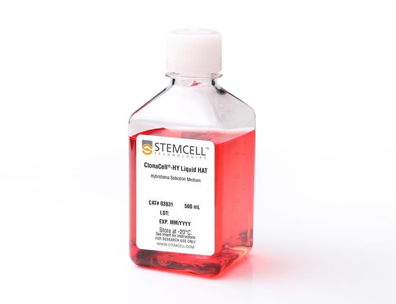
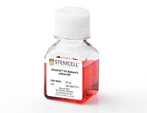
ClonaCell™-HY Liquid HAT Selection Medium

Hybridoma selection medium, with HAT (serum-containing)

ClonaCell™-HY Liquid HAT Selection Medium

Hybridoma selection medium, with HAT (serum-containing)

From: 383 USD
Catalog #
(Select a product)
Hybridoma selection medium, with HAT (serum-containing)
Add to Wish List

Product Advantages


  • Simplifies hybridoma selection in liquid suspension cultures

Overview

ClonaCell™-HY Liquid HAT Selection Medium is a serum-containing medium with hypoxanthine, aminopterin, and thymidine (HAT) for the selection of hybridomas following the fusion of lymphocytes and myeloma cells. This medium has been verified for use in mouse and rat hydridoma development and reportedly is compatible for production and cloning of myelomas and/or hybridomas using lymphocytes from a variety of host animals including human, mouse, rat, and hamster.
Contains
• DMEM
• Serum
• Hypoxanthine, aminopterin, and thymidine (HAT)
• Gentamicin
• 2-Mercaptoethanol
• Phenol red
• L-Glutamine and other supplements
• Other ingredients
Subtype
Specialized Media
Cell Type
Hybridomas
Species
Mouse
Application
Cell Culture, Hybridoma Generation
Brand
ClonaCell
Area of Interest
Antibody Development, Cell Line Development, Drug Discovery and Toxicity Testing, Hybridoma Generation

Protocols and Documentation

Find supporting information and directions for use in the Product Information Sheet or explore additional protocols below.

Document Type
Product Name
Catalog #
Lot #
Language
Catalog #
03831
Lot #
All
Language
English
Document Type
Technical Manual
Catalog #
03831
Lot #
All
Language
English
Document Type
Safety Data Sheet
Catalog #
03831
Lot #
All
Language
English

Applications

This product is designed for use in the following research area(s) as part of the highlighted workflow stage(s). Explore these workflows to learn more about the other products we offer to support each research area.

Resources and Publications

Frequently Asked Questions

Why is there HT (hypoxanthine, thymidine) in Medium E?

Hybridomas are selected using HAT (hypoxanthine, aminopterin, thymidine). Aminopterin blocks the de novo pathway for synthesizing nucleotide precursors for DNA synthesis. The inhibition of the de novo pathway can persist even after the cells are removed from selection. Hypoxanthine and thymidine (HT) provide the necessary nucleotide precursors for hybridoma cells to synthesize DNA using the salvage pathway. Once the cells are growing well in Medium E, they can be gradually switched to Medium A or another medium without HT.

Is the serum in ClonaCell™-HY media heat inactivated?

Yes, all serum used in ClonaCell™-HY media is heat inactivated.

Is there any IgG in clonacell™-HY media?

While we don't add IgG to the ClonaCell™-HY media, we do add serum, which contains an undefined amount of IgG. We selectively use serum lots with low IgG levels in the production of ClonaCell™-HY media, however, levels vary from lot to lot. IgG levels in a specific lot of ClonaCell™-HY medium are available in the lot-specific Certificate of Analysis.

Are there antibiotics in ClonaCell™-HY media?

These products contain gentamycin rather than penicillin/streptomycin/amphotericin B, because gentamycin is more stable and is a broad spectrum antibiotic that is non-toxic to most mammalian cells in culture.

What is the optimal number of colonies per plate?

We recommend 50-150 colonies per plate. An average fusion will result in approximately 1000 colonies per fusion (approx. 100 colonies per plate). Even if the average number of colonies per plate approaches 300, there should still be enough separation between colonies to pick easily.

Why do I have to put my fused cells into liquid medium overnight? Why can't I just plate directly into Medium D?

We recommend waiting up to 24 hours so that all of the fused cells can go through one cell cycle. This will ensure they have a chance to express HPRT (hypoxanthine guanine phosphoribosyltransferase), the enzyme necessary to survive in the presence of aminopterin (present in Medium D). Additionally, fused cells are very fragile immediately after fusion. Waiting a day before mixing the cells with the methylcellulose will improve their survival. Although it is not recommended, fused cells may be plated on the same day as fusion, but the cells should be allowed to recover for several hours in ClonaCell™-HY Medium C prior to plating.

What myeloma and mouse strains should I use?

Myeloma: There are at least two common myeloma cell lines used to generate hybridomas - SP2/0 and P3X63Ag8.683. Both are available from ATCC. Researchers should ensure that the myeloma line is from a reliable source and is negative for mycoplasma. Mycoplasma contamination of the myeloma line can result in decreased efficiency of hybridoma formation. Mouse: We suggest using BALB/c splenocytes and parental myeloma cells of BALB/c for the following reasons: they are highly immune reactive, well characterized and myeloma cells are available from the same genetic strain. Other mouse strains, however, are also compatible with cloning in ClonaCell™-HY media.

Can I grow human/rat/T cell hybridomas in ClonaCell™-HY?

Although we have not tried to generate human, rat or T cell hybridomas during in-house testing, these experiments are expected to be successful using ClonaCell™-HY. The researcher would need to ensure that the cell lines used in the fusion are sensitive to HAT selection and grow well in methylcellulose-based medium.

There are very few colonies growing in my Medium D. Why?

Low numbers of colonies is generally a result of low fusion efficiency, which can have many causes. The fusion efficiency can be affected by the presence of serum during fusion, the presence of mycoplasma, low viability of cells, overexposure to polyethylene glycol or slow-growing myeloma cells prior to fusion.

Why does the ClonaCell™-HY manual suggest two different methods for fusion (A or B)? Can one expect better results with one method over the other?

Which method chosen is a personal preference and there should not be significant differences in efficiency. Method B is faster and has less steps, but Method B requires you to remove all the PEG before the cells are diluted, so you will risk aspirating cells if not very careful. With Method A, you dilute the PEG with Medium B, so you have less opportunity to lose cells.

Why does the ClonaCell™-HY manual suggest two different methods for fusion (A or B)? Can one expect better results with one method over the other?

A: Which method chosen is a personal preference and there should not be significant differences in efficiency. Method B is faster and has less steps, but Method B requires you to remove all the PEG before the cells are diluted, so you will risk aspirating cells if not very careful. With Method A, you dilute the PEG with Medium B, so you have less opportunity to lose cells.

Once I pick the colonies and grow the cells in plates, will the residual methylcellulose interfere with characterization? For example, will I have problems doing an ELISA?

 There will likely be some residual methylcellulose contamination when colonies are picked and transferred to the 96-well plate with the liquid growth medium. The concentration of methylcellulose, however, should be low enough that it should not interfere with most assays.

Is the serum in ClonaCell™-TCS medium heat inactivated?

Yes, all serum used in ClonaCell™ is heat inactivated.

Is there any IgG in ClonaCell™ TCS?

While we don't add IgG to the ClonaCell™ media, we do add serum, which contains an undefined amount of IgG. We selectively use serum lots with low IgG levels in the production of ClonaCell™ media, however, levels vary from lot to lot. IgG levels in a specific lot of ClonaCell™ TCS medium are available in the lot-specific Certificate of Analysis.

Can ClonaCell™-TCS be used with any cell line?

A list of recommended cell lines can be found in the manual. Other cell lines may be compatible with ClonaCell™-TCS. It will be necessary, however, to determine the plating cell density and growth efficiency of the desired cells in ClonaCell™-TCS.

Publications (6)

Characterization of an enhanced antigenic change in the pandemic 2009 H1N1 influenza virus haemagglutinin Garc&iacute et al. Journal of General Virology 2014

Abstract

Murine hybridomas producing neutralizing mAbs specific to the pandemic influenza virus A/California/07/2009 haemagglutinin (HA) were isolated. These antibodies recognized at least two different but overlapping new epitopes that were conserved in the HA of most Spanish pandemic isolates. However, one of these isolates (A/Extremadura/RR6530/2010) lacked reactivity with the mAbs and carried two unique mutations in the HA head (S88Y and K136N) that were required simultaneously to eliminate reactivity with the murine antibodies. This unusual requirement directly illustrates the phenomenon of enhanced antigenic change proposed previously for the accumulation of simultaneous amino acid substitutions at antigenic sites of the influenza A virus HA during virus evolution (Shih et al., Proc Natl Acad Sci USA, 104 , 6283-6288, 2007). The changes found in the A/Extremadura/RR6530/2010 HA were not found in escape mutants selected in vitro with one of the mAbs, which contained instead nearby single amino acid changes in the HA head. Thus, either single or double point mutations may similarly alter epitopes of the new antigenic site identified in this work in the 2009 H1N1 pandemic virus HA. Moreover, this site is relevant for the human antibody response, as shown by competition of mAbs and human post-infection sera for virus binding. The results are discussed in the context of the HA antigenic structure and challenges posed for identification of sequence changes with possible antigenic impact during virus surveillance.
Isolation of mouse pancreatic alpha, beta, duct and acinar populations with cell surface markers Dorrell C et al. Molecular and Cellular Endocrinology 2011 JUN

Abstract

Tools permitting the isolation of live pancreatic cell subsets for culture and/or molecular analysis are limited. To address this, we developed a collection of monoclonal antibodies with selective surface labeling of endocrine and exocrine pancreatic cell types. Cell type labeling specificity and cell surface reactivity were validated on mouse pancreatic sections and by gene expression analysis of cells isolated using FACS. Five antibodies which marked populations of particular interest were used to isolate and study viable populations of purified pancreatic ducts, acinar cells, and subsets of acinar cells from whole pancreatic tissue or of alpha or beta cells from isolated mouse islets. Gene expression analysis showed the presence of known endocrine markers in alpha and beta cell populations and revealed that TTR and DPPIV are primarily expressed in alpha cells whereas DGKB and GPM6A have a beta cell specific expression profile.
Essential role of TNF receptor superfamily 25 (TNFRSF25) in the development of allergic lung inflammation Fang L et al. The Journal of Experimental Medicine 2008 MAY

Abstract

We identify the tumor necrosis factor receptor superfamily 25 (TNFRSF25)/TNFSF15 pair as critical trigger for allergic lung inflammation, which is a cardinal feature of asthma. TNFRSF25 (TNFR25) signals are required to exert T helper cell 2 (Th2) effector function in Th2-polarized CD4 cells and co-stimulate interleukin (IL)-13 production by glycosphingolipid-activated NKT cells. In vivo, antibody blockade of TNFSF15 (TL1A), which is the ligand for TNFR25, inhibits lung inflammation and production of Th2 cytokines such as IL-13, even when administered days after airway antigen exposure. Similarly, blockade of TNFR25 by a dominant-negative (DN) transgene, DN TNFR25, confers resistance to lung inflammation in mice. Allergic lung inflammation-resistant, NKT-deficient mice become susceptible upon adoptive transfer of wild-type NKT cells, but not after transfer of DN TNFR25 transgenic NKT cells. The TNFR25/TL1A pair appears to provide an early signal for Th2 cytokine production in the lung, and therefore may be a drug target in attempts to attenuate lung inflammation in asthmatics.