MethoCult™ H4034 Optimum

Methylcellulose-based medium with recombinant cytokines for human cells

MethoCult™ H4034 Optimum

Methylcellulose-based medium with recombinant cytokines for human cells

From: 660 USD
Catalog #
(Select a product)
Methylcellulose-based medium with recombinant cytokines for human cells
Add to Wish List

Overview

MethoCult™ H4034 Optimum (MethoCult™ GF H4034) is a methylcellulose-based medium for the growth and enumeration of hematopoietic progenitor cells in colony-forming unit (CFU) assays of human bone marrow, mobilized peripheral blood, peripheral blood, and cord blood samples. MethoCult™ H4034 Optimum has been formulated to support optimal growth of erythroid progenitor cells (BFU-E and CFU-E), granulocyte-macrophage progenitor cells (CFU-GM, CFU-G and CFU-M), and multipotential granulocyte, erythroid, macrophage, megakaryocyte progenitor cells (CFU-GEMM). The inclusion of G-CSF in Optimum formulations improves discrimination of myeloid colony subtypes. This formulation is compatible with STEMvision™ software for automated colony counting.

Browse our Frequently Asked Questions (FAQs) on performing the CFU assay and explore its utility as part of the cell therapy workflow.
Contains
• Methylcellulose in Iscove's MDM
• Fetal bovine serum
• Bovine serum albumin
• 2-Mercaptoethanol
• Recombinant human stem cell factor (SCF)
• Recombinant human interleukin 3 (IL-3)
• Recombinant human erythropoietin (EPO)
• Recombinant human granulocyte colony-stimulating factor (G-CSF)
• Recombinant human granulocyte-macrophage colony-stimulating factor (GM-CSF)
• Supplements
Subtype
Semi-Solid Media, Specialized Media
Cell Type
Hematopoietic Stem and Progenitor Cells
Species
Human, Non-Human Primate
Application
Cell Culture, Colony Assay, Functional Assay
Brand
MethoCult
Area of Interest
Drug Discovery and Toxicity Testing, Stem Cell Biology

Data Figures

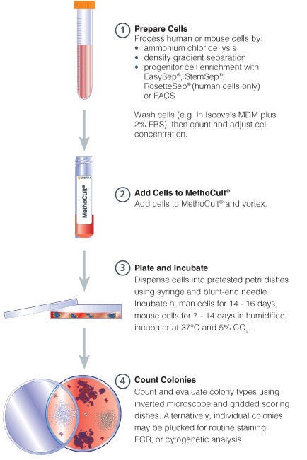
Procedure Summary for Hematopoietic CFC Assays

Figure 1. Procedure Summary for Hematopoietic CFU Assays

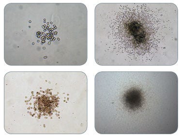
Examples of Colonies Derived from CFU-GM in MethoCult™ H4034 Optimum

Figure 2. Examples of Colonies Derived from CFU-GM in MethoCult™ H4034 Optimum

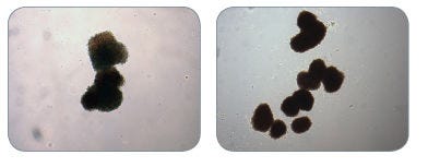
Examples of Colonies Derived from BFU-E in MethoCult™ H4034 Optimum

Figure 3. Examples of Colonies Derived from BFU-E in MethoCult™ H4034 Optimum

Protocols and Documentation

Find supporting information and directions for use in the Product Information Sheet or explore additional protocols below.

Document Type
Product Name
Catalog #
Lot #
Language
Catalog #
04044, 04034
Lot #
All
Language
English
Document Type
Technical Manual
Catalog #
04034
Lot #
All
Language
English
Document Type
Safety Data Sheet
Catalog #
04044, 04034
Lot #
All
Language
English

Applications

This product is designed for use in the following research area(s) as part of the highlighted workflow stage(s). Explore these workflows to learn more about the other products we offer to support each research area.

Resources and Publications

Educational Materials (19)

Frequently Asked Questions

Why use semi-solid media?

Semi-solid media (methylcellulose-based MethoCult™ and collagen-based MegaCult™-C) allow the clonal progeny of a single progenitor cell to remain spatially isolated from other colonies within a culture, so they may be separately identified and counted.

Why use methylcellulose-based media?

Methylcellulose permits better growth of erythroid colonies than other types of semi-solid support systems (eg. agar) while allowing optimal myeloid colony formation. When appropriate cytokines are present, committed progenitor cells of both erythroid and granulocyte/macrophage lineages (CFU-GM, CFU-G, CFU-M) as well as multi-potential progenitor cells (CFU-GEMM), can be assayed simultaneously in the same culture dish.

Is it necessary to add antibiotics to the media?

No, aseptic technique should be sufficient to maintain sterile cultures. However, antibiotics (eg. Penicillin/Streptomycin) or anti-fungals (eg. Amphotericin B) may be added to the methylcellulose medium if desired.

Is there anything I can do if my cultures appear contaminated?

No, once contamination is visible, it is not possible to rescue the cultures by the addition of antibiotics. Bacteria and yeast inhibit colony formation by depleting nutrients or by releasing toxic substances.

Why can't I use a pipette to dispense methylcellulose-based media?

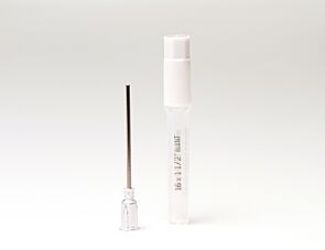
Methylcellulose is a viscous solution that cannot be accurately dispensed using a pipette due to adherence of the medium to the walls of the pipette tip. Blunt-End, 16 Gauge needles (Catalog #28110), in combination with 3 cc Syringes (Catalog #28230) are recommended for accurate dispensing of MethoCult™.

Can I 'pluck' the colonies for individual analysis?

Yes, colonies can be 'plucked' using a pipette with 200 µL sterile pipette tips or using a glass Pasteur pipette with an elongated tip. Individual colonies should be placed in a volume of 25 - 50 µL of medium, and diluted into suitable culture medium for further culture or analysis.

Why are low adherence dishes so important?

Adherent cells such as fibroblasts can cause inhibition of colony growth and obscure visualization of colonies.

Can MethoCult™ products be used for lymphoid progenitor CFU assays?

Human lymphoid progenitors (B, NK and T) seem to require stromal support for growth therefore cannot be grown in MethoCult™. Mouse pre-B clonogenic progenitors can be grown in MethoCult™ M3630 (Catalog #03630).

Is it possible to set up CFU assays in a 24-well plate?

Yes, as long as a plating concentration optimized for the smaller surface area of a well in a 24-well plate (1.9 cm2 as compared to ~9.5 cm2 for a 35 mm dish) is used for these assays. The number of replicate wells required to get an accurate estimation of CFU numbers may also need to be increased.

Can I stain colonies in MethoCult™ medium?

The cells in individual colonies in MethoCult™ can be stained, eg., for analysis of morphology or phenotype, after they are plucked from the dish and washed free of methylcellulose. Colonies grown in collagen-based MegaCult™-C medium can be used for immunohistochemical or enzymatic staining in situ after dehydration and fixation onto glass slides.

Are there differences in colony morphology with serum-free media?

Serum-containing media generally give better overall growth (colonies may appear larger) but there are no large differences in total colony numbers when CFU assays using serum-free media and serum-containing media are compared, provided that identical cytokines are present.

Can MethoCult™ be made with alternate base media?

Yes, this can be done as a 'custom' media order. Please contact techsupport@stemcell.com for more information.

Is there a MethoCult™ formulation suitable for HPP-CFC (high proliferative potential colony forming cell)?

Yes, MethoCult™ H4535 (Catalog #04535) can be used for the HPP-CFC assay as it does not contain EPO. The culture period is usually 28 days. It is not necessary to feed these cultures as growth factors in the medium are present in excess. As HPP-CFCs can be quite large, overplating can be a problem. It is recommended to plate cells at two or more different concentrations.

Publications (41)

Inhibition of inflammation and senescence with azole compound C7 enables expansion of hematopoietic stem and progenitor cells. Z. Kok et al. Stem cells translational medicine 2026 Jun

Abstract

Human hematopoietic stem and progenitor cells (HSPCs) derived from umbilical cord blood (UCB) are often insufficient in quantity for transplantation, necessitating efforts for ex vivo expansion. We previously developed a novel azole-based small molecule, C7, as a structural analog of the p38 mitogen-activated protein kinase inhibitor SB203580. C7 mediated robust ex vivo expansion of UCB HSPCs from unselected mononuclear cells, compared to its structural analogs and other published compounds. However, the molecular underpinnings of C7's potency remained unclear. In this work, we demonstrate that C7 preferentially enhances the proliferation and viability of early HSPC subsets upon direct culture with pre-isolated HSPCs, while limiting mitochondrially-derived reactive oxygen species production linked to oxidative stress. C7 treatment enhances hematopoietic stem cell self-renewal and in vivo multilineage reconstitution without T- or myeloid lineage bias. Unlike existing p38 inhibitors, C7 targets multiple kinases and pathways to achieve superior HSPC expansion through suppression of NFκB and IL-10 inflammatory signaling and attenuation of stress/senescence‑associated markers (eg, SERPINB2 and CDKN1A/p21). This mechanism of action of C7 is distinct from those of other existing compounds used for ex vivo HSPC expansion and has implications for application in other ex vivo culture platforms and clinical settings.
A porcine model of Fanconi anemia B. Hergert et al. PLOS One 2025 Oct

Abstract

Although small animal models of Fanconi anemia (FA) are useful, they do not faithfully replicate many of the clinical features seen in FA patients. We reasoned that a porcine model of FA with its similar physiology and a relatively long lifespan would produce a phenotype more similar to human FA. Targeting FANCA in domestic swine resulted in skeletal abnormalities and extreme sensitivity to interstrand DNA cross-linking agents. In addition, FANCA disruption followed by mitomycin C treatment resulted in a > 10-fold increase in chromosomal radials, a finding that is considered diagnostic for human FA. Bone marrow derived, hematopoietic progenitor cells from a FANCA null pig showed a 75% reduction in colony forming activity compared to wild type. Evaluation of steady state hematopoiesis in the peripheral blood revealed the gradual development of red cell macrocytosis and a reduction in circulating neutrophils. Targeting of FANCD2 failed to produce any biallelic animals demonstrating the loss of FANCD2 function is embryonic lethal in pigs. These results indicate that a porcine model of FANCA holds promise for the development of strategies to prevent the development of bone marrow failure and malignancies in patients with FA.
Continuous map of early hematopoietic stem cell differentiation across human lifetime H. Komic et al. Nature Communications 2025 Mar

Abstract

Uncovering early gene network changes of human hematopoietic stem cells (HSCs) leading to differentiation induction is of utmost importance for therapeutic manipulation. We employed single cell proteo-transcriptomic sequencing to FACS-enriched bone marrow hematopoietic stem and progenitor cells (HSPCs) from 15 healthy donors. Pseudotime analysis reveals four major differentiation trajectories, which remain consistent upon aging, with an early branching point into megakaryocyte-erythroid progenitors. However, young donors suggest a more productive differentiation from HSPCs to committed progenitors of all lineages. tradeSeq analysis depicts continuous changes in gene expression of HSPC-related genes (DLK1, ADGRG6), and provides a roadmap of gene expression at the earliest branching points. We identify CD273/PD-L2 to be highly expressed in a subfraction of immature multipotent HSPCs with enhanced quiescence. Functional experiments confirm the immune-modulatory function of CD273/PD-L2 on HSPCs in regulating T-cell activation and cytokine release. Here, we present a molecular map of early HSPC differentiation across human life. Therapeutic application of hematopoietic stem cells requires a thorough understanding of their in situ differentiation pathways. Here they use single cell proteo-transcriptomic sequencing to identify human HSPC differentiation cues and an immunoregulatory function of CD273/PD-L2 on early HSPCs.