EasySep™ Human CD8+ T Cell Enrichment Kit

Immunomagnetic negative isolation of untouched human CD8+ T cells
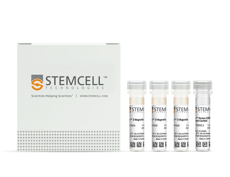
EasySep™ Human CD8+ T Cell Enrichment Kit

Immunomagnetic negative isolation of untouched human CD8+ T cells

From: 969 USD
Catalog #
(Select a product)
Immunomagnetic negative isolation of untouched human CD8+ T cells
Add to Wish List

Product Advantages


  • Fast, easy-to-use and column-free

  • Up to 95% purity

  • Untouched, viable cells

What's Included

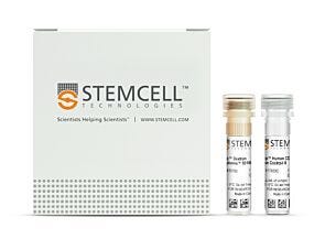
  • EasySep™ Human CD8+ T Cell Enrichment Kit (Catalog #19053)
    • EasySep™ Human CD8 T Cell Enrichment Cocktail, 1 mL
    • EasySep™ D Magnetic Particles, 3 x 1 mL
  • RoboSep™ Human CD8+ T Cell Enrichment Kit with Filter Tips (Catalog #19053RF)
    • EasySep™ Human CD8 T Cell Enrichment Cocktail, 1 mL
    • EasySep™ D Magnetic Particles, 3 x 1 mL
    • RoboSep™ Buffer (Catalog #20104)
    • RoboSep™ Filter Tips (Catalog #20125)
Products for Your Protocol
To see all required products for your protocol, please consult the Protocols and Documentation.

Overview

Easily and efficiently isolate highly purified human CD8+ T cells from fresh or previously frozen human peripheral blood mononuclear cells (PBMCs) or lysed leukapheresis samples by immunomagnetic negative selection, with the EasySep™ Human CD8+ T Cell Enrichment Kit. Widely used in published research for more than 20 years, EasySep™ combines the specificity of monoclonal antibodies with the simplicity of a column-free magnetic system.

In this EasySep™ negative selection procedure, unwanted cells are labeled with antibody complexes and magnetic particles. Unwanted cells expressing the following markers are targeted for removal: CD4, CD14, CD16, CD19, CD20, CD36, CD56, CD66b, TCRgd, and GlyA. The magnetically labeled cells are then separated from the untouched desired CD8+ T cells by using an EasySep™ magnet and simply pouring or pipetting the desired cells into a new tube. Following magnetic cell isolation, the desired CD8+ T cells are ready for downstream applications such as flow cytometry, culture, or DNA/RNA extraction.

For even faster cell isolations, we recommend the EasySep™ Human CD8+ T Cell Isolation Kit (Catalog #17953), which isolates cells in just 8 minutes.

Learn more about how immunomagnetic EasySep™ technology works or how to fully automate immunomagnetic cell isolation with RoboSep™. Alternatively, choose ready-to-use, ethically sourced, primary Human Peripheral Blood CD8+ T Cells, Frozen isolated with EasySep™ Human CD8+ T Cell Enrichment Kit. Explore additional products optimized for your workflow, including culture media, supplements, antibodies, and more.
Magnet Compatibility
• EasySep™ Magnet (Catalog #18000)
• “The Big Easy” EasySep™ Magnet (Catalog #18001)
• Easy 50 EasySep™ Magnet (Catalog #18002)
• EasyPlate™ EasySep™ Magnet (Catalog 18102)
• EasyEights™ EasySep™ Magnet (Catalog #18103)
• RoboSep™-S (Catalog #21000)
Subtype
Cell Isolation Kits
Cell Type
T Cells, T Cells, CD8+
Species
Human
Sample Source
Leukapheresis, PBMC
Selection Method
Negative
Application
Cell Isolation
Brand
EasySep, RoboSep
Area of Interest
Immunology

Data Figures

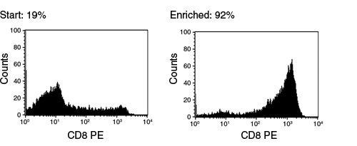
FACS Histogram Results Using EasySep™ Human CD8+ T Cell Enrichment Kit

Figure 1. FACS Histogram Results Using EasySep™ Human CD8+ T Cell Enrichment Kit

Starting with frozen mononuclear cells, the CD8+ cell content of the enriched fraction typically ranges from 84 - 95%

Protocols and Documentation

Find supporting information and directions for use in the Product Information Sheet or explore additional protocols below.

Document Type
Product Name
Catalog #
Lot #
Language
Catalog #
19053
Lot #
All
Language
English
Catalog #
19053RF
Lot #
All
Language
English
Document Type
Safety Data Sheet 1
Catalog #
19053
Lot #
All
Language
English
Document Type
Safety Data Sheet 2
Catalog #
19053
Lot #
All
Language
English
Document Type
Safety Data Sheet 1
Catalog #
19053RF
Lot #
All
Language
English
Document Type
Safety Data Sheet 2
Catalog #
19053RF
Lot #
All
Language
English
Document Type
Safety Data Sheet 3
Catalog #
19053RF
Lot #
All
Language
English

Applications

This product is designed for use in the following research area(s) as part of the highlighted workflow stage(s). Explore these workflows to learn more about the other products we offer to support each research area.

Resources and Publications

Frequently Asked Questions

Can EasySep™ be used for either positive or negative selection?

Yes. The EasySep™ kits use either a negative selection approach by targeting and removing unwanted cells or a positive selection approach targeting desired cells. Depletion kits are also available for the removal of cells with a specific undesired marker (e.g. GlyA).

How does the separation work?

Magnetic particles are crosslinked to cells using Tetrameric Antibody Complexes (TAC). When placed in the EasySep™ Magnet, labeled cells migrate to the wall of the tube. The unlabeled cells are then poured off into a separate fraction.

Which columns do I use?

The EasySep™ procedure is column-free. That's right - no columns!

How can I analyze the purity of my enriched sample?

The Product Information Sheet provided with each EasySep™ kit contains detailed staining information.

Can EasySep™ separations be automated?

Yes. RoboSep™, the fully automated cell separator, automates all EasySep™ labeling and cell separation steps.

Can EasySep™ be used to isolate rare cells?

Yes. We recommend a cell concentration of 2x10⁸ cells/mL and a minimum working volume of 100 µL. Samples containing 2x10⁷ cells or fewer should be suspended in 100 µL of buffer.

Are the EasySep™ magnetic particles FACS-compatible?

Yes, the EasySep™ particles are flow cytometry-compatible, as they are very uniform in size and about 5000X smaller than other commercially available magnetic beads used with column-free systems.

Can the EasySep™ magnetic particles be removed after enrichment?

No, but due to the small size of these particles, they will not interfere with downstream applications.

Can I alter the separation time in the magnet?

Yes; however, this may impact the kit's performance. The provided EasySep™ protocols have already been optimized to balance purity, recovery and time spent on the isolation.

For positive selection, can I perform more than 3 separations to increase purity?

Yes, the purity of targeted cells will increase with additional rounds of separations; however, cell recovery will decrease.

How does the binding of the EasySep™ magnetic particle affect the cells? is the function of positively selected cells altered by the bound particles?

Hundreds of publications have used cells selected with EasySep™ positive selection kits for functional studies. Our in-house experiments also confirm that selected cells are not functionally altered by the EasySep™ magnetic particles.

If particle binding is a key concern, we offer two options for negative selection. The EasySep™ negative selection kits can isolate untouched cells with comparable purities, while RosetteSep™ can isolate untouched cells directly from whole blood without using particles or magnets.

Publications (26)

Plasmacytoid and CD141+ Myeloid Dendritic Cells Cooperation with CD8+ T Cells in Lymph Nodes is Associated with HIV Control. J. Vitallé et al. MedComm 2025 Sep

Abstract

Dendritic cells (DC) are known to modulate antiviral immune responses; however, the knowledge about the role of different DC subsets in antiviral T cell priming in human tissues remains uncompleted. In the context of HIV infection, we determined the phenotype and location of plasmacytoid and CD141+ myeloid DCs (pDCs and mDCs) in lymph nodes of people living with HIV (PLWH). We found an interaction between pDCs and CD141+ mDCs with CD8+ T cells, being associated with participants' viral levels in blood and tissue. Moreover, we demonstrated a higher and more polyfunctional superantigen- and HIV-specific CD8+ T cell response after the coculture with Toll-like receptor (TLR)-primed pDCs and CD141+ mDCs. Last, we showed the potential of programmed cell death-1 (PD-1) blocking using pembrolizumab to further increase antigen-specific CD8+ T cell response along with TLR agonists. Therefore, these results showed a cooperation between pDCs, CD141+ mDCs and CD8+ T cells in lymph nodes of PLWH, which is associated with higher HIV control, highlighting the importance of DC subsets crosstalk to achieve a more potent anti-HIV response and support the use of DC-based immunotherapies for HIV control.
Retinol Binding Protein 4 reactivates latent HIV-1 by triggering canonical NF-κB, JAK/STAT5 and JNK signalling C. Pastorio et al. Signal Transduction and Targeted Therapy 2025 Oct

Abstract

Reactivation of the latent viral reservoirs is crucial for a cure of HIV/AIDS. However, current latency reversing agents are inefficient, and the endogenous factors that have the potential to reactivate HIV in vivo remain poorly understood. To identify natural activators of latent HIV-1, we screened a comprehensive peptide/protein library derived from human hemofiltrate, representing the entire blood peptidome, using J-Lat cell lines harboring transcriptionally silent HIV-1 GFP reporter viruses. Fractions potently reactivating HIV-1 from latency contained human Retinol Binding Protein 4 (RBP4), the carrier of retinol (Vitamin A). We found that retinol-bound holo-RBP4 but not retinol-free apo-RBP4 strongly reactivates HIV-1 in a variety of latently infected T cell lines. Functional analyses indicate that this reactivation involves activation of the canonical NF-κB pathway and is strengthened by JAK/STAT5 and JNK signalling but does not require retinoic acid production. High levels of RBP4 were detected in plasma from both healthy individuals and people living with HIV-1. Physiological concentrations of RBP4 induced significant viral reactivation in latently infected cells from individuals on long-term antiretroviral therapy with undetectable viral loads. As a potent natural HIV-1 latency-reversing agent, RBP4 offers a novel approach to activating the latent reservoirs and bringing us closer to a cure. Subject terms: Preclinical research, Infectious diseases
IFNγ augments TKI efficacy by alleviating protein unfolding stress to promote GSDME-mediated pyroptosis in hepatocellular carcinoma X. Li et al. Cell Death & Disease 2025 Jul

Abstract

Tyrosine kinase inhibitors (TKIs) are the standard treatment for advanced hepatocellular carcinoma (HCC). However, their therapeutic efficacy is often limited by drug resistance, primarily driven by tumoral intrinsic mechanisms. In this study, we demonstrate that IFNγ in the tumor microenvironment can potentiate TKI response, and that ablation of IFNγ receptor on HCC cells leads to TKI resistance in vivo. Mechanistically, IFNγ synergizes with TKI to induce GSDME-mediated pyroptosis of HCC cells. The PERK-mediated unfolded protein response (UPR) protects HCC cells from TKI-induced pyroptosis. IFNγ attenuates PERK activation by inducing the expression of PDIA1, which alleviates the stress of protein unfolding. In vivo, PERK inhibition augments TKI therapy, and elevated PERK expression correlates with poor overall survival of patients with HCC. Moreover, IFNγ-producing CD8+ T cells can potentiate TKI efficacy. Combining PD-1 blockade to activate T-cell response with TKI therapy synergistically suppresses the growth of GSDME-expressing HCC tumors, which is further enhanced by the PERK inhibitor. Our findings reveal how IFNγ signaling modulates TKI response and demonstrate the potential of a sequential combination of ICB-mediated immunotherapy and TKI therapy for patients with GSDME+ HCC. T cell-derived IFNγ enhances TKI-induced pyroptosis in HCC. Mechanistic illustration of IFNγ secreted from CD8+ T cells enhancing TKI-induced GSDME-mediated pyroptosis in hepatocellular carcinoma via suppression of the PERK pathway. Created with BioRender.com.