Erythropoietin (EPO): Properties, Biological Functions, and Measurement
The purpose of this mini-review is to provide an overview of the physical properties and biological functions of EPO, and to describe methods of measuring EPO levels in biological fluids
Request Pricing
Thank you for your interest in this product. Please provide us with your contact information and your local representative will contact you with a customized quote. Where appropriate, they can also assist you with a(n):
Estimated delivery time for your area
Product sample or exclusive offer
In-lab demonstration
- Document # 29013
- Version 4.0.0
- Apr 2015
Introduction
The continuous formation of new red blood cells (RBCs) is regulated by the glycoprotein hormone erythropoietin (EPO). Human EPO was first purified in 1977 from 2500 liters of urine from aplastic anemia patients,1 and the nucleotide sequence of the human EPO cDNA was reported in 1985.2,3 Since then, human EPO has become a major therapeutic agent to treat anemia due to chronic renal failure and other diseases.4 The physiological role of EPO in stimulating the proliferation and survival of erythroid cells has been established for many years. More recently, EPO has also been observed to protect endothelial, neural, cardiac and other cell types against cytotoxic damage. The purpose of this mini-review is to provide an overview of the physical properties and biological functions of EPO, and to describe methods of measuring EPO levels in biological fluids.
Structure of EPO
Human EPO consists of 165 amino acids and has a molecular mass of ~35,000 daltons. Approximately 40% of the molecular mass of the mature molecule is made up of 4 carbohydrate chains (3 N-linked and 1 O-linked).5 The carbohydrate moieties of EPO contain at least 10 molecules of sialic acid, which contribute to the relatively low (~4.4) iso-electric pH of EPO. EPO preparations are heterogeneous and contain multiple isoforms that differ from each other in their carbohydrate structure. Recombinant EPO produced in mammalian cells is fully glycosylated. Two forms of recombinant EPO, Epoietin-alpha and Epoietin-beta, are commonly used in the clinic. These molecules are produced in Chinese Hamster Ovary (CHO) cells and exhibit differences in their carbohydrate structure and pharmacological properties, but not in their respective clinical efficacies.6 The carbohydrate moieties are not essential for biochemical activity of EPO in vitro, indicating that only the polypeptide is involved in binding to the EPO receptor. The in vivo biological activity of EPO, however, is completely abolished when EPO is deglycosylated, suggesting that the carbohydrate moieties are important to prevent degradation and to delay clearance of EPO from the circulation. It is most likely that the carbohydrates are also needed during EPO biosynthesis and secretion.
Functions and Target Cells of EPO
Target cells for EPO have classically been identified by their ability to form hemoglobinized erythroid colonies. This ability is evaluated by using clonal assays performed in methylcellulose-based media, such as MethoCult™, or other semi-solid media, following exposure of bone marrow or spleen cells to EPO.7 The earliest purely erythroid progenitor, the burst-forming unit erythroid (BFU-E), has a low sensitivity for EPO and requires additional factors for survival and proliferation, in particular stem cell factor (SCF).8 The more differentiated erythroid colony-forming unit (CFU-E) has a more limited proliferative potential than its BFU-E precursor, but is highly sensitive to EPO. The CFU-E and its progeny, down to the morphologically recognizable erythroblast stage, are absolutely dependent on EPO for survival, proliferation and differentiation. In some myeloproliferative disorders, in particular polycythemia vera (PV), erythroid progenitor cells have acquired the capacity to proliferate, differentiate and survive in the absence of EPO.9 In conjuction with SCF and other cytokines, EPO can promote massive proliferation and erythroid differentiation of immature progenitors in serum-free liquid culture media (e.g. StemSpan™ Erythroid Expansion Supplement, Catalog #02692), resulting in almost pure erythroblast cultures. Upon switching to appropriate differentiation media, these cultures can mature into functional RBCs.10,11
EPO exerts its actions on RBC development through a single, high-affinity receptor12 that is expressed at relatively low levels (<1000 molecules) on erythroid cells, typically between the BFU-E and normoblast stages, but is absent from mature RBCs.13 The EPO receptor (EPO-R) belongs to the class I cytokine receptor superfamily of class I transmembrane proteins, the members of which share certain structural features, in particular four conserved cysteine residues and the WSxWS motif in their extracellular domain. Other receptors of this group include the receptors for IL-2, IL-3, IL-6, G-CSF and thrombopoietin. Binding of EPO to EPO-R triggers dimerization, which induces tyrosine phosphorylation, activation of the intracellular JAK2 tyrosine kinase and a cascade of signaling events leading to induction of proliferation and other biological effects mediated by EPO. For reviews of signal transduction by EPO-R and related cytokine receptors, please see references 14 and 15.
EPO-R expression has been reported in many non-hematopoietic cells including myocytes, neuronal cells and endothelial cells.16-18 EPO is believed to have a physiological role in cardiac and brain development, as well as in protecting heart and brain tissue against inflammatory and ischemic damage, possibly through direct stimulation of cardiomyocytes and neural cells or mobilization of endothelial progenitor cells and promotion of neo-vascularization.
The actions of EPO and expression of functional EPO-Rs on non-hematopoietic cells are not as clear as the role of EPO in erythropoiesis. Some studies reported EPO-R expression on endothelial cells at levels as high as 27,000 molecules per cell.16 Other studies, however, reported that functional EPO-Rs were expressed at much lower levels in endothelial, neural and other non-hematopoietic cells, such that expression was not detectable using standard methods.19,20 One study reported that the tissue-protective effect of EPO in mice is not mediated through homodimerization of the EPO-R, but through a hetero-receptor containing the EPO-R and the common beta-subunit of the IL-3, IL-5 and GM-CSF receptors (γc).21 Other studies, however, showed that the cytoprotective effect of EPO on neural and myocardial cells is mediated by the classical homodimeric EPO-R and does not require expression of the γc subunit, which was not detectable on these cells.19,22,23
EPO-R expression and EPO-mediated effects on cell survival and growth have also been reported on primary human tumors and cultured tumor cell lines. This is a potential concern for the use of EPO to treat anemia in cancer patients. Reports of EPO-R expression in tumor tissues and cell lines, however, have been inconsistent and dependent on the types of reagents and detection methods used (see reference 24 for a recent review). In a recent study in which more than 200 human tumor cell lines were examined, EPO-R mRNA was detectable in most cell lines, but at low levels. Few cell lines had detectable EPO-R protein expression (>100 molecules per cells), as measured using a monoclonal anti-EPO-R antibody or radiolabeled human recombinant EPO. EPO-Rs in those lines with detectable EPO-Rs also appeared to be non-functional, even after exposure to levels of EPO that were >100-fold higher than those needed to stimulate EPO-dependent hematopoietic cells.25 In this respect, non-hematopoietic and tumor cells appear to be relatively insensitive to EPO and in most studies exposure to very high EPO levels (between 0.5 U/mL and over 100 U/mL) was required for detectable effects on cell growth and survival24 In contrast, stimulation of the proliferation and differentiation of erythroid progenitor cells can be detected at much lower EPO levels, typically between 1 mU/mL and 1 U/mL, depending on the differentiation stage of the cells.7
EPO Production and Hypoxia
Most EPO is produced by peritubular interstitial cells in the renal cortex.26 Small amounts of EPO mRNA have also been detected in the spleen, liver, lung, testis and brain. EPO production increases in response to tissue hypoxia, low arterial oxygen concentration and increased oxygen affinity of RBCs. The hypoxic induction of EPO production is regulated at the transcriptional level by a transcription factor, hypoxia-inducible factor (HIF-1), which binds to hypoxia response elements downstream of the EPO gene. HIF-1 is unstable in the presence of oxygen and can only be detected when oxygen concentration is low.27 HIF-1 thus acts as an oxygen sensor, which ensures that EPO production is increased when oxygen supply to the tissues is low and the demand for new RBCs is high, and that EPO production is shut down when red blood cell numbers and/or tissue oxygen supply returns to normal. This negative feedback mechanism ensures that RBC production is high enough to prevent anemia and maintain tissue oxygen supply at adequate levels, but not so high as to lead to polycythemia, elevated blood viscosity and cardiovascular risks.
EPO Levels Under Physiological and Pathological Conditions
Anemia
The normal range of EPO levels in human serum or plasma is approximately 5 - 25 mU/mL, but EPO levels can be increased 100- to 1000-fold in response to hypoxia or blood loss. In healthy individuals and patients with various types of anemia (e.g. caused by blood loss, hemolysis, iron deficiency, aplastic bone marrow or nutritional deficiencies), EPO levels are inversely correlated with hematocrit and hemoglobin levels and reflect the reciprocal relationship between oxygen supply and EPO production rate. EPO levels can, however, be highly variable between patients at comparable degrees of anemia, as their anemic state is also determined by the rate of EPO consumption and by the activity of the erythroid progenitor compartment. In some disorders, EPO levels are much lower than expected from the degree of anemia. One such disorder is chronic renal disease, in which impaired renal EPO production is the primary cause of the anemia.28 EPO levels are also disproportionately low in anemic patients with chronic disorders such as rheumatoid arthritis, AIDS and cancer. The mechanism of anemia in these disorders is complex, but inhibition of EPO production and erythroid progenitor proliferation by inflammatory cytokines, such as IL-1 and TNF, is thought to be a major cause. Treatment with recombinant EPO has been shown to be very effective in correcting the EPO deficiency and restoring RBC production in patients with chronic renal failure. Dependent on the underlying disease and endogenous EPO levels, EPO treatment has also been shown to be effective in correcting anemia and reducing the need for blood transfusions in cancer and other diseases.
Polycythemia
Polycythemia vera (PV), or primary polycythemia, is a myeloproliferative disorder that is characterized by increased numbers of mature RBCs.29 Hematopoietic stem and progenitor cells of most PV patients have an acquired mutation of the JAK2 tyrosine kinase (JAK2V617F). The kinase activity of the mutant JAK2 protein is dysregulated, resulting in EPO independence of erythroid progenitor growth.30 EPO levels in PV patients are either low or within the normal range. In contrast to PV, secondary polycythemia or erythrocytosis is caused by EPO overproduction, which results in increased proliferation and maturation of normal erythroid progenitor cells. EPO overproduction can be a normal physiological response to decreases in arterial oxygen saturation caused by respiratory or cardiac disorders, increased oxygen affinity of abnormal forms of hemoglobin, or low oxygen tension at high altitudes. EPO overproduction can also occur in the absence of hypoxia as a result of obstruction of local blood flow at the sites of EPO production in the kidney, or by EPO-producing renal or extra-renal tumors. EPO levels in the blood of erythrocytosis patients are usually elevated, but can also be within the normal range, possibly as a result of higher EPO turnover rates and consumption by the expanded erythroid progenitor compartment. Erythrocytosis in healthy individuals can be the result of exposure to low oxygen tension at high altitude, or in some cases as a result of injections with recombinant EPO or EPO analogs to boost athletic performance in sports (see below).
EPO Assays
Assays to measure EPO levels in serum, plasma and other biological fluids are important tools for diagnostic purposes and for research into the role of EPO in various forms of anemia and polycythemia. There are three different types of erythropoietin assays: in vivo bioassays, in vitro bioassays and immunological assays.
In Vivo Bioassays
The classical in vivo bioassays for EPO are based on the stimulation of RBC formation by exogenous EPO in rats or mice in which the erythroid progenitor compartment has been expanded by hypoxia, bleeding or cytotoxic drugs. The erythroid response to injected EPO in these animals is typically measured by radioactive iron incorporation into spleen or blood cells. One of these assays, the exhypoxic polycythemic mouse assay,31 is still used as the reference standard for testing and calibrating EPO preparations and EPO assays. In vivo bioassays are laborious, require many animals and are generally not very sensitive. Their accuracy is also influenced by other compounds that can modify erythroid responses in vivo. These include steroid hormones, prostaglandins, metals (e.g. cobalt), carbon monoxide and various other compounds. Because of the interference by non-EPO substances that stimulate erythropoiesis, in vivo bioassays are not useful for measuring EPO levels in serum, plasma or other biological fluids.
In Vitro Bioassays
In vitro bioassays for EPO measure EPO-induced stimulation of proliferation and/or differentiation of EPO-responsive cells in short-term cultures. Some assays measure erythroid colony formation in semi-solid culture media using bone marrow as the source of EPO-responsive cells. Other assays measure EPO-induced stimulation of proliferation (usually measured by 3H-thymidine incorporation) or differentiation (measured by radioactive iron incorporation into hemoglobin) in suspension cultures in liquid media. Erythroid cells used in these cultures are typically derived from the spleens of mice during the erythroid regeneration phase after injection with phenylhydrazine or other cytotoxic drugs, but bone marrow and fetal liver cells have been used as well.32,33 More recently, EPO-responsive tumor cell lines (e.g. the TF1 cell line, or cell lines engineered to express EPO-receptor cDNA) have been used as more convenient alternatives to primary cells.34,35 In vitro EPO bioassays are generally less laborious and more sensitive than in vivo assays. Like in vivo assays, they are not very suitable for measuring EPO levels in biological fluids, since the EPO response of the cell can be modified by other components of serum and plasma. In one approach, this specificity problem has been addressed by selectively and quantitatively purifying EPO from test and standard samples by immunoaffinity fractionation using an anti- EPO monoclonal antibody immobilized on magnetic particles followed by testing of the eluted EPO in a proliferation assay.36 The immunoaffinity method can also be used to concentrate EPO from large sample volumes in order to increase the detection sensitivity and enable accurate measurement of normal and subnormal levels of biologically active EPO in biological fluids.
Immunological Assays
Immunoassays measure the binding of EPO to anti-EPO polyclonal or monoclonal antibodies. Their specificity and sensitivity are dependent on the specificity and affinity of the antibodies for EPO and on the assay format. Early EPO immunoassays used the radioimmunoassay format in which EPO in samples was measured by competition with radioactively labeled EPO for binding to polyclonal rabbit anti-EPO antibodies. These assays were usually quite specific and sensitive, but also technically challenging due to the dependence on radioactively labeled EPO and the difficulty in separating bound and unbound EPO in a quantitative and reproducible manner.
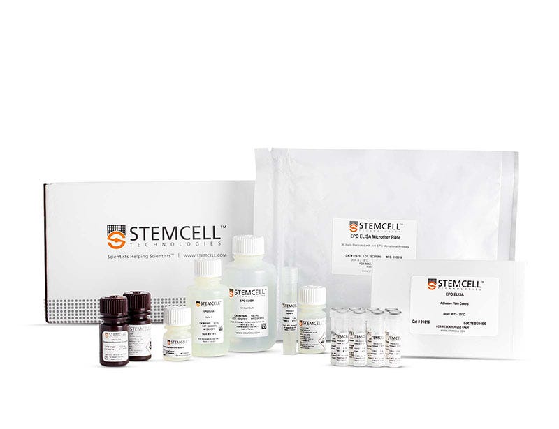
Two-sided or sandwich immunoassays, which use antibodies against two different epitopes on the antigen, have now largely replaced the radioimmunoassays. In these assays, one antibody is immobilized on the surface of microwells or particles and the other is conjugated to an enzyme or other label to detect the captured antigen. Depending on the type of label and substrate, the amount of EPO in test and standard preparations is usually measured by a colorimetric or chemiluminescent reaction. An example of this type of assay is the EPO ELISA kit provided by STEMCELL Technologies (Catalog #01630).
Interpretation of EPO Measurements
In most EPO assays, the EPO concentration is measured by comparison to a standard curve constructed from preparations with known EPO concentrations that are evaluated at the same time as the test sample. EPO standards are calibrated against reference EPO preparations, in particular, the Second International Reference Preparation of Human Urinary EPO or the International Standard for Recombinant-Derived EPO that are supplied by the World Health Organization or the National Institute for Biological Standards and Control.37 The unit of EPO is defined on the basis of EPO biological activity determined by in vivo bioassay (see the EPO International Unit box below for more information). Conventionally, EPO concentrations are expressed as milliunits per milliliter (mU/mL), even if immunoassays are used. Different forms of EPO have different specific activities, and these differences can cause discrepancies between expected and measured results if comparisons are made between EPO preparations of different origin or results of different types of EPO assays. Human urinary EPO typically has a specific activity of ~70,000 U/mg of protein, while values reported for human recombinant EPO preparations are usually higher, ranging between 100,000 - 200,000 U/mg. The variability is mainly caused by differences in the carbohydrate composition of EPO from various sources, which affects the stability and thus the activity of the EPO molecules in vivo, but not in vitro.38 The carbohydrate structure of EPO does not affect the reactivity of EPO with anti-EPO monoclonal antibodies as the dose-response curves of human urinary and recombinant EPO in an EPO ELISA are parallel.39
Immunoassays can be used to detect non-human EPO, but only if the non-human EPO is recognized by the anti-EPO antibodies used in the assay. Differences in immuno-reactivity of non-human EPO and human EPO may result in non-parallel dose response curves. In such cases, human EPO standards do not give reliable values for non-human EPO levels in test samples, and calibrated EPO standards from the same species as the test EPO should be used to get valid results.
Application of Anti-EPO Monoclonal Antibodies to Isolate and Detect Recombinant and Natural EPO
The ability of recombinant EPO to boost RBC production and thus increase aerobic capacity has made it attractive as a performance-enhancing drug in competitive sports. Methods to detect EPO doping must distinguish exogenous recombinant EPO from endogenous natural EPO in human blood and urine. As EPO disappears rapidly from the circulation, these methods also need to be very sensitive and able to detect trace amounts of recombinant EPO before it completely disappears after cessation of treatment. The current EPO immunoassays detect natural and recombinant EPO with similar efficiency and thus cannot distinguish between recombinant and natural EPO. Recombinant EPO produced in CHO cells, however, is less acidic than endogenous EPO as a result of differences in carbohydrate structure. This difference forms the basis of tests to identify recombinant EPO and EPO analogs in blood and urine samples by isoelectric focusing, immunoblotting and other analytical methods.40 Immunoaffinity isolation of EPO from test samples using immobilized anti-EPO monoclonal antibodies (e.g. EPO Immunoaffinity Isolation Plate, Catalog #01640) can increase the specificity and sensitivity of such detection methods by removing serum components that interfere with the accuracy of detection and by concentrating trace amounts of EPO from large sample volumes.41,42
EPO International Unit
The International Unit (U) of EPO activity is based on EPO measurements obtained using the exhypoxic polycythemic mouse assay or similar in vivo assays, and is defined as the amount of EPO that gives the same amount of erythroid stimulation as 5 micromoles of cobalt. Usually, EPO preparations are calibrated in bioassays or immunoassays against reference preparations, in particular the Second International Reference Preparation of urinary EPO, and the newer International Standard for Recombinant DNA-derived EPO (NIBSC-code 87-684).
Other Erythropoietic Molecules
Since the isolation and cloning of human EPO, several different types of new erythropoiesis-stimulating molecules have been developed. Darbepoetin is an engineered form of EPO that contains 5 N-linked glycosylation sites, compared to three for EPO, and as a result is more extensively glycosylated than EPO.43 These molecules have a much longer half-life in the blood circulation than EPO itself and can be administered less frequently than EPO to be effective in patients.44
Other chemical modifications to lysine residues in EPO through a process called carbamoylation completely abolish the erythropoietic effects of EPO in vitro and in vivo. Carbamoylated EPO, however, retains its ability to protect neural, cardiac, kidney and other tissues against hypoxia and other
injuries.45-47
In addition to modified forms of the EPO protein, several small peptide and non-peptide EPO mimetics have been developed that do not share any structural homology to EPO. These molecules can bind to and activate EPO-R and are effective in vivo.48-50 Another way to stimulate erythropoiesis in vivo is through stimulation of endogenous EPO production using compounds that stabilize HIF-1 or increase its binding to DNA through inhibition of the GATA-2 transcription factor. Compounds that stimulate EPO production via this mechanism increase transcription of the EPO gene, even in the presence of normal oxygen concentrations, and have been shown to effectively promote erythropoiesis in vivo.51,52 EPO immunoassays and culture assays for hematopoietic and non-hematopoietic cells provide essential tools to test novel molecules and examine their stimulatory and potentially inhibitory actions or toxicity on erythroid and non-erythroid cell populations.
Related Products
References
- Miyake T, et al. J Biol Chem 252: 5558-5564, 1977
- Jacobs K, et al. Nature 313: 806-810, 1985
- Lin FK, et al. Proc Natl Acad Sci U S A 82: 7580-7584, 1985
- Eschbach JW, et al. N Engl J Med 316: 73-78, 1987 5. Takeuchi M, et al. Glycobiology 1: 337-346, 1991
- Storring PL, et al. Br J Haematol 100: 79-89, 1998
- Gregory CJ, J Cell Physiol 89: 289-301, 1976
- Dai CH, et al. Blood 78: 2493-2497, 1991
- Weinberg RS, Semin Hematol 34: 64-69, 1997
- Giarratana MC, et al. Nat Biotechnol 23: 69-74, 2005
- Miharada K, et al. Nat Biotechnol 24: 1255-1256, 2006
- D’Andrea AD, et al. Cell 57: 277-285, 1989
- Wognum AW, et al. Blood 76: 697-705, 1990
- Damen JE, et al. Exp Hematol 24: 1455-1459, 1996
- Ihle JN, et al. Ann N Y Acad Sci 865: 1-611, 1998
- Anagnostou A, et al. Proc Natl Acad Sci U S A 87: 5978-5982, 1990
- Ogilvie M, et al. J Biol Chem 275: 39754-69761, 2000
- Yu X, et al. Development 129: 505-516, 2002
- Um M, et al. Cell Signal 19: 634-645, 2007
- Sinclair AM, et al. Blood 115: 4264-4272, 2010
- Brines M, et al. Proc Natl Acad Sci U S A 101: 14907-14912, 2004
- Sanchez PE, et al. J Comp Neurol 514: 403-414, 2009
- Kanellakis P, et al. Br J Pharmacol 160: 2085-2096, 2010
- Jelkmann W, et al. Crit Rev Oncol Hematol 67: 39-61, 2008
- Swift S, et al. Blood 115: 4254-4263, 2010
- Lacombe C, et al. Haematologica 83: 724-732, 1998
- Bunn HF, et al. J Exp Biol 201: 1197-1201, 1998
- Spivak JL, Lancet 355: 1707-1712, 2000
- Spivak JL, Blood 100: 4272-4290, 2002
- Kota J et al. Leukemia 22: 1828-1840, 2008
- Cotes PM, et al. Nature 191: 1065-1068, 1961
- de Klerk G, et al. Blood 52: 560-568, 1978
- Krystal G, Exp Hematol 11: 649-660, 1983
- Kitamura T, et al. J Cell Physiol 140: 323-334, 1989
- Damen J, et al. Blood 80: 1923-1932, 1992
- Wognum AW, et al. Blood 76: 1323-1329, 1990
- Storring PL, et al. J Endocrinol 134: 459-484, 1992
- Yuen CT, et al. Br J Haematol 121: 511-526, 2003
- Wognum AW, et al. Blood 74: 622-628, 1989
- Reichel C. Br J Sports Med 48: 842847, 2014
- Kohler M, et al. Int J Sports Med 29: 1-6, 2008
- Reichel C, et al. Drug Test Analysis 1: 43-50, 2009
- Egrie JC, et al. Exp Hematol 31: 290-299, 2003
- Locatelli F, et al. J Nephrol 26: 1114-1121, 2013
- Leist M, et al. Science 305: 239-242, 2004
- Fantacci M, et al. Proc Natl Acad Sci U S A 103: 17531-17536, 2006
- Thomas TJ et al. Brain Res 1502: 55-70, 2013
- Wrighton NC, et al. Science 273: 458-463, 1996
- Qureshi SA, et al. Proc Natl Acad Sci U S A 96: 12156-12161, 1999
- MacDougall IC, et al. N Engl J Med 368: 320-332, 2013
- Nakano Y, et al. Blood 104: 4300-4307, 2004
- Bernhardt WM, et al. J Am Soc Nephrol 21: 2151-2156, 2010


