Optimization of Human T Cell Expansion Protocol: Effects of Early Cell Dilution
In this Technical Bulletin, we highlight the importance of cell density on T cell expansion. Our findings suggest that maintaining T cells at lower cell densities improves T cell growth and viability, especially at early stages of cell expansion.
- Document # 27143
- Version 1.0.0
- Oct 2018
Adoptive T cell therapy is a rapidly progressing field, and optimized protocols for the scalable manufacture of T cells are essential to maximize the therapeutic potential of patient-derived T cells. A single administration of adoptive T cell therapy can require billions of expanded T cells. Large-scale production of human T cells for cellular therapy is a complex, multi-step process that presents many opportunities for optimization in order to obtain maximum yield while retaining desired end phenotype and function. In this Technical Bulletin, we highlight the importance of cell density on T cell expansion. Our findings suggest that maintaining T cells at lower cell densities improves T cell growth and viability, especially at early stages of cell expansion. With optimized culture protocols, up to 800-fold expansion of human T cells with > 85% cell viability was achieved over 10 - 14 days in static culture. This culture strategy can also promote overall cell growth using the Xuri™ Cell Expansion System W25 (GE Healthcare). The protocol described here can be easily implemented in a T cell expansion workflow to ensure optimal viability and total fold expansion.
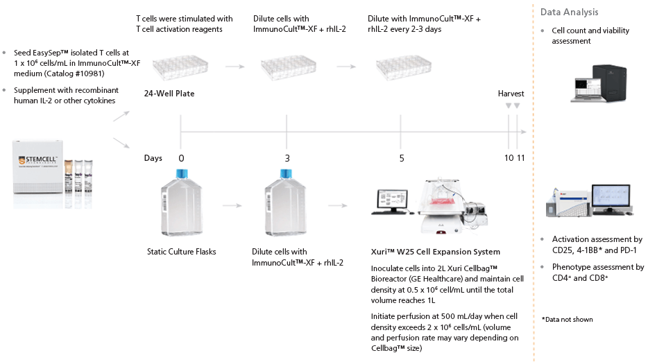
Figure 1. T Cell Isolation, Activation, and Expansion Workflow
Purified T cells were seeded at 1 x 10⁶ cells/mL in ImmunoCult™-XF T Cell Expansion Medium (Catalog #10981) supplemented with 10 ng/mL Human Recombinant IL-2 (rhIL-2). ImmunoCult™ Human CD3/CD28/CD2 or CD3/CD28 T Cell Activators (Catalog # 10970 and 10971, respectively) were added at 25 μL/mL on day 0. On days 3, 5, 7, and/or 10, cells were passaged according to Figure 1. Cell count and viability assessment were conducted using Nucleocounter® NC-250™ (ChemoMetec), and flow cytometry was performed using CytoFLEX™ S (Beckman Coulter).
To investigate the effects of early cell dilution on T cell expansion, T cells were activated with ImmunoCult™ Human T Cell Activator in ImmunoCult™-XF T Cell Expansion Medium supplemented with rhIL-2 at a cell density of 1 x 10⁶ cells/mL. On day 3, T cell culture volume was increased by 2-, 4-, 8-, 16-, or 32-fold by the addition of fresh ImmunoCult™-XF T Cell Expansion Medium supplemented with rhIL-2. On days 5 and 7, T cell culture volume was increased by 4-fold. Where indicated, T cells were transferred into a 2L Xuri™ Cellbag™ Bioreactors, and maintained in the Xuri™ Cell Expansion System W25 on day 5. T cells were harvested for analysis on day 10 or 11 as indicated.
Results
Effect of Early Cell Dilution on Human T Cell Expansion
To investigate the effect of cell density on human T cell expansion, we activated T cells on day 0 using ImmunoCult™ Human CD3/CD28/CD2 T Cell Activator or ImmunoCult™ Human CD3/28 T Cell Activator. On day 3, we increased the total culture volume by 2-, 4-, 8-, 16- or 32-fold, and determined the effects on total expansion. The 4- and 8-fold culture volume increases resulted in the highest total fold expansion and cell viability without any significant effects on the activation phenotype of expanded cells (Figure 2).
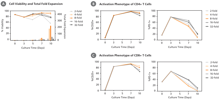
Figure 2. Total Cell Expansion is Affected by the Culturing Protocol Three Days Following Initial T Cell Activation
(A) Optimal viable T cell expansion at day 10 was observed with 4- and 8-fold culture volume increases on day 3 of culture. Total fold expansion of 176 and 222 was observed in 4- and 8-fold culture volume increases, respectively. (B) CD4+ and (C) CD8+ T cell subsets had subtle differences in activation marker expression (CD25 and PD-1) that did not correlate with fold expansion or the subculture protocol on day 3 (data shown for one representative donor).
Comparison of T Cell Activators
The 8-fold volume increase resulted in significantly higher cell expansion compared to the 4-fold volume increase for cells activated using ImmunoCult™ Human CD3/CD28/CD2 T Cell Activator (Figure 3A). On the contrary, the 8-fold volume increase did not cause a statistically significant increase in expansion compared to 4-fold volume increase for cells activated using ImmunoCult™ Human CD3/CD28 T Cell Activator (Figure 3B).
Recommended Human T Cell Expansion Protocol
The findings above have led us to revise our human T cell expansion protocol with optimized cell dilutions (Figure 3C). It is optimal to dilute cells by increasing volume 8-fold or maintaining cell density at 1 - 2.5 x 10 5 cells/mL at day 3 post-activation. We further dilute the expanding cells by increasing culture volume 4-fold at days 5 and 7 post-activation.
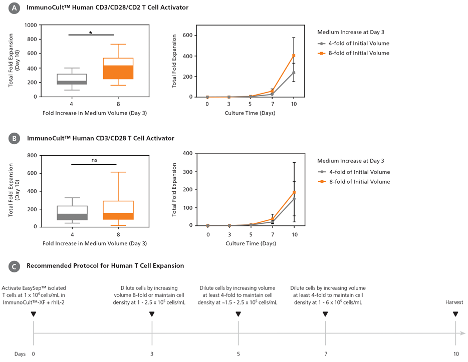
Figure 3. Comparison of ImmunoCult™ Human T Cell Activators with Optimal Culture Protocols
T cells were stimulated on day 0 using ImmunoCult™ Human CD3/CD28/CD2 T Cell Activator or ImmunoCult™ Human CD3/CD28 T Cell Activator in ImmunoCult™-XF T Cell Expansion Medium supplemented with rhIL-2 in static culture. Total culture volumes were increased by 4- or 8-fold using fresh medium on day 3. Total culture volumes were further increased by 4-fold on days 5 and 7 until day 10 of culture. Cumulative fold expansion during culture and total fold expansion at day 10 were analyzed relative to the initial cell seeding number. (A) With ImmunoCult™ Human CD3/CD28/CD2 T Cell Activator, increasing the culture volume by 8-fold on day 3 resulted in high cell growth, and an overall 405 ± 174 total fold expansion (mean ± SD, n = 14). Increasing the culture volume by 4-fold on day 3 resulted in a total fold expansion of 240 ± 90. (*p < 0.0001, paired t-test). (B) With ImmunoCult™ Human CD3/CD28 T Cell Activator, increasing the culture volume by 8-fold or 4-fold resulted in total fold expansions of 150 ± 95 and 186 ± 165, respectively (mean ± SD, n = 14) (ns indicates no significant difference, paired t-test). (C) The recommended human T cell expansion protocol with ImmunoCult™ reagents.
Human T Cell Expansion Using the Xuri™ Cell Expansion System
Obtaining billions of T cells for cell therapy requires large volumes of cells to be expanded in a controlled environment. The optimized T cell expansion protocol above can be adapted for use with the Xuri™ Cell Expansion System W25 for T cell expansion in a closed system for cell therapy manufacturing.
T cells were initially seeded in cell culture flasks as outlined in Figure 1. On day 3 of culture, fresh medium was added to increase the total culture volume by 2-, 4-, or 8-fold. Cells were then transferred into a 2 L Xuri™ Cellbag™ Bioreactors on day 5. We found that the 8-fold culture volume increase on day 3 resulted in the highest total fold expansion (Figure 4).
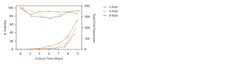
Figure 4. T Cell Expansion in Xuri™ Cell Expansion System W25 Also Benefits from Early Cell Dilution for Optimal T Cell Expansion
Purified T cells were seeded at 1 x 10⁶ cells/mL in cell culture flasks and stimulated with the ImmunoCult™ Human CD3/CD28 T Cell Activator. On day 3 of culture, fresh medium was added to increase the total culture volume by 2-, 4-, and 8-fold. On day 5 of culture, the cells were transferred to 2 L Xuri™ Cellbag™ Bioreactors. Perfusion with fresh medium was initiated on day 8 as outlined in Figure 1. Following 11 days of culture, the highest total fold expansion (263) was observed with the 8-fold culture volume increase on day 3. Viability was similar across the different cell dilutions.
Phenotype of Expanded T Cells
The T cell phenotype plays a critical role in the efficacy of cellular therapy products in addition to cell number. For example, studies in mouse models suggest that minimally differentiated T cells (naïve T cells, stem cell memory T cells, or central memory T cells) exhibit superior antitumor activity compared to highly differentiated T cell subsets.1
The majority of T cells expanded using ImmunoCult™ reagents in static culture alone or in the Xuri™ Cell Expansion System had a central memory phenotype as determined by their co-expression of CD62L and CD45RO (Figure 5).
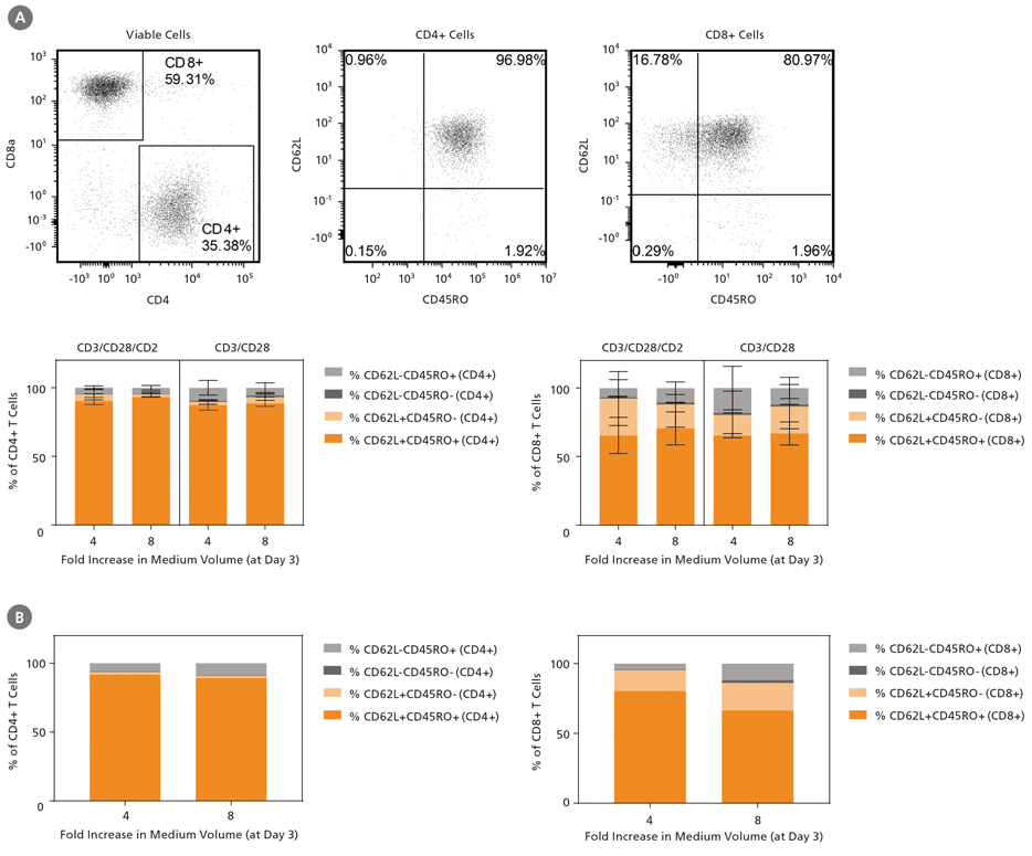
Figure 5. The Majority of Expanded T Cells had a Central Memory T Cell Phenotype (CD62L+CD45RO+) Following Stimulation and Expansion with ImmunoCult™ Reagents
Purified T cells were stimulated with ImmunoCult™ Human T Cell Activators on day 0 and expanded for 10 days in ImmunoCult™-XF T Cell Expansion Medium supplemented
with rhIL-2. Summary of the resulting subset composition in CD4+ or CD8+ T cells using a (A) static culture alone (n = 2) with both
ImmunoCult™ Human T Cell Activators (the representative FACS plots show T cells stimulated with ImmunoCult֭™ Human CD3/CD28/CD2 T Cell Activator)
or (B) GE Xuri Cell Expansion System W25 (n = 1) with
ImmunoCult™ Human CD3/CD28 T Cell Activator. There are subtle differences in phenotype marker expression with
various activators and expansion protocols.
Discussion
The implication of having an optimized method to produce T cells for cell therapy is significant. As described in this Technical Bulletin, we have identified cell density following T cell activation as a key factor in optimizing T cell expansion.
We show that maintaining T cells at lower cell densities during early T cell expansion improves T cell growth and viability. Our study identifies day 3 as a key timepoint for culture volume adjustment when optimizing T cell expansion protocols. At day 3 post-activation, cultured T cells display high levels of cell activation, as indicated by the expression of CD25 and PD-1, but have not significantly expanded (Figure 2). We speculate that lowering cell density at this time point improves T cell expansion by increasing the amount of available nutrients and space per cell after they have received their activation signals. On the other hand, lowering cell density too much may reduce the concentration of autocrine survival factors or cell-cell contact necessary to support optimal fold expansion and cell viability.
One variable that needs to be taken into account when researchers optimize their cell expansion protocol is genetic modification. Producing T cells for therapy often requires genetic modification of the T cells, which can be achieved using viral vectors 2 or gene-editing nucleases.3 How these methods may affect optimal culture dilutions remain to be investigated.
The protocol described here provides a basic framework for testing the effects of other variables that can affect T cell expansion, including
starting cell populations (T cell subsets), cytokines, and growth factors. The optimal culture volume increases will vary depending on these
variables, so we recommend that researchers find the culture volume increases that work best with their specific protocols. Ultimately,
having a well-validated, fully optimized protocol can contribute to solving some of the major challenges of T cell therapy, including lack of
standardization.
Optimized T Cell Activation and Expansion Reagents
The T cell culture reagents used in this Technical Bulletin were developed to enable T cell therapy research and contribute to bringing T cell therapy research from bench to bedside. Learn more:
-
EasySep™ Release Human CD3 Positive Selection Kit
Immunomagnetic positive selection of particle-free human CD3+ cells -
ImmunoCult™-XF T Cell Expansion Medium
Serum-free and xeno-free medium for the expansion of human T cells -
ImmunoCult™ Human CD3/CD28 T Cell Activator
Human T cell activation and expansion reagent -
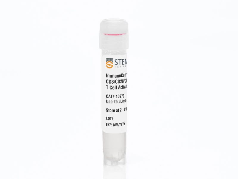
ImmunoCult™ Human CD3/CD28/CD2 T Cell Activator
Human T cell activation and expansion reagent
Featured Resources
On-Demand Course on Human T Cell Expansion
Learn how to optimize your T cell culture and expansion protocol by incorporating ImmunoCult™ media and activators in this free self-paced course.

Resources for T Cell Therapy Research and Development
Discover comprehensive resources to support and enhance your translational T cell research and T cell therapy development, including webinars, technical bulletins, and more.
Resources for T Cell Therapy Research and Production


References
- Gattinoni L et al. (2011) A human memory T cell subset with stem cell-like properties. Nat Med 17(10): 1290–7.
- Li G et al. (2017) Gammaretroviral production and T cell transduction to genetically retarget primary T cells against cancer. Methods Mol Biol 1514: 111–8.
- Osborn MJ et al. (2016) Evaluation of TCR gene editing achieved by TALENs, CRISPR/Cas9, and megaTAL nucleases. Mol Ther 24(3): 570–81.
Request Pricing
Thank you for your interest in this product. Please provide us with your contact information and your local representative will contact you with a customized quote. Where appropriate, they can also assist you with a(n):
Estimated delivery time for your area
Product sample or exclusive offer
In-lab demonstration


