STEMdiff™-XF RPE Maturation Medium

Serum-free and xeno-free medium for generating fully functional and mature hPSC-derived retinal pigment epithelium

STEMdiff™-XF RPE Maturation Medium

Serum-free and xeno-free medium for generating fully functional and mature hPSC-derived retinal pigment epithelium

From: 289 USD
Catalog #
(Select a product)
Serum-free and xeno-free medium for generating fully functional and mature hPSC-derived retinal pigment epithelium
Add to Wish List

Product Advantages


  • Generate mature RPE with key functionality in just 35 days

  • Obtain highly pure RPE without manual selection or cell enrichment

  • Achieve efficient, reproducible differentiation of hPSCs into highly pure, functional RPE cells

  • Streamline RPE cultures with a simple, scalable workflow

Overview

Rapidly mature highly pure functional human pluripotent stem cell (hPSC)-derived retinal pigment epithelial (RPE) cells without the need for manual selection or cell enrichment.

STEMdiff™-XF RPE Maturation Medium is a xeno- and serum-free culture medium that supports the generation of mature and functional RPE from immature RPE in 35 days. Mature RPE cells, generated with STEMdiff™-XF RPE Maturation Medium, express maturity markers (Figure 2) and display key functionalities comparable to primary RPE cells, including pigmentation, polygonal shape, polarization, and phagocytosis.

This product is optimized for the maturation of iPSC-derived RPE cells generated with STEMdiff™-ACF RPE Differentiation Kit.
STEMdiff™-XF RPE Maturation Medium can be used as part of a complete, optimized workflow with the following compatible STEMdiff™ RPE products:

STEMdiff™-ACF RPE Plating Supplement
STEMdiff™-ACF RPE Differentiation Kit

Please contact us if you intend to use this product for commercial or clinical applications.
Cell Type
Pluripotent Stem Cells
Application
Cell Culture
Brand
STEMdiff
Area of Interest
Disease Modeling, Drug Discovery and Toxicity Testing, Neuroscience, Cell Therapy Development

Data Figures

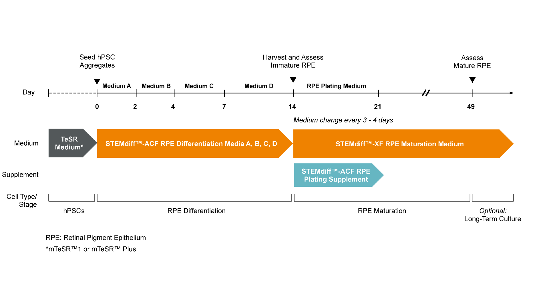
Figure 1. Workflow for the Differentiation of hPSCs into Retinal Pigment Epithelium (RPE) with STEMdiff™ RPE Culture System

hPSC colonies, previously harvested and seeded as clumps, are put directly into the medium provided in STEMdiff™-ACF RPE Differentiation Kit. Seeding into STEMdiff™ RPE Differentiation Medium A induces cells toward immature RPE. A full medium change is performed on Day 1 with fresh STEMdiff™ RPE Differentiation Medium A and then on Day 2 with STEMdiff™ RPE Differentiation Medium B. On Days 4 and 6, medium changes are performed with fresh STEMdiff™ RPE Differentiation Medium C. On Days 7 and every second day thereafter a medium change is performed with fresh STEMdiff™ RPE Differentiation Medium D. On Day 14, immature RPE cells are enzymatically harvested and subcultured in STEMdiff™-XF RPE Maturation Medium with the addition of STEMdiff™-ACF RPE Plating Supplement from Days 14 to 21 to improve survival after passaging. RPE cells begin to mature over the course of 5 weeks of culture in STEMdiff™-XF RPE Maturation Medium and become fully matured by Day 49, possessing key characteristics such as polygonal shape, polarization, pigmentation and phagocytosis.

Figure 2 RPE Kit

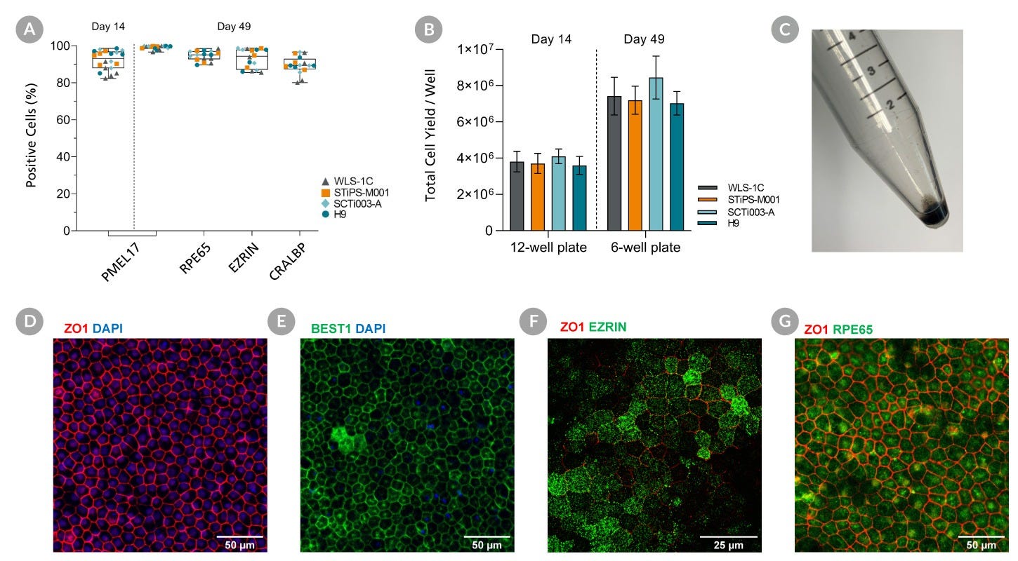
Figure 2. Robust and Rapid Generation of Mature Retinal Pigment Epithelial Cells (RPE) across multiple PSC Cell Lines with the STEMdiff™-ACF RPE Differentiation Kit

hPSCs were cultured for 14 Days using STEMdiff™-ACF RPE Differentiation Kit and subsequently subcultured in STEMdiff™-XF RPE Maturation Medium. Flow cytometry expression of RPE markers are shown at Day 14 and Day 49. (A) The percentage of cells expressing PMEL17, RPE65, EZRIN, and CRALBP and (B) Viable cell yields for 4 hPSC cell lines. PMEL17 is expressed at Day 14 and 49 while the other markers are only present at Day 49. Data are reported as mean + SEM; n = 16 -20. (C) A cell pellet of mature RPE cells demonstrates the pigmentation. Maturation is further demonstrated with immunohistochemistry for expression of RPE markers at Day 49. (D, E, F, G) Mature RPE display tight junctions marked by localization of ZO1 and BEST1 to cell junctions. Mature RPE are polarized, expressing EZRIN apically and ZO1 subapically and express proteins required for the visual cycle (RPE65).

Mature Retinal Pigment Epithelial (RPE) Cells Display Key Functionalities Corresponding to RPE Behaviour

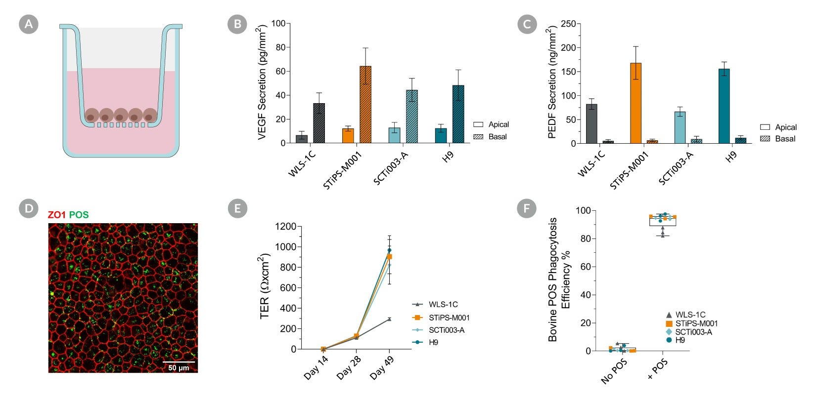
Figure 3. Mature Retinal Pigment Epithelial (RPE) Cells Display Key Functionalities Corresponding to RPE Behaviour

hPSC’s were cultured for 14 Days using STEMdiff™-ACF RPE Differentiation Kit and subsequently subcultured on cell culture inserts in STEMdiff™-XF RPE Maturation Medium for 5 weeks. Apical and basal conditioned medium were collected from Mature RPE, and a sandwich ELISA was performed to quantify Vascular Endothelial Growth Factor (VEGF) and Pigment Epithelial Derived Growth Factor (PEDF) secretion. (A, B) Mature RPE secreted more basal VEGF and apical PEDF demonstrating RPE display correct apicobasal polarity. Data shown as mean + SEM; n = 3. (C) Mature RPE were able to generate a strong barrier with high transepithelial resistance (TER). Data shown as mean + SEM; n = 3-6. (D) Mature RPE were fed FITC-labelled bovine photoreceptor outer segments (POS) for 4 to 5 hours prior to being enzymatically dissociated for flow cytometry analysis or fixed with paraformaldehyde for immunostaining. (E) Mature RPE efficiently internalize bovine POS. Data shown as mean + SEM; n = 3. (F) A cross-sectional schematic of the cell insert culture system.

Protocols and Documentation

Find supporting information and directions for use in the Product Information Sheet or explore additional protocols below.

Document Type
Product Name
Catalog #
Lot #
Language
Catalog #
100-1365
Lot #
All
Language
English
Document Type
Safety Data Sheet
Catalog #
100-1365
Lot #
All
Language
English

Applications

This product is designed for use in the following research area(s) as part of the highlighted workflow stage(s). Explore these workflows to learn more about the other products we offer to support each research area.