RosetteSep™ Human B Cell Enrichment Cocktail

Immunodensity negative selection cocktail

New look, same high quality and support! You may notice that your instrument or reagent packaging looks slightly different from images displayed on the website, or from previous orders. We are updating our look but rest assured, the products themselves and how you should use them have not changed. Learn more

RosetteSep™ Human B Cell Enrichment Cocktail

Immunodensity negative selection cocktail

From: 240 USD
Catalog #
(Select a product)
Immunodensity negative selection cocktail
Add to Wish List

Product Advantages


  • Fast and easy-to-use

  • Requires no special equipment or training

  • Isolated cells are untouched

  • Can be combined with SepMate™ for consistent, high-throughput sample processing

What's Included

  • RosetteSep™ Human B Cell Enrichment Cocktail (Catalog #15024)
    • RosetteSep™ Human B Cell Enrichment Cocktail, 2 mL
  • RosetteSep™ Human B Cell Enrichment Cocktail (Catalog #15064)
    • RosetteSep™ Human B Cell Enrichment Cocktail, 5 x 2 mL
Products for Your Protocol

Overview

The RosetteSep™ Human B Cell Enrichment Cocktail is designed to isolate B cells from whole blood by negative selection. Unwanted cells are targeted for removal with Tetrameric Antibody Complexes recognizing non-B cells and glycophorin A on red blood cells (RBCs). When centrifuged over a buoyant density medium such as RosetteSep™ DM-L (Catalog #15705) or Lymphoprep™ (Catalog #07801), the unwanted cells pellet along with the RBCs. The purified B cells are present as a highly enriched population at the interface between the plasma and the buoyant density medium.
Subtype
Cell Isolation Kits
Cell Type
B Cells
Species
Human
Sample Source
Buffy Coat, Whole Blood
Selection Method
Negative
Application
Cell Isolation
Brand
RosetteSep
Area of Interest
Immunology

Data Figures

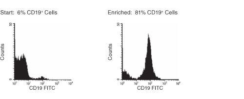
FACS Histogram Results With RosetteSep™ Human B Cell Enrichment Cocktail

Figure 1. FACS Histogram Results With RosetteSep™ Human B Cell Enrichment Cocktail

Starting with fresh whole blood, the CD19+ cell content of the enriched fraction typically ranges from 81% - 83%.

Protocols and Documentation

Find supporting information and directions for use in the Product Information Sheet or explore additional protocols below.

Document Type
Product Name
Catalog #
Lot #
Language
Catalog #
15064, 15024
Lot #
All
Language
English
Document Type
Safety Data Sheet
Catalog #
15064, 15024
Lot #
All
Language
English

Applications

This product is designed for use in the following research area(s) as part of the highlighted workflow stage(s). Explore these workflows to learn more about the other products we offer to support each research area.

Resources and Publications

Frequently Asked Questions

What is RosetteSep™?

RosetteSep™ is a rapid cell separation procedure for the isolation of purified cells directly from whole blood, without columns or magnets.

How does RosetteSep™ work?

The antibody cocktail crosslinks unwanted cells to red blood cells (RBCs), forming rosettes. The unwanted cells then pellet with the free RBCs when centrifuged over a density centrifugation medium (e.g. Ficoll-Paque™ PLUS, Lymphoprep™).

What factors affect cell recovery?

The temperature of the reagents can affect cell recovery. All reagents should be at room temperature (sample, density centrifugation medium, PBS, centrifuge) before performing the isolations. Layering can also affect recovery so be sure to carefully layer the sample to avoid mixing with the density centrifugation medium as much as possible. Be sure to collect the entire enriched culture without disturbing the RBC pellet. A small amount of density centrifugation medium can be collected without worry.

Which cell samples can RosetteSep™ be used with?

RosetteSep™ can be used with leukapheresis samples, bone marrow or buffy coat, as long as: the concentration of cells does not exceed 5 x 10⁷ per mL (can dilute if necessary); and there are at least 100 RBCs for every nucleated cell (RBCs can be added if necessary).

Can RosetteSep™ be used with previously frozen or cultured cells?

Yes. Cells should be re-suspended at 2 - 5 x 10⁷ cells / mL in PBS + 2% FBS. Fresh whole blood should be added at 250 µL per mL of sample, as a source of red cells.

Can RosetteSep™ be used to enrich progenitors from cord blood?

Yes. Sometimes cord blood contains immature nucleated red cells that have a lower density than mature RBCs. These immature red cells do not pellet over Ficoll™, which can lead to a higher RBC contamination than peripheral blood separations.

Does RosetteSep™ work with mouse cells?

No, but we have developed EasySep™, a magnetic-based cell isolation system which works with mouse and other non-human species.

Which anticoagulant should be used with RosetteSep™?

Peripheral blood should be collected in heparinized Vacutainers. Cord blood should be collected in ACD.

Should the anticoagulant be washed off before using RosetteSep™?

No, the antibody cocktail can be added directly to the sample.

Publications (39)

Differential Expression of IgM and IgD Discriminates Two Subpopulations of Human Circulating IgM+IgD+CD27+ B Cells That Differ Phenotypically, Functionally, and Genetically. D. Bautista et al. Frontiers in immunology 2020

Abstract

The origin and function of blood IgM+IgD+CD27+ B cells is controversial, and they are considered a heterogeneous population. Previous staining of circulating B cells of healthy donors with rotavirus fluorescent virus-like particles allowed us to differentiate two subsets of IgM+IgD+CD27+: IgMhi and IgMlo B cells. Here, we confirmed this finding and compared the phenotype, transcriptome, in vitro function, and Ig gene repertoire of these two subsets. Eleven markers phenotypically discriminated both subsets (CD1c, CD69, IL21R, CD27, MTG, CD45RB, CD5, CD184, CD23, BAFFR, and CD38) with the IgMhi phenotypically resembling previously reported marginal zone B cells and the IgMlo resembling both na{\{i}}ve and memory B cells. Transcriptomic analysis showed that both subpopulations clustered close to germinal center-experienced IgM only B cells with a Principal Component Analysis but differed in expression of 78 genes. Moreover IgMhi B cells expressed genes characteristic of previously reported marginal zone B cells. After stimulation with CpG and cytokines significantly (p {\textless} 0.05) higher frequencies (62.5{\%}) of IgMhi B cells proliferated compared with IgMlo B cells (35.37{\%}) and differentiated to antibody secreting cells (14.22{\%} for IgMhi and 7.19{\%} for IgMlo). IgMhi B cells had significantly (p {\textless} 0.0007) higher frequencies of mutations in IGHV and IGKV regions IgMlo B cells had higher usage of IGHJ6 genes (p {\textless} 0.0001) and both subsets differed in their HCDR3 properties. IgMhi B cells shared most of their shared IGH clonotypes with IgM only memory B cells and IgMlo B cells with IgMhi B cells. These results support the notion that differential expression of IgM and IgD discriminates two subpopulations of human circulating IgM+IgD+CD27+ B cells with the IgMhi B cells having similarities with previously described marginal zone B cells that passed through germinal centers and the IgMlo B cells being the least differentiated amongst the IgM+CD27+ subsets."
Mechanism for IL-15-Driven B Cell Chronic Lymphocytic Leukemia Cycling: Roles for AKT and STAT5 in Modulating Cyclin D2 and DNA Damage Response Proteins. R. Gupta et al. Journal of immunology (Baltimore, Md. : 1950) 2019 may

Abstract

Clonal expansion of B cell chronic lymphocytic leukemia (B-CLL) occurs within lymphoid tissue pseudofollicles. IL-15, a stromal cell-associated cytokine found within spleens and lymph nodes of B-CLL patients, significantly boosts in vitro cycling of blood-derived B-CLL cells following CpG DNA priming. Both IL-15 and CpG DNA are elevated in microbe-draining lymphatic tissues, and unraveling the basis for IL-15-driven B-CLL growth could illuminate new therapeutic targets. Using CpG DNA-primed human B-CLL clones and approaches involving both immunofluorescent staining and pharmacologic inhibitors, we show that both PI3K/AKT and JAK/STAT5 pathways are activated and functionally important for IL-15→CD122/ɣc signaling in ODN-primed cells expressing activated pSTAT3. Furthermore, STAT5 activity must be sustained for continued cycling of CFSE-labeled B-CLL cells. Quantitative RT-PCR experiments with inhibitors of PI3K and STAT5 show that both contribute to IL-15-driven upregulation of mRNA for cyclin D2 and suppression of mRNA for DNA damage response mediators ATM, 53BP1, and MDC1. Furthermore, protein levels of these DNA damage response molecules are reduced by IL-15, as indicated by Western blotting and immunofluorescent staining. Bioinformatics analysis of ENCODE chromatin immunoprecipitation sequencing data from cell lines provides insight into possible mechanisms for STAT5-mediated repression. Finally, pharmacologic inhibitors of JAKs and STAT5 significantly curtailed B-CLL cycling when added either early or late in a growth response. We discuss how the IL-15-induced changes in gene expression lead to rapid cycling and possibly enhanced mutagenesis. STAT5 inhibitors might be an effective modality for blocking B-CLL growth in patients.
Computational analysis of the evolutionarily conserved Missing In Metastasis/Metastasis Suppressor 1 gene predicts novel interactions, regulatory regions and transcriptional control. P. Petrov et al. Scientific reports 2019 mar

Abstract

Missing in Metastasis (MIM), or Metastasis Suppressor 1 (MTSS1), is a highly conserved protein, which links the plasma membrane to the actin cytoskeleton. MIM has been implicated in various cancers, however, its modes of action remain largely enigmatic. Here, we performed an extensive in silico characterisation of MIM to gain better understanding of its function. We detected previously unappreciated functional motifs including adaptor protein (AP) complex interaction site and a C-helix, pointing to a role in endocytosis and regulation of actin dynamics, respectively. We also identified new functional regions, characterised with phosphorylation sites or distinct hydrophilic properties. Strong negative selection during evolution, yielding high conservation of MIM, has been combined with positive selection at key sites. Interestingly, our analysis of intra-molecular co-evolution revealed potential regulatory hotspots that coincided with reduced potentially pathogenic polymorphisms. We explored databases for the mutations and expression levels of MIM in cancer. Experimentally, we focused on chronic lymphocytic leukaemia (CLL), where MIM showed high overall expression, however, downregulation on poor prognosis samples. Finally, we propose strong conservation of MTSS1 also on the transcriptional level and predict novel transcriptional regulators. Our data highlight important targets for future studies on the role of MIM in different tissues and cancers.
New look, same high quality and support! You may notice that your instrument or reagent packaging looks slightly different from images displayed on the website, or from previous orders. We are updating our look but rest assured, the products themselves and how you should use them have not changed. Learn more