Collagenase/Hyaluronidase

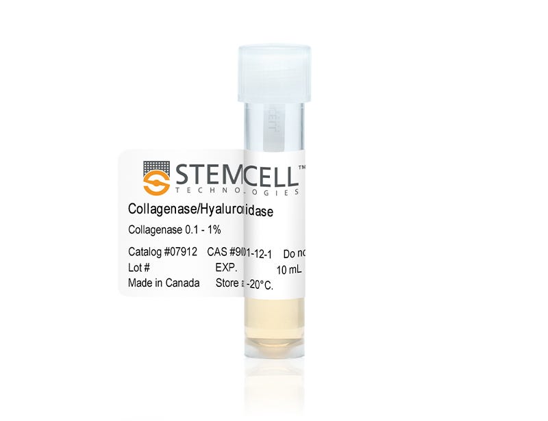
10X Collagenase/hyaluronidase in DMEM

Collagenase/Hyaluronidase

10X Collagenase/hyaluronidase in DMEM

From: 293 USD
Catalog #
(Select a product)
10X Collagenase/hyaluronidase in DMEM
Add to Wish List

Product Advantages


  • Effectively dissociate human mammary tissue and prostate epithelial cells

Overview

Enzymatically dissociate human mammary cells with 10X Collagenase/Hyaluronidase in Dulbecco’s Modified Eagle’s Medium (DMEM). This protease/polysaccharidase combination digests native collagen fibrils and hydrolizes hyaluronic acids in connective tissues for effective tissue dissociation. Collagenase/Hyluronidase can also been used for enzymatic dissociation of prostate epithelial cells.
Contains
• 3000 U/mL Collagenase
• 1000 U/mL Hyaluronidase
• DMEM (1000 mg D-glucose/L)
Subtype
Enzymatic
Cell Type
Mammary Cells, Other, Prostate Cells
Species
Human, Mouse, Non-Human Primate, Other, Rat
Application
Cell Culture
Area of Interest
Epithelial Cell Biology

Protocols and Documentation

Find supporting information and directions for use in the Product Information Sheet or explore additional protocols below.

Document Type
Product Name
Catalog #
Lot #
Language
Catalog #
07912
Lot #
All
Language
English
Document Type
Safety Data Sheet
Catalog #
07912
Lot #
All
Language
English

Applications

This product is designed for use in the following research area(s) as part of the highlighted workflow stage(s). Explore these workflows to learn more about the other products we offer to support each research area.

Resources and Publications

Publications (1)

Silica bioreplication preserves three-dimensional spheroid structures of human pluripotent stem cells and HepG2 cells. Lou Y-R et al. Scientific reports 2015 SEP

Abstract

Three-dimensional (3D) cell cultures produce more in vivo-like multicellular structures such as spheroids that cannot be obtained in two-dimensional (2D) cell cultures. Thus, they are increasingly employed as models for cancer and drug research, as well as tissue engineering. It has proven challenging to stabilize spheroid architectures for detailed morphological examination. Here we overcome this issue using a silica bioreplication (SBR) process employed on spheroids formed from human pluripotent stem cells (hPSCs) and hepatocellular carcinoma HepG2 cells cultured in the nanofibrillar cellulose (NFC) hydrogel. The cells in the spheroids are more round and tightly interacting with each other than those in 2D cultures, and they develop microvilli-like structures on the cell membranes as seen in 2D cultures. Furthermore, SBR preserves extracellular matrix-like materials and cellular proteins. These findings provide the first evidence of intact hPSC spheroid architectures and similar fine structures to 2D-cultured cells, providing a pathway to enable our understanding of morphogenesis in 3D cultures.