MammoCult™ Human Medium Kit

For culture of human mammospheres and tumorspheres

MammoCult™ Human Medium Kit

For culture of human mammospheres and tumorspheres

From: 410 USD
Catalog #
(Select a product)
For culture of human mammospheres and tumorspheres
Add to Wish List

What's Included

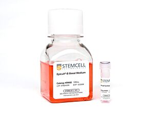
  • MammoCult™ Basal Medium (Human), 450 mL
  • MammoCult™ Proliferation Supplement (Human), 50 mL
Products for Your Protocol
To see all required products for your protocol, please consult the Protocols and Documentation.

Overview

MammoCult™ Medium (Human) is a serum-free, estrogen and progesterone-free culture medium optimized for the culture of mammospheres from normal human primary breast tissues and tumorspheres from human breast cancer cell lines. For preparation of complete MammoCult™ Medium, Hydrocortisone Stock Solution (Catalog #07925) and Heparin Solution (Catalog #07980) are also required.
Subtype
Specialized Media
Cell Type
Cancer Cells and Cell Lines, Mammary Cells
Species
Human
Application
Cell Culture, Maintenance, Spheroid Culture
Brand
MammoCult
Area of Interest
Cancer, Epithelial Cell Biology
Formulation Category
Serum-Free

Data Figures

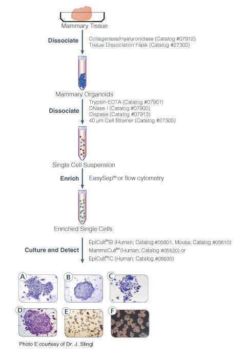
Protocol for isolation and identification of human and mouse mammary epithelial progenitor cells

Figure 1. Protocol for Isolation and Identification of Human and Mouse Mammary Epithelial Progenitor Cells

Phase contrast photographs of (A) a pure human myoepithelial cell colony, (B) a pure human luminal cell colony, and (C) a mixed human colony. (D) is a mouse colony. Unlike human mammary CFC colonies, subtypes of mouse mammary epithelial cell colonies are not easily identifiable. All colonies were cultured in either EpiCult™-B (Human: Catalog #05601) or EpiCult™-B (Mouse:Catalog #5610) in the presence of an irradiated NIH 3T3 feeder layer. Colonies were visualized by staining with Wright"s Giemsa. (E) is a picture of mammospheres obtained from primary human mammary epithelial cells and (F) is an image of tumorspheres obtained from MCF7 human breast cancer cell line.

Protocols and Documentation

Find supporting information and directions for use in the Product Information Sheet or explore additional protocols below.

Document Type
Product Name
Catalog #
Lot #
Language
Catalog #
05620
Lot #
All
Language
English
Document Type
Safety Data Sheet 1
Catalog #
05620
Lot #
All
Language
English
Document Type
Safety Data Sheet 2
Catalog #
05620
Lot #
All
Language
English

Applications

This product is designed for use in the following research area(s) as part of the highlighted workflow stage(s). Explore these workflows to learn more about the other products we offer to support each research area.

Resources and Publications

Publications (124)

Tumor-Suppressive microRNA Therapy Inhibits Growth of Glioblastoma Multiforme Xenografts. E. Biltekin et al. Cancers 2026 May

Abstract

Glioblastoma multiforme (GBM) is defined by rapid progression, high invasiveness, and a poor prognosis, with a median survival of only ≅13 months despite current treatments. Its marked genetic heterogeneity, high mutational burden, and cancer stem cell population make GBM exceptionally difficult to treat, highlighting the urgent need for more effective, multitargeted therapies. Non-coding RNAs, particularly tumor suppressor microRNAs (miRNAs), have gained attention for suppressing key oncogenic processes that drive tumorigenesis, metastasis, and drug resistance, positioning them as promising tools for targeting multiple oncogenic pathways. We recently found that FOXM1/AXL-eEF2K collaboratively drive GBM cell proliferation, survival, and invasion through the formation of a signaling hub complex. In this study, we employed miRNA prediction algorithms to identify a specific miRNA, in vitro functional assays and in vivo GBM flank model to target GBM tumorigenesis by distrupting the FOXM1/AXL-eEF2K signaling hub. Our results indicated that FOXM1, AXL, and eEF2K are overexpressed in GBM patient tumors. To target the FOXM1/AXL-eEF2K signaling hub, we identified miR-449b-5p, miR-329-3p, and miR-518c as potential co-inhibitors of FOXM1/AXL-eEF2K and suppressors of cell proliferation, migration-invasion, and spheroid formation. Furthermore, the combination of miR-449b-5p, miR-329-3p, and miR-518c treatments with temozolomide led to synergistic enhancements in cell proliferation suppression and the induction of apoptosis and ferroptosis. More importantly, in vivo miR-329-3p treatment led to remarkable suppression of GBM tumor xenografts. These findings indicate that miR-329-3p-based tumor suppressor therapy may offer a multitargeted approach for GBM treatment.
Developing a Phosphodiesterase 10A Inhibitor as a Novel Therapeutic Agent for Triple-Negative Breast Cancer. M. Biswas et al. Cells 2026 Jul

Abstract

Triple-negative breast cancer (TNBC) is a highly aggressive subtype of breast cancer with limited therapeutic options for patients at high risk of disease recurrence and metastasis. The cyclic nucleotide-degrading enzyme, phosphodiesterase 10A (PDE10), that hydrolyzes both cAMP and cGMP has been previously reported to be expressed in multiple cancers and regulates key cellular signaling pathways involved in cancer cell proliferation, survival, and maintenance of stem cell-like properties. We found that PDE10 overexpression was associated with poor relapse-free survival of TNBC patients and identified its potential as a therapeutic target for TNBC using a novel inhibitor, ADT-030. Our results showed that ADT-030 inhibited the growth of TNBC cells, reduced colony-forming efficiency and enhanced the therapeutic efficacy of paclitaxel. A TNBC mouse model demonstrated that oral administration of ADT-030 significantly suppressed syngeneic tumor growth and enhanced the antitumor efficacy of paclitaxel. ADT-030 treatment altered differentially expressed genes (DEGs), signaling pathways, and cellular processes. Overall, our findings suggest that ADT-030, as a monotherapy or in combination with standard-of-care chemotherapy, may be an effective therapeutic approach for TNBC. Further studies are warranted to better understand the oncogenic role of PDE10 in TNBC and the mechanisms by which ADT-030 modulates the tumor microenvironment (TME) and enhances chemotherapy response.
CYR61 Expression Is Induced by IGF1 and Promotes the Proliferation of Prostate Cancer Cells Through the PI3/AKT Signaling Pathway G. Ortiz-Hernández et al. International Journal of Molecular Sciences 2025 Sep

Abstract

Cysteine-rich angiogenic inducer 61 (CYR61) promotes prostate cancer (PCa) cell growth, but its role in disease progression remains unclear. Given its insulin-like growth factor (IGF)-binding domain and the known involvement of insulin-like growth factor-1 (IGF1) in PCa, we investigated the molecular interplay between CYR61 and IGF1. CYR61 was silenced using small interfering RNA (siRNA) in prostate carcinoma 3 (PC3), lymph node carcinoma of the prostate (LNCaP), and androgen receptor (AR)-positive 22Rv1 cells, followed by assessments of their proliferation, viability, colony formation, migration, and signaling pathway activation. CYR61 knockdown significantly reduced cell growth, viability, prostasphere formation, and migration across all three cell lines. Mechanistically, CYR61 silencing inhibited PI3K/AKT signaling but had no effect on MAPK activation. In addition, treatment with recombinant IGF1 induced CYR61 expression in a time-dependent manner, and the inhibition of PI3K/AKT signaling suppressed both CYR61 expression and cell proliferation. These findings suggest that IGF1 promotes PCa progression through CYR61 and that CYR61 may serve as a potential therapeutic target for limiting tumor growth and metastasis.