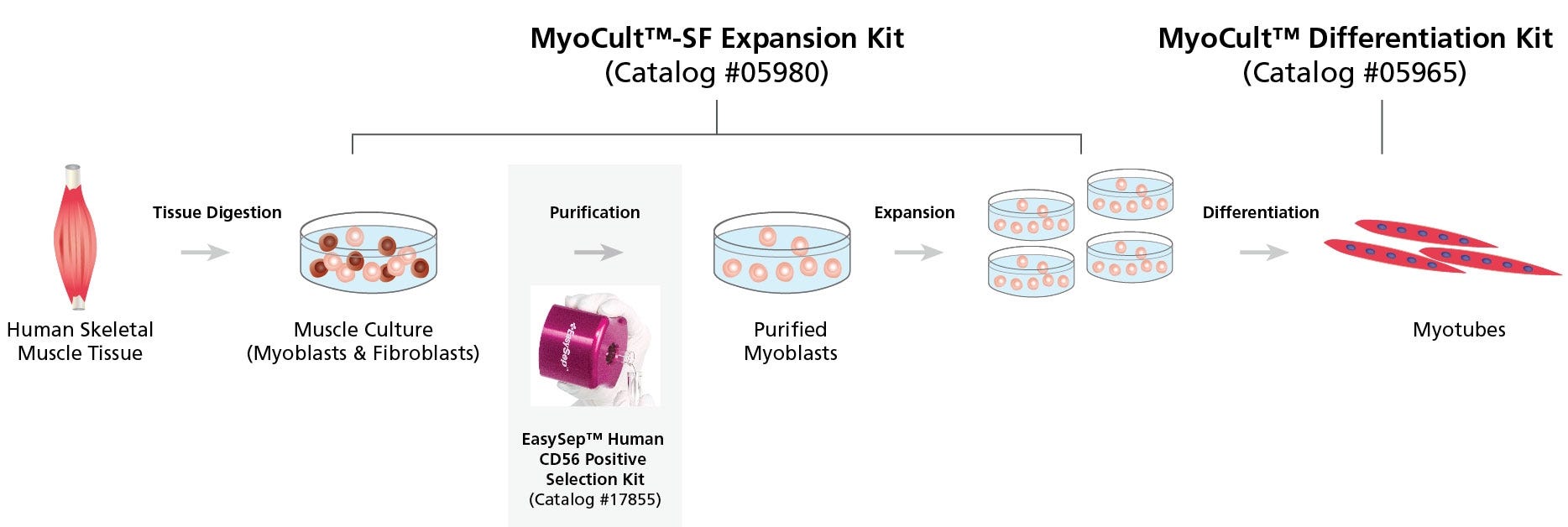
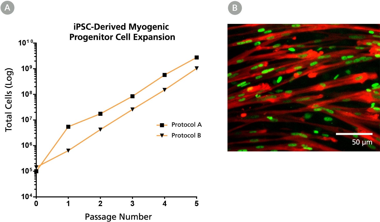
MyoCult™-SF Expansion Supplement Kit (Human)
Serum-free supplement and attachment substrate for the derivation and expansion of human skeletal muscle progenitor cells (myoblasts)
Request Pricing
Thank you for your interest in this product. Please provide us with your contact information and your local representative will contact you with a customized quote. Where appropriate, they can also assist you with a(n):
Estimated delivery time for your area
Product sample or exclusive offer
In-lab demonstration
-
 Collagenase A, ACF
Animal component-free collagenase for the digestion of native collagen fibrils
-
 DMEM with 1000 mg/L D-Glucose
Dulbecco's Modified Eagle's Medium (DMEM) with 1000 mg/L D-glucose
-
 Culture Dish, Non-Treated
Sterile, flat-bottom, clear polystyrene non-treated dish with lid; 35, 60, or 100 mm formats
Overview
Data Figures
Protocols and Documentation
Find supporting information and directions for use in the Product Information Sheet or explore additional protocols below.
Applications
This product is designed for use in the following research area(s) as part of the highlighted workflow stage(s). Explore these workflows to learn more about the other products we offer to support each research area.
Resources and Publications
Educational Materials (4)


Publications (3)
Abstract
Abstract
Abstract
Related Products

PRODUCTS ARE FOR RESEARCH USE ONLY AND NOT INTENDED FOR HUMAN OR ANIMAL DIAGNOSTIC OR THERAPEUTIC USES UNLESS OTHERWISE STATED. FOR ADDITIONAL INFORMATION ON QUALITY AT STEMCELL, REFER TO WWW.STEMCELL.COM/COMPLIANCE.
