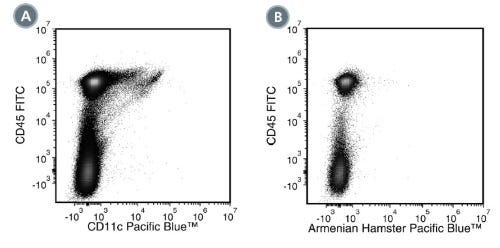
Anti-Mouse CD11c Antibody, Clone N418
Hamster (Armenian) monoclonal IgG antibody against mouse CD11c
Request Pricing
Thank you for your interest in this product. Please provide us with your contact information and your local representative will contact you with a customized quote. Where appropriate, they can also assist you with a(n):
Estimated delivery time for your area
Product sample or exclusive offer
In-lab demonstration
Overview
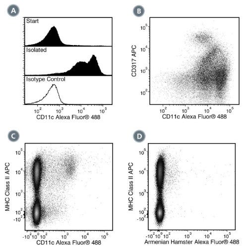
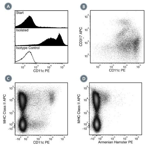
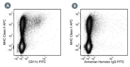
Data Figures
Protocols and Documentation
Find supporting information and directions for use in the Product Information Sheet or explore additional protocols below.
Applications
This product is designed for use in the following research area(s) as part of the highlighted workflow stage(s). Explore these workflows to learn more about the other products we offer to support each research area.
Resources and Publications
Educational Materials (3)


Publications (1)
Abstract
Related Products

Alexa Fluor and Pacific Blue are trademarks of Life Technologies Corporation. Antibodies conjugated to Alexa Fluor® or Pacific Blue™ are licensed for internal research use only and sale is expressly conditioned on the buyer not using the antibody for manufacturing, performing a service or medical test, or otherwise generating revenue. For use other than research, contact Life Technologies Corporation, 5791 Van Allen Way, Carlsbad, CA 92008 USA or outlicensing@lifetech.com.
PRODUCTS ARE FOR RESEARCH USE ONLY AND NOT INTENDED FOR HUMAN OR ANIMAL DIAGNOSTIC OR THERAPEUTIC USES UNLESS OTHERWISE STATED. FOR ADDITIONAL INFORMATION ON QUALITY AT STEMCELL, REFER TO WWW.STEMCELL.COM/COMPLIANCE.
