MethoCult™ SF M3236

Serum-free methylcellulose-based medium without cytokines for mouse cells

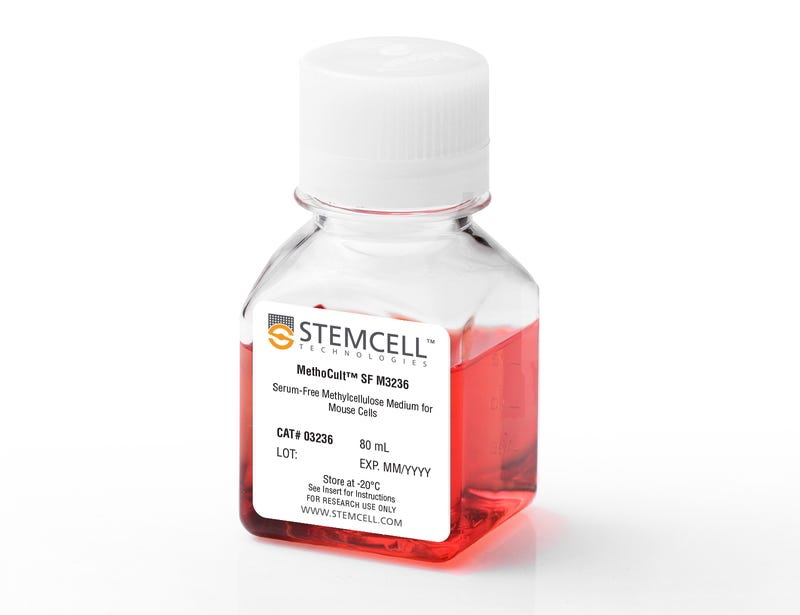
MethoCult™ SF M3236

Serum-free methylcellulose-based medium without cytokines for mouse cells

From: 439 USD
Catalog #
(Select a product)
Serum-free methylcellulose-based medium without cytokines for mouse cells
Add to Wish List

Overview

MethoCult™ SF M3236 is a serum-free, incomplete medium formulation that contains methylcellulose in Iscove's MDM. MethoCult™ SF M3236 is suitable for the growth and enumeration of hematopoietic progenitor cells in colony-forming unit (CFU) assays of mouse bone marrow, spleen, and fetal liver, when the appropriate growth factors and supplements are added. This formulation does not contain serum or cytokines and is ideal for testing cytokine effects where the presence of fetal bovine serum is not desired.

Browse our Frequently Asked Questions (FAQs) on performing the CFU assay.
Contains
• Methylcellulose in Iscove's MDM
• Bovine serum albumin
• Recombinant human insulin
• Human transferrin (iron-saturated)
• 2-Mercaptoethanol
• Supplements
Subtype
Semi-Solid Media, Specialized Media
Cell Type
Hematopoietic Stem and Progenitor Cells
Species
Mouse
Application
Cell Culture, Colony Assay, Functional Assay
Brand
MethoCult
Area of Interest
Stem Cell Biology
Formulation Category
Serum-Free

Protocols and Documentation

Find supporting information and directions for use in the Product Information Sheet or explore additional protocols below.

Document Type
Product Name
Catalog #
Lot #
Language
Catalog #
03236
Lot #
All
Language
English
Document Type
Technical Manual
Catalog #
03236
Lot #
All
Language
English
Document Type
Safety Data Sheet
Catalog #
03236
Lot #
All
Language
English

Applications

This product is designed for use in the following research area(s) as part of the highlighted workflow stage(s). Explore these workflows to learn more about the other products we offer to support each research area.

Resources and Publications

Frequently Asked Questions

Why use semi-solid media?

Semi-solid media (methylcellulose-based MethoCult™ and collagen-based MegaCult™-C) allow the clonal progeny of a single progenitor cell to remain spatially isolated from other colonies within a culture, so they may be separately identified and counted.

Why use methylcellulose-based media?

Methylcellulose permits better growth of erythroid colonies than other types of semi-solid support systems (eg. agar) while allowing optimal myeloid colony formation. When appropriate cytokines are present, committed progenitor cells of both erythroid and granulocyte/macrophage lineages (CFU-GM, CFU-G, CFU-M) as well as multi-potential progenitor cells (CFU-GEMM), can be assayed simultaneously in the same culture dish.

Is it necessary to add antibiotics to the media?

No, aseptic technique should be sufficient to maintain sterile cultures. However, antibiotics (eg. Penicillin/Streptomycin) or anti-fungals (eg. Amphotericin B) may be added to the methylcellulose medium if desired.

Is there anything I can do if my cultures appear contaminated?

No, once contamination is visible, it is not possible to rescue the cultures by the addition of antibiotics. Bacteria and yeast inhibit colony formation by depleting nutrients or by releasing toxic substances.

Why can't I use a pipette to dispense methylcellulose-based media?

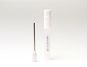
Methylcellulose is a viscous solution that cannot be accurately dispensed using a pipette due to adherence of the medium to the walls of the pipette tip. Blunt-End, 16 Gauge needles (Catalog #28110), in combination with 3 cc Syringes (Catalog #28230) are recommended for accurate dispensing of MethoCult™.

Can I 'pluck' the colonies for individual analysis?

Yes, colonies can be 'plucked' using a pipette with 200 µL sterile pipette tips or using a glass Pasteur pipette with an elongated tip. Individual colonies should be placed in a volume of 25 - 50 µL of medium, and diluted into suitable culture medium for further culture or analysis.

Why are low adherence dishes so important?

Adherent cells such as fibroblasts can cause inhibition of colony growth and obscure visualization of colonies.

Can MethoCult™ products be used for lymphoid progenitor CFU assays?

Human lymphoid progenitors (B, NK and T) seem to require stromal support for growth therefore cannot be grown in MethoCult™. Mouse pre-B clonogenic progenitors can be grown in MethoCult™ M3630 (Catalog #03630).

Is it possible to set up CFU assays in a 24-well plate?

Yes, as long as a plating concentration optimized for the smaller surface area of a well in a 24-well plate (1.9 cm2 as compared to ~9.5 cm2 for a 35 mm dish) is used for these assays. The number of replicate wells required to get an accurate estimation of CFU numbers may also need to be increased.

Can I stain colonies in MethoCult™ medium?

The cells in individual colonies in MethoCult™ can be stained, eg., for analysis of morphology or phenotype, after they are plucked from the dish and washed free of methylcellulose. Colonies grown in collagen-based MegaCult™-C medium can be used for immunohistochemical or enzymatic staining in situ after dehydration and fixation onto glass slides.

Are there differences in colony morphology with serum-free media?

Serum-containing media generally give better overall growth (colonies may appear larger) but there are no large differences in total colony numbers when CFU assays using serum-free media and serum-containing media are compared, provided that identical cytokines are present.

Can MethoCult™ be made with alternate base media?

Yes, this can be done as a 'custom' media order. Please contact techsupport@stemcell.com for more information.

Is there a MethoCult™ formulation suitable for HPP-CFC (high proliferative potential colony forming cell)?

Yes, MethoCult™ H4535 (Catalog #04535) can be used for the HPP-CFC assay as it does not contain EPO. The culture period is usually 28 days. It is not necessary to feed these cultures as growth factors in the medium are present in excess. As HPP-CFCs can be quite large, overplating can be a problem. It is recommended to plate cells at two or more different concentrations.

Publications (7)

Smad4 binds Hoxa9 in the cytoplasm and protects primitive hematopoietic cells against nuclear activation by Hoxa9 and leukemia transformation. Qu&eacute et al. Blood 2011 JUN

Abstract

We studied leukemic stem cells (LSCs) in a Smad4(-/-) mouse model of acute myelogenous leukemia (AML) induced either by the HOXA9 gene or by the fusion oncogene NUP98-HOXA9. Although Hoxa9-Smad4 complexes accumulate in the cytoplasm of normal hematopoietic stem cells and progenitor cells (HSPCs) transduced with these oncogenes, there is no cytoplasmic stabilization of HOXA9 in Smad4(-/-) HSPCs, and as a consequence increased levels of Hoxa9 is observed in the nucleus leading to increased immortalization in vitro. Loss of Smad4 accelerates the development of leukemia in vivo because of an increase in transformation of HSPCs. Therefore, the cytoplasmic binding of Hoxa9 by Smad4 is a mechanism to protect Hoxa9-induced transformation of normal HSPCs. Because Smad4 is a potent tumor suppressor involved in growth control, we developed a strategy to modify the subcellular distribution of Smad4. We successfully disrupted the interaction between Hoxa9 and Smad4 to activate the TGF-β pathway and apoptosis, leading to a loss of LSCs. Together, these findings reveal a major role for Smad4 in the negative regulation of leukemia initiation and maintenance induced by HOXA9/NUP98-HOXA9 and provide strong evidence that antagonizing Smad4 stabilization by these oncoproteins might be a promising novel therapeutic approach in leukemia.
Hypoxia-inducible factor-1 deficiency results in dysregulated erythropoiesis signaling and iron homeostasis in mouse development. Yoon D et al. The Journal of biological chemistry 2006 SEP

Abstract

Hypoxia-inducible factor-1 (HIF-1) regulates the transcription of genes whose products play critical roles in energy metabolism, erythropoiesis, angiogenesis, and cell survival. Limited information is available concerning its function in mammalian hematopoiesis. Previous studies have demonstrated that homozygosity for a targeted null mutation in the Hif1alpha gene, which encodes the hypoxia-responsive alpha subunit of HIF-1, causes cardiac, vascular, and neural malformations resulting in lethality by embryonic day 10.5 (E10.5). This study revealed reduced myeloid multilineage and committed erythroid progenitors in HIF-1alpha-deficient embryos, as well as decreased hemoglobin content in erythroid colonies from HIF-1alpha-deficient yolk sacs at E9.5. Dysregulation of erythropoietin (Epo) signaling was evident from a significant decrease in mRNA levels of Epo receptor (EpoR) in Hif1alpha-/- yolk sac as well as Epo and EpoR mRNA in Hif1alpha-/- embryos. The erythropoietic defects in HIF-1alpha-deficient erythroid colonies could not be corrected by cytokines, such as vascular endothelial growth factor and Epo, but were ameliorated by Fe-SIH, a compound delivering iron into cells independently of iron transport proteins. Consistent with profound defects in iron homeostasis, Hif1alpha-/- yolk sac and/or embryos demonstrated aberrant mRNA levels of hepcidin, Fpn1, Irp1, and frascati. We conclude that dysregulated expression of genes encoding Epo, EpoR, and iron regulatory proteins contributes to defective erythropoiesis in Hif1alpha-/- yolk sacs. These results identify a novel role for HIF-1 in the regulation of iron homeostasis and reveal unexpected regulatory differences in Epo/EpoR signaling in yolk sac and embryonic erythropoiesis.
Kinase domain mutants of Bcr-Abl exhibit altered transformation potency, kinase activity, and substrate utilization, irrespective of sensitivity to imatinib. Griswold IJ et al. Molecular and cellular biology 2006 AUG

Abstract

Kinase domain (KD) mutations of Bcr-Abl interfering with imatinib binding are the major mechanism of acquired imatinib resistance in patients with Philadelphia chromosome-positive leukemia. Mutations of the ATP binding loop (p-loop) have been associated with a poor prognosis. We compared the transformation potency of five common KD mutants in various biological assays. Relative to unmutated (native) Bcr-Abl, the ATP binding loop mutants Y253F and E255K exhibited increased transformation potency, M351T and H396P were less potent, and the performance of T315I was assay dependent. The transformation potency of Y253F and M351T correlated with intrinsic Bcr-Abl kinase activity, whereas the kinase activity of E255K, H396P, and T315I did not correlate with transforming capabilities, suggesting that additional factors influence transformation potency. Analysis of the phosphotyrosine proteome by mass spectroscopy showed differential phosphorylation among the mutants, a finding consistent with altered substrate specificity and pathway activation. Mutations in the KD of Bcr-Abl influence kinase activity and signaling in a complex fashion, leading to gain- or loss-of-function variants. The drug resistance and transformation potency of mutants may determine the outcome of patients on therapy with Abl kinase inhibitors.