MyoCult™-SF Attachment Substrate
Serum-free attachment substrate for the derivation and expansion of human skeletal muscle progenitor cells (myoblasts)
Request Pricing
Thank you for your interest in this product. Please provide us with your contact information and your local representative will contact you with a customized quote. Where appropriate, they can also assist you with a(n):
Estimated delivery time for your area
Product sample or exclusive offer
In-lab demonstration
Overview
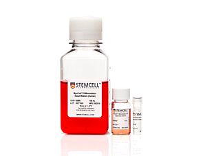
MyoCult™-SF Attachment Substrate is also available as part of the MyoCult™-SF Expansion Supplement Kit (Human), which includes MyoCult™-SF Expansion 10X Supplement (Human). The supplement is combined with a basal medium to prepare MyoCult™-SF Expansion Medium for the culture of primary human and hPSC-derived myoblasts. Myoblasts cultured using MyoCult™-SF Expansion Medium can be differentiated into multinucleated myotubes using MyoCult™ Differentiation Kit (Human).
For more information on protocols for the derivation and expansion of human skeletal muscle progenitor cells with MyoCult™, please explore the Product Information Sheet.
Data Figures

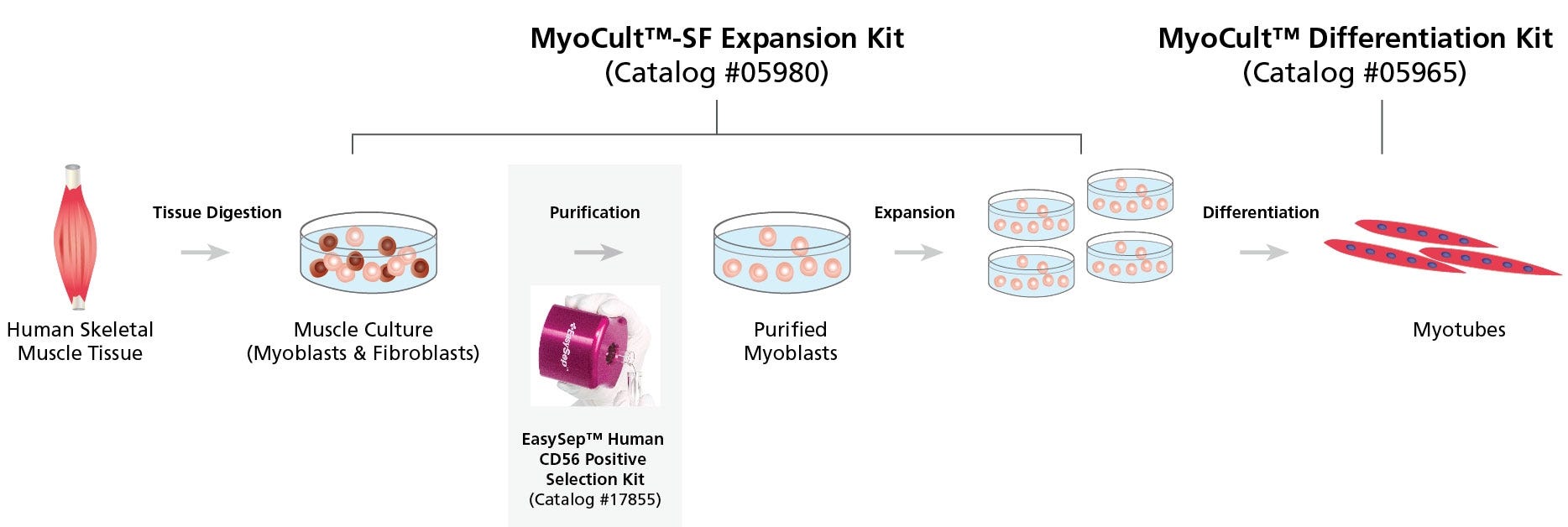
Figure 1. Workflow for the Derivation, Expansion and Differentiation of Human Myogenic Progenitor Cells
Human skeletal muscle tissue is digested into a single cell suspension and plated into MyoCult™-SF Expansion Medium. Myoblasts are then enriched by cell sorting or using a selection kit such as the EasySep™ Human CD56 Positive Selection Kit (Catalog #17855). Purified myoblasts are then culture-expanded or differentiated into myotubes for further studies. Myoblasts can be derived and expanded, using the MyoCult™-SF Expansion Kit (Catalog #05980) and then differentiated using the MyoCult™ Differentiation Kit (Catalog #05965).

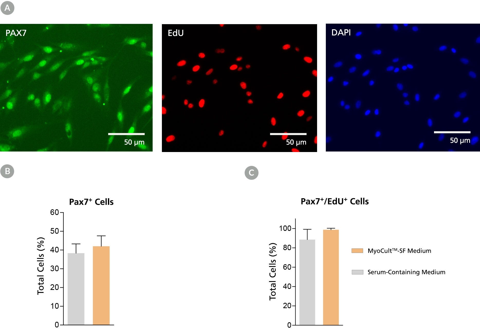
Figure 2. Derivation of PAX7+ Myogenic Progenitor Cells in MyoCult™-SF Expansion Medium from Human Skeletal Muscle Tissue
Myogenic progenitor cells were derived from human skeletal muscle tissue and culture-expanded using the MyoCult™-SF Expansion Kit or a serum-containing medium. (A) Following 6 days of expansion, myoblasts were fixed and immunostained for PAX7 (green), a thymine analogue (EdU; red) and nuclei (DAPI; blue). Total percentage of cells expressing (B) PAX7 or (C) PAX7 and EdU were quantified (n = 4). Data was generated and used with permission by Dr. Penny M. Gilbert’s Lab, Department of Biochemistry, University of Toronto.

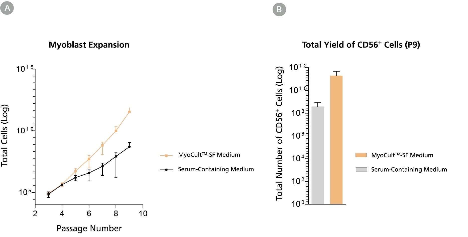
Figure 3. Long-Term Expansion of Myoblasts were Observed When Using MyoCult™-SF Expansion Kit
Myoblasts were culture-expanded in MyoCult™-SF Expansion Medium or in serum-containing medium. (A) Myoblasts culture-expanded in MyoCult™-SF Expansion Medium displayed superior expansion rate than when cells were expanded in serum-containing medium (n=3). (B) Expansion of myoblasts in MyoCult™-SF Expansion Medium generates a greater yield of CD56+ cells after 9 passages compared to myoblasts cultured in serum-containing medium (P9; n = 2). Data was generated and used with permission by Dr. Penny M. Gilbert’s Lab, Department of Biochemistry, University of Toronto.

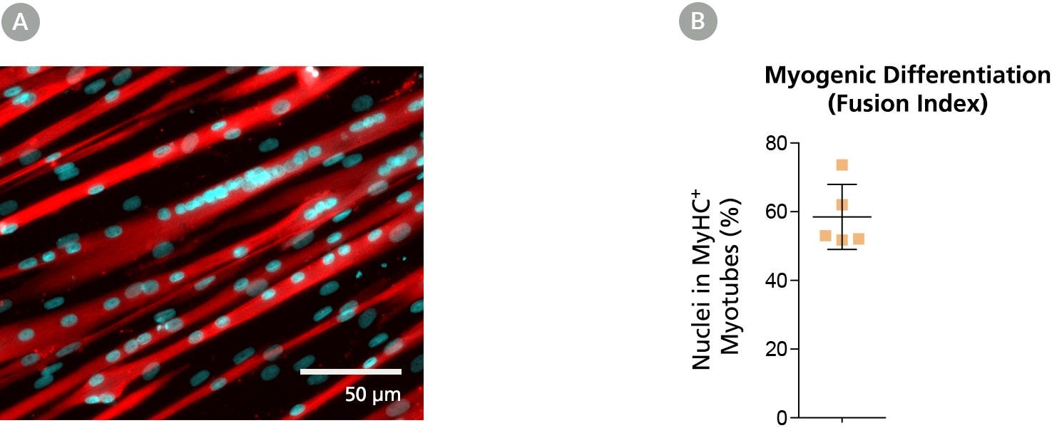
Figure 4. Myoblasts Culture-Expanded in MyoCult™-SF Expansion Medium Maintain Differentiation and Transplantation Potential in Mice
Myoblasts derived and expanded in MyoCult™-SF Expansion Medium were further differentiated into myotubes using the MyoCult™ Differentiation Kit (Catalog #05965) at passage 5. (A) Myotubes differentiated from myoblasts were immunostained for myosin heavy chain (MyHC; red) and nuclei (DAPI; blue). (B) Fusion index displaying approximately 60% of total nuclei were found within myotubes expressing MyHC. Each donor is indicated by a yellow square. Data from 5 independent donors.

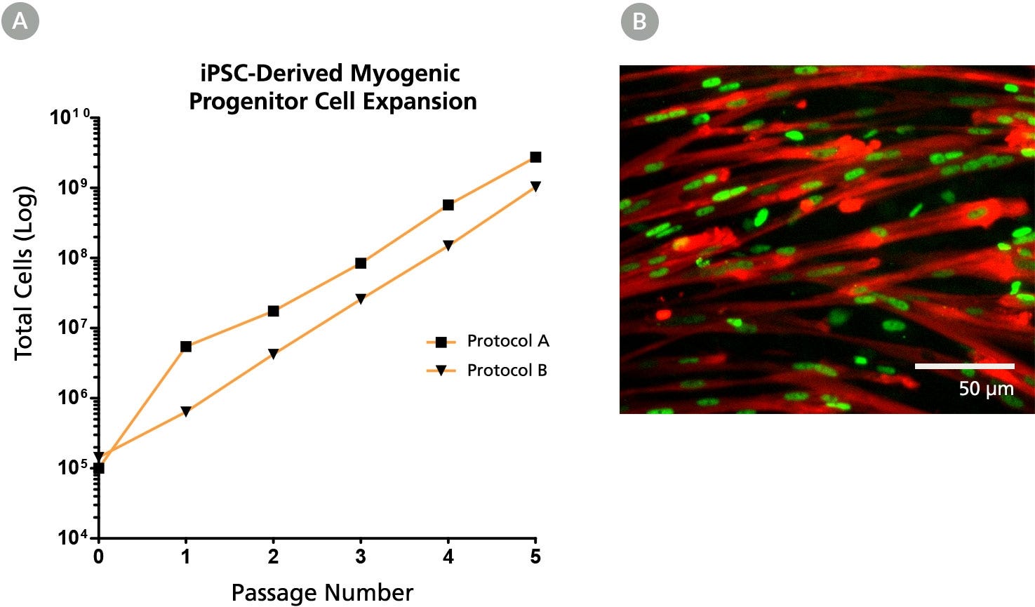
Figure 5. The MyoCult™-SF Expansion Medium Supports Expansion of iPSC-Derived Myogenic Progenitor Cells
(A) iPSC-derived myogenic progenitor cells displayed long-term expansion when using the MyoCult™ Expansion Kit. iPSC-derived myogenic progenitor cells were generated using Protocol A (Chal et al. from Nature Protocols (2016)) and Protocol B (Xi et al. Cell Rep (2017)). (B) Culture-expanded iPSC-derived myogenic progenitor cells expressed MyHC (red) and MyoD (green).
Protocols and Documentation
Find supporting information and directions for use in the Product Information Sheet or explore additional protocols below.
Applications
This product is designed for use in the following research area(s) as part of the highlighted workflow stage(s). Explore these workflows to learn more about the other products we offer to support each research area.
Resources and Publications
Educational Materials (4)
Publications (3)
Abstract
Abstract
Abstract
Quality Statement:
PRODUCTS ARE FOR RESEARCH USE ONLY AND NOT INTENDED FOR HUMAN OR ANIMAL DIAGNOSTIC OR THERAPEUTIC USES UNLESS OTHERWISE STATED. FOR ADDITIONAL INFORMATION ON QUALITY AT STEMCELL, REFER TO WWW.STEMCELL.COM/COMPLIANCE.