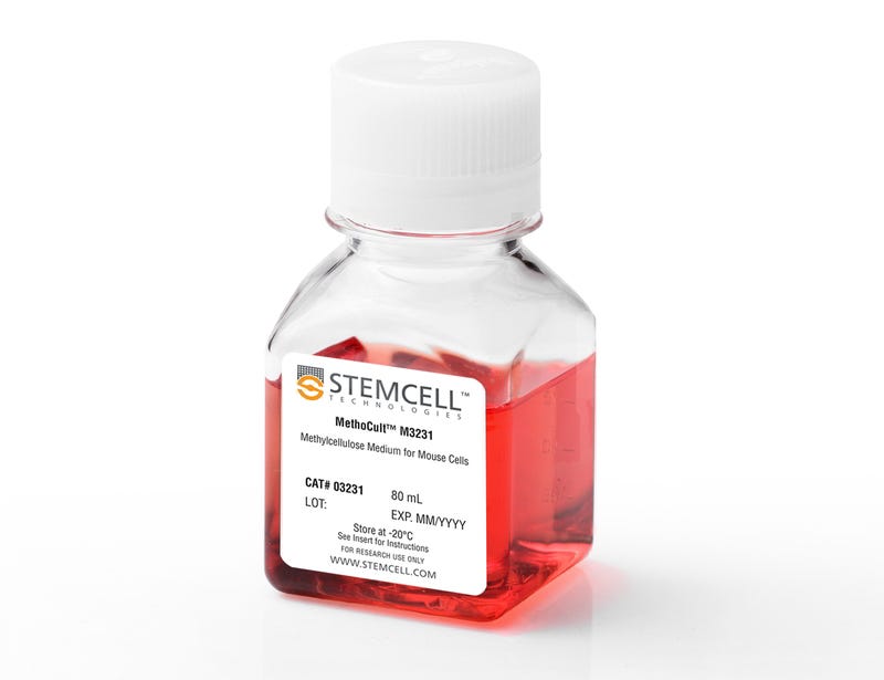
MethoCult™ M3231

Methylcellulose-based medium without cytokines for mouse cells

MethoCult™ M3231

Methylcellulose-based medium without cytokines for mouse cells

From: 375 USD
Catalog #
(Select a product)
Methylcellulose-based medium without cytokines for mouse cells
Add to Wish List

Overview

MethoCult™ M3231 is an incomplete medium formulation that contains methylcellulose in Iscove's MDM. MethoCult™ M3231 is suitable for the growth and enumeration of hematopoietic progenitor cells in colony-forming unit (CFU) assays of mouse bone marrow, spleen, peripheral blood, and fetal liver, when the appropriate growth factors and supplements are added. This formulation does not contain erythropoietin or other cytokines.

Browse our Frequently Asked Questions (FAQs) on performing the CFU assay.
Contains
• Methylcellulose in Iscove's MDM
• Fetal bovine serum
• Bovine serum albumin
• 2-Mercaptoethanol
• Supplements
Subtype
Semi-Solid Media, Specialized Media
Cell Type
Hematopoietic Stem and Progenitor Cells
Species
Mouse
Application
Cell Culture, Colony Assay, Functional Assay
Brand
MethoCult
Area of Interest
Stem Cell Biology
Formulation Category
Methylcellulose-Based

Protocols and Documentation

Find supporting information and directions for use in the Product Information Sheet or explore additional protocols below.

Document Type
Product Name
Catalog #
Lot #
Language
Catalog #
03231
Lot #
All
Language
English
Document Type
Technical Manual
Catalog #
03231
Lot #
All
Language
English
Document Type
Safety Data Sheet
Catalog #
03231
Lot #
All
Language
English

Applications

This product is designed for use in the following research area(s) as part of the highlighted workflow stage(s). Explore these workflows to learn more about the other products we offer to support each research area.

Resources and Publications

Frequently Asked Questions

Why use semi-solid media?

Semi-solid media (methylcellulose-based MethoCult™ and collagen-based MegaCult™-C) allow the clonal progeny of a single progenitor cell to remain spatially isolated from other colonies within a culture, so they may be separately identified and counted.

Why use methylcellulose-based media?

Methylcellulose permits better growth of erythroid colonies than other types of semi-solid support systems (eg. agar) while allowing optimal myeloid colony formation. When appropriate cytokines are present, committed progenitor cells of both erythroid and granulocyte/macrophage lineages (CFU-GM, CFU-G, CFU-M) as well as multi-potential progenitor cells (CFU-GEMM), can be assayed simultaneously in the same culture dish.

Is it necessary to add antibiotics to the media?

No, aseptic technique should be sufficient to maintain sterile cultures. However, antibiotics (eg. Penicillin/Streptomycin) or anti-fungals (eg. Amphotericin B) may be added to the methylcellulose medium if desired.

Is there anything I can do if my cultures appear contaminated?

No, once contamination is visible, it is not possible to rescue the cultures by the addition of antibiotics. Bacteria and yeast inhibit colony formation by depleting nutrients or by releasing toxic substances.

Why can't I use a pipette to dispense methylcellulose-based media?

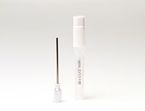
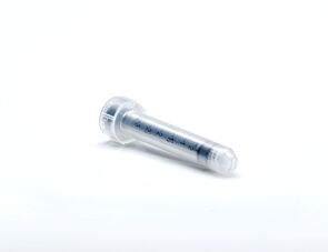
Methylcellulose is a viscous solution that cannot be accurately dispensed using a pipette due to adherence of the medium to the walls of the pipette tip. Blunt-End, 16 Gauge needles (Catalog #28110), in combination with 3 cc Syringes (Catalog #28230) are recommended for accurate dispensing of MethoCult™.

Can I 'pluck' the colonies for individual analysis?

Yes, colonies can be 'plucked' using a pipette with 200 µL sterile pipette tips or using a glass Pasteur pipette with an elongated tip. Individual colonies should be placed in a volume of 25 - 50 µL of medium, and diluted into suitable culture medium for further culture or analysis.

Why are low adherence dishes so important?

Adherent cells such as fibroblasts can cause inhibition of colony growth and obscure visualization of colonies.

Can MethoCult™ products be used for lymphoid progenitor CFU assays?

Human lymphoid progenitors (B, NK and T) seem to require stromal support for growth therefore cannot be grown in MethoCult™. Mouse pre-B clonogenic progenitors can be grown in MethoCult™ M3630 (Catalog #03630).

Is it possible to set up CFU assays in a 24-well plate?

Yes, as long as a plating concentration optimized for the smaller surface area of a well in a 24-well plate (1.9 cm2 as compared to ~9.5 cm2 for a 35 mm dish) is used for these assays. The number of replicate wells required to get an accurate estimation of CFU numbers may also need to be increased.

Can I stain colonies in MethoCult™ medium?

The cells in individual colonies in MethoCult™ can be stained, eg., for analysis of morphology or phenotype, after they are plucked from the dish and washed free of methylcellulose. Colonies grown in collagen-based MegaCult™-C medium can be used for immunohistochemical or enzymatic staining in situ after dehydration and fixation onto glass slides.

Are there differences in colony morphology with serum-free media?

Serum-containing media generally give better overall growth (colonies may appear larger) but there are no large differences in total colony numbers when CFU assays using serum-free media and serum-containing media are compared, provided that identical cytokines are present.

Can MethoCult™ be made with alternate base media?

Yes, this can be done as a 'custom' media order. Please contact techsupport@stemcell.com for more information.

Is there a MethoCult™ formulation suitable for HPP-CFC (high proliferative potential colony forming cell)?

Yes, MethoCult™ H4535 (Catalog #04535) can be used for the HPP-CFC assay as it does not contain EPO. The culture period is usually 28 days. It is not necessary to feed these cultures as growth factors in the medium are present in excess. As HPP-CFCs can be quite large, overplating can be a problem. It is recommended to plate cells at two or more different concentrations.

Publications (41)

Twins with different personalities: STAT5B-but not STAT5A-has a key role in BCR/ABL-induced leukemia. S. Kollmann et al. Leukemia 2019 jul

Abstract

Deregulation of the Janus kinase/signal transducers and activators of transcription (JAK/STAT) signaling pathway is found in cancer with STAT5A/B controlling leukemic cell survival and disease progression. As mutations in STAT5B, but not STAT5A, have been frequently described in hematopoietic tumors, we used BCR/ABL as model systems to investigate the contribution of STAT5A or STAT5B for leukemogenesis. The absence of STAT5A decreased cell survival and colony formation. Even more drastic effects were observed in the absence of STAT5B. STAT5B-deficient cells formed BCR/ABL+ colonies or stable cell lines at low frequency. The rarely evolving Stat5b-/- cell lines expressed enhanced levels of BCR/ABL oncoprotein compared to wild-type cells. In line, Stat5b-/- leukemic cells induced leukemia with a significantly prolonged disease onset, whereas Stat5a-/- cells rapidly caused a fatal disease superimposable to wild-type cells. RNA-sequencing (RNA-seq) profiling revealed a marked enhancement of interferon (IFN)-alpha and IFN-gamma signatures in Stat5b-/- cells. Inhibition of IFN responses rescued BCR/ABL+ colony formation of Stat5b-/--deficient cells. A downregulated IFN response was also observed in patients suffering from leukemia carrying STAT5B mutations. Our data define STAT5B as major STAT5 isoform driving BCR/ABL+ leukemia. STAT5B enables transformation by suppressing IFN-alpha/gamma, thereby facilitating leukemogenesis. Our findings might help explain the high frequency of STAT5B mutations in hematopoietic tumors.
Targeting the RNA m6A Reader YTHDF2 Selectively Compromises Cancer Stem Cells in Acute Myeloid Leukemia. J. Paris et al. Cell stem cell 2019 jul

Abstract

Acute myeloid leukemia (AML) is an aggressive clonal disorder of hematopoietic stem cells (HSCs) and primitive progenitors that blocks their myeloid differentiation, generating self-renewing leukemic stem cells (LSCs). Here, we show that the mRNA m6A reader YTHDF2 is overexpressed in a broad spectrum of human AML and is required for disease initiation as well as propagation in mouse and human AML. YTHDF2 decreases the half-life of diverse m6A transcripts that contribute to the overall integrity of LSC function, including the tumor necrosis factor receptor Tnfrsf2, whose upregulation in Ythdf2-deficient LSCs primes cells for apoptosis. Intriguingly, YTHDF2 is not essential for normal HSC function, with YTHDF2 deficiency actually enhancing HSC activity. Thus, we identify YTHDF2 as a unique therapeutic target whose inhibition selectively targets LSCs while promoting HSC expansion.
Genomic subtyping and therapeutic targeting of acute erythroleukemia. I. Iacobucci et al. Nature genetics 2019

Abstract

Acute erythroid leukemia (AEL) is a high-risk leukemia of poorly understood genetic basis, with controversy regarding diagnosis in the spectrum of myelodysplasia and myeloid leukemia. We compared genomic features of 159 childhood and adult AEL cases with non-AEL myeloid disorders and defined five age-related subgroups with distinct transcriptional profiles: adult, TP53 mutated; NPM1 mutated; KMT2A mutated/rearranged; adult, DDX41 mutated; and pediatric, NUP98 rearranged. Genomic features influenced outcome, with NPM1 mutations and HOXB9 overexpression being associated with a favorable prognosis and TP53, FLT3 or RB1 alterations associated with poor survival. Targetable signaling mutations were present in 45{\%} of cases and included recurrent mutations of ALK and NTRK1, the latter of which drives erythroid leukemogenesis sensitive to TRK inhibition. This genomic landscape of AEL provides the framework for accurate diagnosis and risk stratification of this disease, and the rationale for testing targeted therapies in this high-risk leukemia.