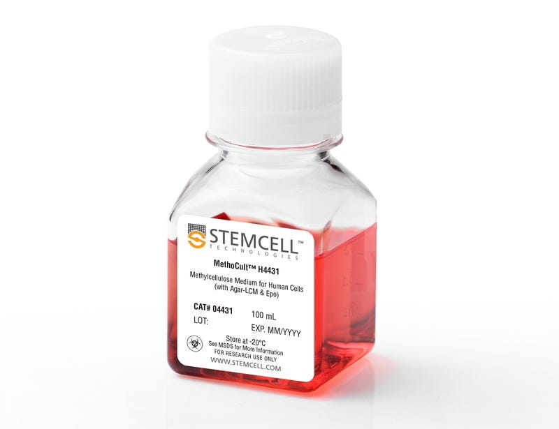
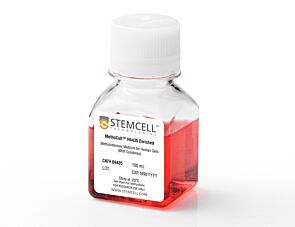
MethoCult™ H4431

Methylcellulose-based medium with agar-LCM and EPO for human cells

MethoCult™ H4431

Methylcellulose-based medium with agar-LCM and EPO for human cells

From: 721 USD
Catalog #
(Select a product)
Methylcellulose-based medium with agar-LCM and EPO for human cells
Add to Wish List

Overview

MethoCult™ H4431 is a methylcelluose-based medium for the growth and enumeration of hematopoietic progenitor cells in colony-forming unit (CFU) assays of bone marrow, mobilized peripheral blood, peripheral blood, and cord blood samples. MethoCult™ H4431 supports the growth of erythroid progenitor cells (BFU-E and CFU-E), granulocyte-macrophage progenitor cells (CFU-GM, CFU-G and CFU-M), and multipotential granulocyte, erythroid, macrophage, megakaryocyte progenitor cells (CFU-GEMM). This formulation contains agar leukocyte conditioned medium (agar-LCM) as a source of colony-stimulating factors, plus recombinant human erythropoietin (EPO). It is suitable for use as a control medium for detection of “EPO-independent” erythroid progenitor cells using MethoCult™ H4531 (Catalog #04531).

Browse our Frequently Asked Questions (FAQs) on performing the CFU assay and explore its utility as part of the cell therapy workflow.
Contains
• Methylcellulose in Iscove's MDM
• Fetal bovine serum
• Bovine serum albumin
• 2-Mercaptoethanol
• Agar-LCM
• Recombinant human erythropoietin (EPO)
• Supplements
Subtype
Semi-Solid Media, Specialized Media
Cell Type
Hematopoietic Stem and Progenitor Cells
Species
Human
Application
Cell Culture, Colony Assay, Functional Assay
Brand
MethoCult
Area of Interest
Stem Cell Biology

Data Figures

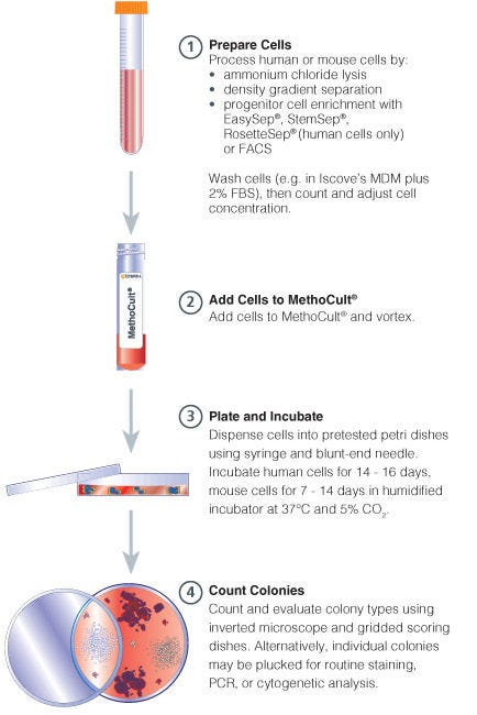
Procedure Summary for Hematopoietic CFU Assays

Figure 1. Procedure Summary for Hematopoietic CFU Assays

Protocols and Documentation

Find supporting information and directions for use in the Product Information Sheet or explore additional protocols below.

Document Type
Product Name
Catalog #
Lot #
Language
Catalog #
04431
Lot #
All
Language
English
Document Type
Technical Manual
Catalog #
04431
Lot #
All
Language
English
Document Type
Safety Data Sheet
Catalog #
04431
Lot #
All
Language
English

Applications

This product is designed for use in the following research area(s) as part of the highlighted workflow stage(s). Explore these workflows to learn more about the other products we offer to support each research area.

Resources and Publications

Frequently Asked Questions

Why use semi-solid media?

Semi-solid media (methylcellulose-based MethoCult™ and collagen-based MegaCult™-C) allow the clonal progeny of a single progenitor cell to remain spatially isolated from other colonies within a culture, so they may be separately identified and counted.

Why use methylcellulose-based media?

Methylcellulose permits better growth of erythroid colonies than other types of semi-solid support systems (eg. agar) while allowing optimal myeloid colony formation. When appropriate cytokines are present, committed progenitor cells of both erythroid and granulocyte/macrophage lineages (CFU-GM, CFU-G, CFU-M) as well as multi-potential progenitor cells (CFU-GEMM), can be assayed simultaneously in the same culture dish.

Is it necessary to add antibiotics to the media?

No, aseptic technique should be sufficient to maintain sterile cultures. However, antibiotics (eg. Penicillin/Streptomycin) or anti-fungals (eg. Amphotericin B) may be added to the methylcellulose medium if desired.

Is there anything I can do if my cultures appear contaminated?

No, once contamination is visible, it is not possible to rescue the cultures by the addition of antibiotics. Bacteria and yeast inhibit colony formation by depleting nutrients or by releasing toxic substances.

Why can't I use a pipette to dispense methylcellulose-based media?

Methylcellulose is a viscous solution that cannot be accurately dispensed using a pipette due to adherence of the medium to the walls of the pipette tip. Blunt-End, 16 Gauge needles (Catalog #28110), in combination with 3 cc Syringes (Catalog #28230) are recommended for accurate dispensing of MethoCult™.

Can I 'pluck' the colonies for individual analysis?

Yes, colonies can be 'plucked' using a pipette with 200 µL sterile pipette tips or using a glass Pasteur pipette with an elongated tip. Individual colonies should be placed in a volume of 25 - 50 µL of medium, and diluted into suitable culture medium for further culture or analysis.

Why are low adherence dishes so important?

Adherent cells such as fibroblasts can cause inhibition of colony growth and obscure visualization of colonies.

Can MethoCult™ products be used for lymphoid progenitor CFU assays?

Human lymphoid progenitors (B, NK and T) seem to require stromal support for growth therefore cannot be grown in MethoCult™. Mouse pre-B clonogenic progenitors can be grown in MethoCult™ M3630 (Catalog #03630).

Is it possible to set up CFU assays in a 24-well plate?

Yes, as long as a plating concentration optimized for the smaller surface area of a well in a 24-well plate (1.9 cm2 as compared to ~9.5 cm2 for a 35 mm dish) is used for these assays. The number of replicate wells required to get an accurate estimation of CFU numbers may also need to be increased.

Can I stain colonies in MethoCult™ medium?

The cells in individual colonies in MethoCult™ can be stained, eg., for analysis of morphology or phenotype, after they are plucked from the dish and washed free of methylcellulose. Colonies grown in collagen-based MegaCult™-C medium can be used for immunohistochemical or enzymatic staining in situ after dehydration and fixation onto glass slides.

Are there differences in colony morphology with serum-free media?

Serum-containing media generally give better overall growth (colonies may appear larger) but there are no large differences in total colony numbers when CFU assays using serum-free media and serum-containing media are compared, provided that identical cytokines are present.

Can MethoCult™ be made with alternate base media?

Yes, this can be done as a 'custom' media order. Please contact techsupport@stemcell.com for more information.

Is there a MethoCult™ formulation suitable for HPP-CFC (high proliferative potential colony forming cell)?

Yes, MethoCult™ H4535 (Catalog #04535) can be used for the HPP-CFC assay as it does not contain EPO. The culture period is usually 28 days. It is not necessary to feed these cultures as growth factors in the medium are present in excess. As HPP-CFCs can be quite large, overplating can be a problem. It is recommended to plate cells at two or more different concentrations.

Publications (10)

Clonal analysis of deletions on chromosome 20q and JAK2-V617F in MPD suggests that del20q acts independently and is not one of the predisposing mutations for JAK2-V617F. Schaub FX et al. Blood 2009 FEB

Abstract

We developed a real-time copy number polymerase chain reaction assay for deletions on chromosome 20q (del20q), screened peripheral blood granulocytes from 664 patients with myeloproliferative disorders, and identified 19 patients with del20q (2.9%), of which 14 (74%) were also positive for JAK2-V617F. To examine the temporal relationship between the occurrence of del20q and JAK2-V617F, we performed colony assays in methylcellulose, picked individual burst-forming units-erythroid (BFU-E) and colony-forming units-granulocyte (CFU-G) colonies, and genotyped each colony individually for del20q and JAK2-V617F. In 2 of 9 patients, we found that some colonies with del20q carried only wild-type JAK2, whereas other del20q colonies were JAK2-V617F positive, indicating that del20q occurred before the acquisition of JAK2-V617F. However, in colonies from 3 of 9 patients, we observed the opposite order of events. The lack of a strict temporal order of occurrence makes it doubtful that del20q represents a predisposing event for JAK2-V617F. In 2 patients with JAK2-V617F and 1 patient with MPL-W515L, microsatellite analysis revealed that del20q affected chromosomes of different parental origin and/or 9pLOH occurred at least twice. The fact that rare somatic events, such as del20q or 9pLOH, occurred more than once in subclones from the same patients suggests that the myeloproliferative disorder clone carries a predisposition to acquiring such genetic alterations.
Distinct signals control the hematopoiesis of lymphoid-related dendritic cells. Galy A et al. Blood 2000 JAN

Abstract

The molecular and cellular requirements for the development of different populations of human dendritic cells (DC) were studied. Conditions were defined that support DC production from lymphoid progenitors but that fail to induce DC formation from peripheral monocytes. The production of these lymphoid-related DC was severely blocked when hematopoietic progenitors overexpressed Ik7, a mutant dominant-negative Ikaros protein. In contrast, Ik7 did not block the formation of DC in conditions supporting the development of monocyte-derived DC. Furthermore, Ik7 did not block the formation of monocyte/macrophages and enhanced granulopoiesis. One of the molecular mechanisms mediated by Ik7 appears to be down-regulation of the flt3-receptor mRNA. Thus, distinct signals control the formation of DC demonstrating that some aspects of DC diversity are determined in part by distinct molecular cues at the hematopoietic level. (Blood. 2000;95:128-137)
High levels of lymphoid expression of enhanced green fluorescent protein in nonhuman primates transplanted with cytokine-mobilized peripheral blood CD34(+) cells. Donahue RE et al. Blood 2000 JAN

Abstract

We have used a murine retrovirus vector containing an enhanced green fluorescent protein complimentary DNA (EGFP cDNA) to dynamically follow vector-expressing cells in the peripheral blood (PB) of transplanted rhesus macaques. Cytokine mobilized CD34(+) cells were transduced with an amphotropic vector that expressed EGFP and a dihydrofolate reductase cDNA under control of the murine stem cell virus promoter. The transduction protocol used the CH-296 recombinant human fibronectin fragment and relatively high concentrations of the flt-3 ligand and stem cell factor. Following transplantation of the transduced cells, up to 55% EGFP-expressing granulocytes were obtained in the peripheral circulation during the early posttransplant period. This level of myeloid marking, however, decreased to 0.1% or lower within 2 weeks. In contrast, EGFP expression in PB lymphocytes rose from 2%-5% shortly following transplantation to 10% or greater by week 5. After 10 weeks, the level of expression in PB lymphocytes continued to remain at 3%-5% as measured by both flow cytometry and Southern blot analysis, and EGFP expression was observed in CD4(+), CD8(+), CD20(+), and CD16/56(+) lymphocyte subsets. EGFP expression was only transiently detected in red blood cells and platelets soon after transplantation. Such sustained levels of lymphocyte marking may be therapeutic in a number of human gene therapy applications that require targeting of the lymphoid compartment. The transient appearance of EGFP(+) myeloid cells suggests that transduction of a lineage-restricted myeloid progenitor capable of short-term engraftment was obtained with this protocol. (Blood. 2000;95:445-452)