Make more informed purchasing decisions with our new product availability and delivery estimate feature, now available on all product pages, in your cart, and during checkout.
Sign In
New to STEMCELL?
Register for an account to quickly and easily purchase products online and for one-click access to all educational content.
Thank you for your interest in this product.
Please provide us with your contact information and your local representative
will contact you with a customized quote. Where appropriate, they can also assist you with a(n):
Estimated delivery time for your area
Product sample or exclusive offer
In-lab demonstration
By submitting this form, you are providing your consent to STEMCELL Technologies Canada Inc. and its subsidiaries and affiliates (“STEMCELL”) to collect and use your information, and send you newsletters and emails in accordance with our privacy policy. Please contact us with any questions that you may have. You can unsubscribe or change your email preferences at any time.
Streamline your assays with ready-to-use, ethically sourced, primary mononuclear cells, including lymphocytes, monocytes, hematopoietic stem and progenitor cells, and mesenchymal stromal cells. With personalized service, custom products, flexible delivery times, and the option to reserve entire lots to prescreen cells for applications, we help you get the cells you need.
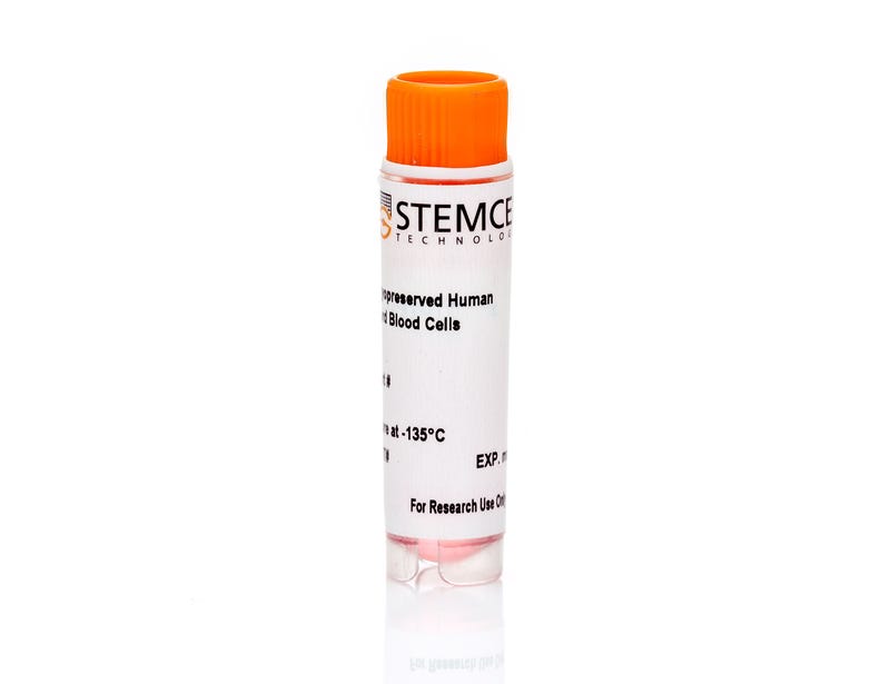
Isolated from umbilical cord blood using density gradient separation and cryopreserved in animal component-free CryoStor®CS10 medium (Catalog #07930), cells are collected using Institutional Review Board (IRB) or other regulatory authority-approved consent forms and protocols. Additional documentation and high-resolution HLA typing (Class I and Class II alleles and CMV status) are available upon request. Citrate-phosphate-dextrose (CPD) is added as an anticoagulant. Donor specifications (e.g. BMI category, smoking status, ethnicity, etc.) can be requested in the comment box above, after selecting from the product options. Donors are screened for HIV-1, HIV-2, hepatitis B, and hepatitis C.
Certain products are only available in select territories. Please contact your local sales representative or Product & Scientific Support at techsupport@stemcell.com for further information.
This product is designed for use in the following research area(s) as part
of the highlighted workflow stage(s). Explore these workflows to learn more about the other products we
offer to support each research area.
A phenotypic screening approach in cord blood-derived mast cells to identify anti-inflammatory compounds.
Kaur R et al.
Journal of biomolecular screening 2013 DEC
Abstract
Mast cells are unique hematopoietic cells that are richly distributed in the skin and mucosal surfaces of the respiratory and gastrointestinal tract. They play a key role in allergic inflammation by releasing a cocktail of granular constituents, including histamine, serine proteases, and various eicosanoids and cytokines. As such, a number of drugs target either inhibition of mast cell degranulation or the products of degranulation. To identify potential novel drugs and mechanisms in mast cell biology, assays were developed to identify inhibitors of mast cell degranulation and activation in a phenotypic screen. Due to the challenges associated with obtaining primary mast cells, cord blood-derived mononuclear cells were reproducibly differentiated to mast cells and assays developed to monitor tryptase release and prostaglandin D2 generation. The tryptase assay was particularly sensitive, requiring only 500 cells per data point, which permitted a set of approximately 12,000 compounds to be screened robustly and cost-effectively. Active compounds were tested for concomitant inhibition of prostaglandin D2 generation. This study demonstrates the robustness and effectiveness of this approach in the identification of potential novel compounds and mechanisms targeting mast cell-driven inflammation, to enable innovative drug discovery efforts to be prosecuted.
ETV6-RUNX1 promotes survival of early B lineage progenitor cells via a dysregulated erythropoietin receptor.
Torrano V et al.
Blood 2011 NOV
Abstract
ETV6-RUNX1 gene fusion is usually an early, prenatal event in childhood acute lymphoblastic leukemia (ALL). Transformation results in the generation of a persistent (> 14 years) preleukemic clone, which postnatally converts to ALL after the acquisition of necessary secondary genetic alterations. Many cancer cells show some expression of the erythropoietin receptor (EPOR) gene, although the functionality" of any EPOR complexes and their relevant signaling pathways in nonerythroid cells has not been validated. EPOR mRNA is selectively and ectopically expressed in ETV6-RUNX1(+) ALL but the presence of a functional EPOR on the cell surface and its role in leukemogenesis driven by ETV6-RUNX1 remains to be identified. Here we show that ETV6-RUNX1 directly binds the EPOR promoter and that expression of ETV6-RUNX1 alone in normal pre-B cells is sufficient to activate EPOR transcription. We further reveal that murine and human ETV6-RUNX1(+) cells expressing EPOR mRNA have EPO ligand binding activity that correlates with an increased cell survival through activation of the JAK2-STAT5 pathway and up-regulation of antiapoptotic BCL-XL. These data support the contention that ETV6-RUNX1 directly activates ectopic expression of a functional EPOR and provides cell survival signals that may contribute critically to persistence of covert premalignant clones in children.
Methylation profiling using degenerated oligonucleotide primer-PCR specific for genome-wide amplification of bisulfite-modified DNA.
Yang W-H et al.
Analytical biochemistry 2007 OCT
Abstract
DNA methylation is one of the essential epigenetic processes that play a role in regulating gene expression. Aberrant methylation of CpG-rich promoter regions has been associated with many forms of human cancers. The current method for determining the methylation status relies mainly on bisulfite treatment of genomic DNA, followed by methylation-specific PCR (MSP). The difficulty in acquiring a methylation profiling often is limited by the amount of genomic DNA that can be recovered from a given sample, whereas complex procedures of bisulfite treatment further compromise the effective template for PCR analysis. To circumvent these obstacles, we developed degenerated oligonucleotide primer (DOP)-PCR to enable amplification of bisulfite-modified genomic DNA at a genome-wide scale. A DOP pair was specially designed as follows: first 3' DOP, CTCGAGCTGHHHHHAACTAC, where H is a mixture of base consisting of 50% A, 25% T, and 25% C; and second 5' DOP, CTCGAGCTGDDDDDGTTTAG, where D is a mixture of base consisting of 50% T, 25% G, and 25% A. Our results showed that bisulfite-modified DNAs from a cell line, cord blood cells, or cells obtained by laser capture microdissection were amplified by up to 1000-fold using this method. Subsequent MSP analysis using these amplified DNAs on nine randomly selected cancer-related genes revealed that the methylation status of these genes remained identical to that derived from the original unamplified template.
PRODUCTS ARE FOR RESEARCH USE ONLY AND NOT INTENDED FOR HUMAN OR ANIMAL DIAGNOSTIC OR THERAPEUTIC USES UNLESS OTHERWISE STATED. FOR ADDITIONAL INFORMATION ON QUALITY AT STEMCELL, REFER TO WWW.STEMCELL.COM/COMPLIANCE.