Assessing Morphology of hPSCs Passaged As Aggregates
Technical tip from our dedicated team of Product and Scientific Support specialists
Routine culture of human pluripotent stem cells (hPSCs) presents some unique challenges when compared to culturing other adherent cells. Careful monitoring and handling techniques are critical to ensure maintenance and expansion of healthy cells in the undifferentiated state over long periods of time.
Though visual assessments can be subjective, visible changes are sensitive indicators of hPSC quality. Cultures should be examined daily using a light microscope under phase contrast to monitor the quality of the cells, as well as to check whether they are ready to be passaged.
It should be noted that not all changes in appearance indicate a decrease in quality. It is normal to see variations due to a number of factors, including the experience level of the end user. This Tech Tip describes expected morphology of high-quality cells passaged as aggregates, some common normal variations, and other variations that may indicate poor cell quality.
Healthy Colony Morphology and Normal Variations
Assessing hPSC quality can be difficult for the first few days after passaging, and becomes easier as the colonies mature. This typically happens approximately 48 hours prior to the optimal passaging time. Under low magnification, healthy and high-quality hPSCs should exhibit the following characteristics:
- Relatively round colonies
- Tightly packed cells with a high nucleus:cytoplasm ratio, where the nucleus practically inhabits the entire cell
- Prominent nucleoli
- Colony centers become very dense and may appear as phase brightness under a phase contrast microscope
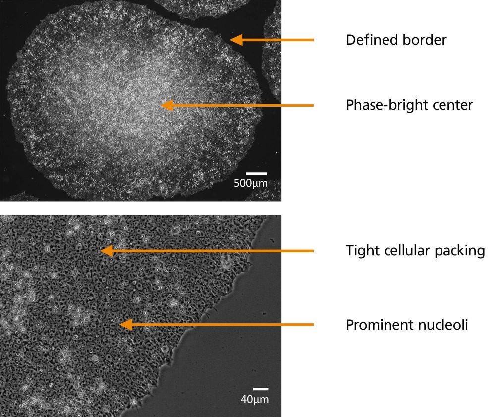
Figure 1. Aggregate-Passaged Culture. High-Quality hPSC Colonies Near the Optimal Time of Passaging
Some factors that may affect the visual appearance of a colony include days since last passage or feed, cell line used, medium used, passaging method, and matrix used. Here are some variations that you may notice, which are considered normal and not necessarily indicators of poor cell quality:
- “Spiky” colony edges and looser packing in the initial days post-passage
For up to 4 days after plating, colonies may exhibit looser packing as the colonies spread out and become established. At this point, spiky colony edges should not be a cause for concern. The density and robustness of the colonies increases rapidly after this timepoint and the morphology changes significantly in the last 1 - 2 days before passaging.
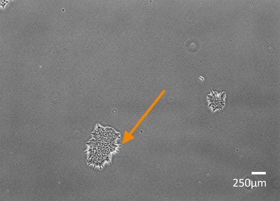
Figure 2. Human Embryonic Stem (ES) (H9) Cells Two Days After Passaging with Spiky Colony Edges (Orange Arrow)
- Asymmetrical and/or merging colonies
Colonies may appear slightly asymmetric and merging, especially toward the time of passaging. This is not typically a cause for concern. Merging colonies may also occur when aggregates are seeded at low density and are not well dispersed. Again, this is not usually a cause for concern. Symmetry of colonies can vary not only between cell lines but also between matrices, with hPSCs cultured in Vitronectin XF™ typically appearing more compact and symmetric.
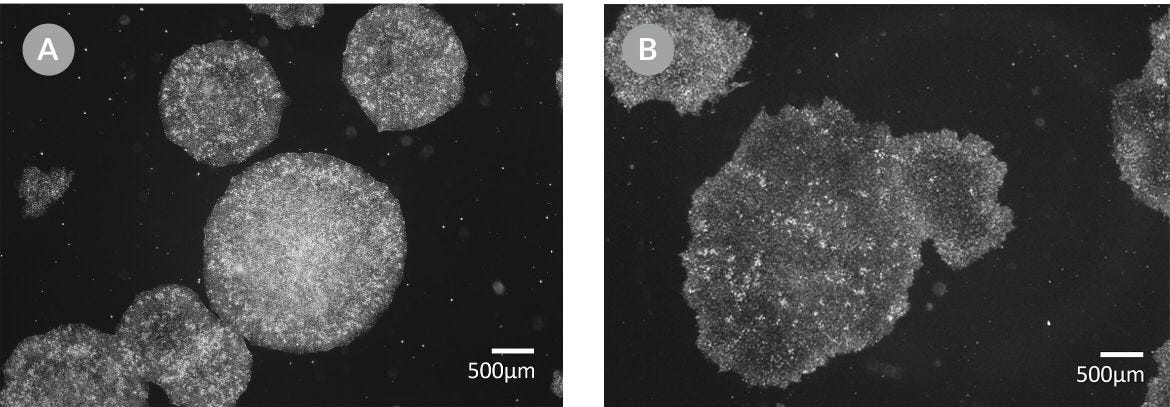
Figure 3. (A) Human induced Pluripotent Stem (iPS) Cells (WLS-1C) on Vitronectin XF™ and (B) Human ES Cells (H9) on Corning® Matrigel®
The next section includes some examples of morphology to watch for, that indicate decreased cell quality.
Variations in Morphology Indicating Poor Cell Quality
- Areas of differentiation
- It is normal for healthy hPSC colonies to exhibit a low amount (5-10%) of spontaneous differentiation. However, an increase in areas of differentiation is indicative of poor-quality hPSCs. Notably, visible signs of differentiation can appear prior to a decrease in marker expression.
- Here are some characteristics of poor colony morphology that indicate decreased cell quality:
- An increase (> 10%) in spontaneous differentiation
- A loss of uniformity
- Phase-brightness or multilayering that appears “mottled”, sporadic, and not at the center of the colony
- Loss of border integrity
- Loose cellular packing
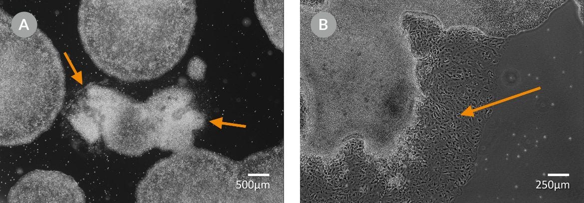
Figure 4. (A) Areas of Spontaneous Differentiation and (B) Loss of Colony Border Integrity and Differentiated Cells (Orange Arrows) in STiPS-M001 Colonies
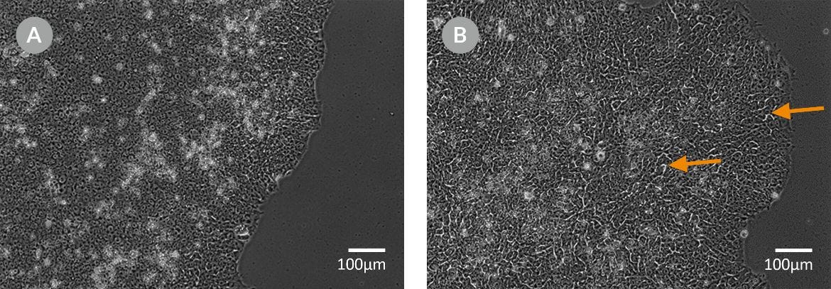
Figure 5. (A) Tightly Packed Cells vs. (B) Loosely Packed Cells With Phase-Bright Gaps Visible Between Cells in hPSC Colonies (Orange Arrows)
For more information, refer to the Technical Manuals for Maintenance of Human Pluripotent Stem Cells in mTeSR™ Plus (Document #10000005507) or mTeSR™1 (Document #10000005505) and if in doubt about your hPSC morphology and quality, reach out to our Product and Scientific Support team (techsupport@stemcell.com), who will be happy to assist.
Learn more about PSC quality and maintenance, including morphology assessment, by taking our training courses.

On-Demand Training
Learn the techniques and protocols to move your research forward. Recorded lectures, step-by-step instructional videos, and libraries of curated resources will help guide you through entire workflows. Topics include hPSC maintenance and cell quality, neural induction, and expansion of hPSCs in 3D suspension culture. From passaging to cryopreservation, learn fundamental laboratory skills from the comfort of your own home.

Live Virtual Training
Join from anywhere in the world to watch real-time video demonstrations, participate in online workshops, and learn from our team of scientific experts in our interactive, virtual training courses. Learn techniques and protocols to successfully derive and maintain high-quality human iPS cells from somatic cells and differentiate them toward specialized cell types in a virtual and interactive setting.
Learn more at www.stemcell.com/psc-training
Understanding the ISSCR Standards for Human Stem Cell Use in Research
Learn how STEMCELL can help you apply the Standards to improve the quality and reproducibility of your research with a curated collection of resources and more standardized practices for genomic characterization. From webinars, to interviews, to articles, you will find the information you need to achieve higher-quality results and more standardized practices. www.stemcell.com/ISSCR-Standards
Request Pricing
Thank you for your interest in this product. Please provide us with your contact information and your local representative will contact you with a customized quote. Where appropriate, they can also assist you with a(n):
Estimated delivery time for your area
Product sample or exclusive offer
In-lab demonstration


