Enzymatic Dissociation of Adult Mouse or Rat CNS Tissue into Single-Cell Suspension
The protocol below describes how to prepare a single-cell suspension from adult mouse or rat central nervous system (CNS) tissue using NeuroCult™ Enzymatic Dissociation Kit for Adult CNS Tissue (Mouse and Rat).
This procedure has been optimized so that it is reproducible, fast, and yields high cell numbers and viabilities. The resulting single-cell suspension is ready for immediate use in downstream applications, such as culture and expansion of neural stem and progenitor cells in the neurosphere or adherent monolayer system using NeuroCult™ Proliferation Kit (Mouse & Rat).
Materials
- NeuroCult™ Enzymatic Dissociation Kit for Adult CNS Tissue (Mouse and Rat, Catalog #05715). Kit components (not for individual sale) include:
- Tissue Collection Solution, 500 mL
- Dissociation Solution, 30 mL
- Inhibition Solution, 30 mL
- Resuspension Solution, 500 mL
- Scalpel
- 37 µm reversible strainer, large (e.g. Catalog #27250)
- 100 mm tissue culture-treated dish (e.g. Catalog #38046)
- 14 mL round-bottom tube (e.g. Falcon® Round-Bottom Tubes, Catalog #38008)
- 50 mL conical tube (e.g. Falcon® Conical Tubes, Catalog #38010)
- Hemocytometer
Protocol
This procedure is optimized for processing subventricular tissue isolated from up to 8 adult mouse or 6 adult rat brains. If more brains are used, set up more tubes for the additional tissue and thaw additional aliquots of Dissociation Solution.
Important Notes:
- The incubation times outlined in the procedure are crucial for performance; it is important to observe the incubation times precisely and to use an accurate lab timer.
- The mechanical dissociation of tissue involves gently pipetting the tissue suspension in a consistent rhythm up and down through the plastic disposable tip pressed against the bottom of the tube—without introducing any air bubbles. Air bubbles will result in high cell death.
- The microdissection of different regions of the adult mouse or rat brain is not described in this protocol. Perform microdissections according to Gritti et al.1 or use routine dissection procedures recommended by your institution.
- Add 10 mL Tissue Collection Solution to a 100 mm tissue culture-treated dish.
- Thaw a 3 mL aliquot of Dissociation Solution at room temperature (15 - 25°C).
- Perform dissections on the CNS tissue region of interest from adult mouse or rat brains and transfer dissected tissue pieces to the 100 mm dish containing Tissue Collection Solution.
- Remove all the Tissue Collection Solution from the dish and mince tissue by chopping in a quick rhythm with a sterile scalpel for approximately 1 minute.
Note: Divide the tissues into two roughly equal piles in the same 100 mm dish and mince each pile separately. This ensures that the tissue is minced into the smallest pieces possible and can be pipetted through disposable plastic tips.
- Dispense 1 mL Dissociation Solution into the dish containing the minced tissue. Resuspend the tissue pieces, and transfer the suspension into a 14 mL round-bottom tube. Repeat twice, using the entire 3 mL of Dissociation Solution.
Note: It is very important that no air bubbles are introduced into the tissue suspension during the transfer steps.
- Incubate the minced tissue at 37°C for 7 minutes, preferably in a water bath (or in a beaker containing pre-warmed water that is placed in a 37°C incubator).
- Remove the tube containing the minced tissue from the water bath or incubator.
- Add 3 mL of Inhibition Solution and mix the tissue suspension gently, avoiding air bubbles.
- Centrifuge the suspension at 100 x g for 7 minutes.
- Discard the supernatant and resuspend the pellet with 150 µL Resuspension Solution.
- Mechanically dissociate (triturate) the digested tissue using a plastic disposable tip attached to a 200 µL pipettor that has been set to 180 µL, by pipetting up and down ~5 - 10 times until a smooth and “creamy” suspension is achieved. The suspension should be able to pass through the plastic tip bore without getting lodged in the tip.
- Once a smooth suspension has been achieved, add 100 µL Resuspension Solution to bring the volume to approximately 300 µL.
- Pipette the suspension approximately 5 more times with a disposable plastic tip attached to a 1 mL pipettor (set to 280 µL) to achieve a homogeneous cell suspension without any clumps of tissue.
- Add Resuspension Solution to the cell suspension to a final volume of 10 mL. Mix thoroughly.
- Centrifuge at 100 x g for 7 minutes. Discard the supernatant.
- Add 200 µL Resuspension Solution and resuspend the pellet by pipetting up and down 5 times using a plastic disposable tip attached to a 200 µL pipettor.
- Add Resuspension Solution to a final volume of 10 mL. Mix thoroughly. Centrifuge at 100 x g for 7 minutes. Discard the supernatant.
- Repeat steps 16 & 17 for a total of 3 washes with Resuspension Solution.
- Resuspend the final pellet in 0.5 - 1 mL of medium (volume dependent on the size of the pellet and number of brains dissected) appropriate for subsequent experiments. For example:
- For experiments involving antibody labeling for FACS or cell separation, resuspend the pellet in Resuspension Solution
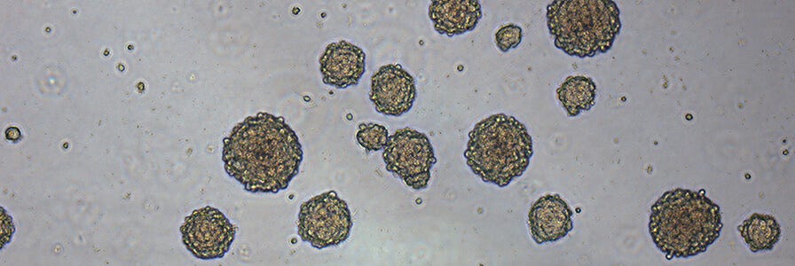
- For generating neurospheres from adult mouse or rat CNS cells, resuspend the pellet in Complete NeuroCult™ Proliferation Medium
- Place a sterile 37 µm strainer on a sterile 50 mL conical tube. Gently dispense the cell suspension (from step 19) through the strainer. Allow the cell suspension to flow through the strainer by gravity (do not force the cells through).
- Measure the volume of the flow-through and count cells using trypan blue dye exclusion (1 in 5 or 1 in 10 dilution) and a hemocytometer.
Note: Counting cells can be difficult because of the small pieces of debris, damaged blood vessels, and myelin. However, the viable cell yield should be approximately 5 x 10^4 cells from the subventricular zone (SVZ) of one mouse brain.
- Document #PR00043
- Version 1.0.0
- May 2021
Request Pricing
Thank you for your interest in this product. Please provide us with your contact information and your local representative will contact you with a customized quote. Where appropriate, they can also assist you with a(n):
Estimated delivery time for your area
Product sample or exclusive offer
In-lab demonstration

