CRISPR-Cas9 Genome Editing of Human Liver Organoids
Liver organoids are miniature three-dimensional (3D) cell culture systems that serve as a valuable model for studying liver cell biology. These organoids retain many features of in vivo hepatocytes and can recapitulate donor heterogeneity. As an alternative to conventional two-dimensional (2D) cell cultures, including primary human hepatocytes, they provide a more physiologically relevant model for drug screening and the study of hepatic development and disease.
A key benefit of liver organoids is that they are amenable to genome editing, such as by CRISPR-Cas9, an RNA-guided genome editing technology that is revolutionizing cell biology with its ease and efficiency. By combining CRISPR-Cas9 genome editing and the ability of organoids to model tissues in vitro, researchers can develop better in vitro human disease models and study organ development.
Below, we describe a protocol for CRISPR-Cas9 genome editing of liver organoids cultured in HepatiCult™ Organoid Growth Medium (Human) (Catalog #100-0385) using the ArciTect™ CRISPR-Cas9 ribonucleoprotein (RNP)-based system and STEMCELL’s Guide RNA Design Tool.
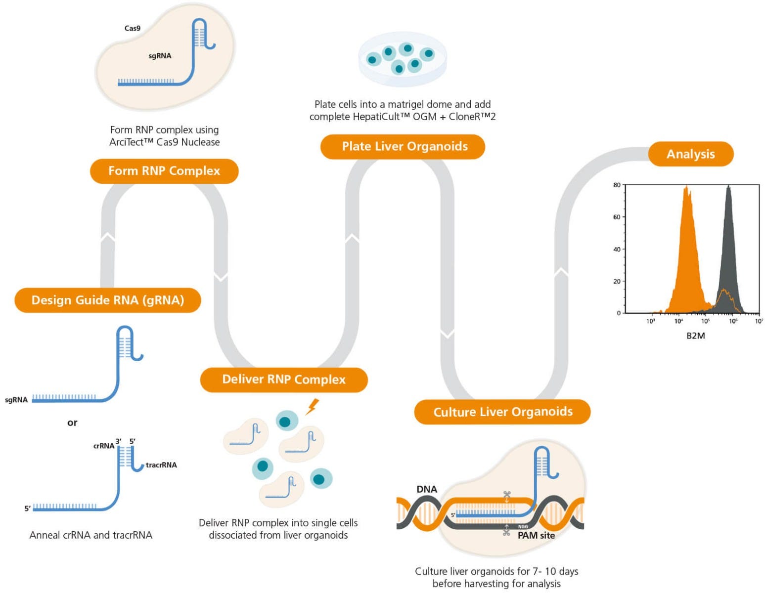
Figure 1. Experimental Workflow for Liver Organoid Genome Editing
The guide RNA (gRNA) sequence is designed once a target locus for editing is identified. ArciTect™ single guide RNA (sgRNA) or CRISPR RNA (crRNA) can be designed using STEMCELL’s CRISPR Design Tool. The ArciTect™ CRISPR-Cas9 ribonucleoprotein (RNP) complex is then prepared and delivered into single cells dissociated from liver organoids using electroporation. Cells are then plated into a matrigel dome with complete HepatiCult™ OGM supplemented with CloneR™. Editing efficiency can be analyzed after 7 - 10 days using ArciTect™ T7 Endonuclease I Kit (Catalog #76021) or via flow cytometry.
Materials
- ArciTect™ Cas9 Nuclease (Catalog #76002/76004)
- ArciTect™ sgRNA† (Catalog #200-0013)
- HepatiCult™ Organoid Growth Medium (Human) (Catalog #100-0385)
- HepatiCult™ Organoid Basal Medium (Human), 95 mL (Catalog #100-0387)
- HepatiCult™ Organoid Growth Supplement (Human), 5 mL (Catalog #100-0389)
- Advanced DMEM/F-12 (Thermo Fisher 12634028)
- 25% Bovine serum albumin (BSA), 100g in phosphate-buffered saline (PBS)
- Corning® Matrigel® Growth Factor Reduced (GFR) Basement Membrane Matrix, Phenol Red-free, LDEV-free (≥ 8 mg/mL protein) (Corning 356231)
- D-PBS (Without Ca++ and Mg++) (Catalog #37350)
The materials required are indicated as per-well, adjust these amounts based on the actual number of wells in your experiment.
Multiple guide RNA (gRNA) targeting sequences are often tested when targeting a new gene, as different gRNAs can exhibit a range of targeting/editing efficiencies at both on- and off-target sites. Single guide RNA (sgRNA) can be designed using the Guide RNA Design Tool. This tool uses best practices and the latest computational tools to design optimal targeting sequences for every gene in the human and mouse genomes.
Protocol
Part I: Preparation of ArciTect™ sgRNA Working Solution
- Briefly centrifuge the vials before opening.
- Add nuclease-free water to each vial to give a final concentration of 100 μM single guide RNA (sgRNA), as indicated in Table 1.
Table 1. Resuspension Volume for 100 μM* ArciTect™ sgRNA
AMOUNT OF ArciTect™ sgRNA (nmol)VOLUME OF NUCLEASE-FREE WATER (µL)440*100 µM is equal to 100 pmol/µL
- Mix thoroughly. If not used immediately, aliquot and store at -20°C for up to 6 months or at -80°C for longer than 6 months. After thawing the aliquots, use immediately. Do not re-freeze.
Part II: Preparation of Complete HepatiCult™ Organoid Growth Medium + CloneR™2 (OGM + CloneR™2)
Use sterile technique to prepare complete HepatiCult™ OGM (HepatiCult™ Organoid Basal Medium + HepatiCult™ Organoid Growth Supplement + antibiotics). The following example is for preparing 100 mL of complete medium. If preparing other volumes, adjust accordingly.
- Thaw HepatiCult™ Organoid Growth Supplement overnight at 2 - 8°C. Mix thoroughly.
Note: If not used immediately, aliquot and store at -20°C. Do not exceed the shelf life of the supplement. After thawing aliquots, use immediately. Do not re-freeze.
- Add 5 mL of Growth Supplement to 95 mL of Basal Medium. Add antibiotics (e.g. final concentration 50 µg/mL gentamicin). Mix thoroughly. Warm to room temperature (15 - 25°C) before use.
Note: If not used immediately, store complete HepatiCult™ OGM at 2 - 8°C for up to 2 weeks.
- Add 2.5 mL of CloneR™2 to 22.5 mL of complete HepatiCult™ OGM (final concentration 1X CloneR™2). Mix thoroughly. Warm to room temperature (15 - 25°C) before use.
Part III: Preparation of Liver Organoid Single-Cell Suspension for Electroporation
- Place a 24-well tissue culture-treated plate in a 37°C incubator for at least 1 hour prior to plating Matrigel® domes.
- Thaw ~40 µL of Corning® Matrigel® on ice for each dome to be seeded. Keep Matrigel® on ice when handling to prevent it from solidifying.
- Warm complete HepatiCult™ OGM + CloneR™ 2 (prepared in part II) to room temperature.
- Prepare 50 mL of Advanced DMEM/F-12 + 1% BSA as follows:
a. Combine 48 mL of Advanced DMEM/F-12 and 2 mL of 25% BSA in water. Mix thoroughly.
b. Store on ice and use cold.Note: This is sufficient volume to passage one full 24-well plate. If not used immediately, store at 2 - 8°C for up to 1 month. - Without touching the dome, aspirate and discard the medium in each well to be passaged.
- Add 1mL of GCDR on top of the exposed dome in each well. Use a 2 mL serological pipette to dislodge the domes from the wells and gently triturate 5 - 10 times.
- Place the plate in an incubator at 37°C and 5% CO2 for 10 minutes to begin recovery of organoids from Matrigel®.
- Check to see if most organoids are removed from Matrigel®. Transfer organoids from up to 4 wells to one pre-rinsed Advanced DMEM/F-12 + 1% BSA 15 mL Falcon tube.
- Add 1 mL cold Advanced DMEM/F-12 +1% BSA to each well and transfer this volume to the same tube(s).
- Centrifuge the tube(s) at 300 x g for 5 minutes.
- Remove the supernatant and add 1mL of ACF Enzymatic Dissociation Solution to the pellet.
- Triturate the organoid suspension 10 times, then incubate at 37°C for 5 minutes. Triturate 10 times again, and incubate at 37°C for 5 more minutes, repeating this a third time if needed.
- Add 1 mL of ACF Enzyme Inhibition Solution to the cell suspension.
- Centrifuge the tube at 300 x g for 5 minutes.
- Remove supernatant and resuspend cell pellets in 1 mL of Advanced DMEM/ F-12 + 1% BSA.
Note: If more than 4 wells from the same treatment condition/donor were dissociated, cells from all processed wells can be pooled into the same 15 mL tube at this stage.
- Perform a live cell count.
- Prepare 1 x 10⁵ live cells per electroporation reaction and centrifuge at 300 x g for 5 minutes.
- Proceed immediately to part IV.
Part IV: Preparation of ArciTect™ CRISPR-Cas9 RNP Complex Mix for Electroporation
- To prepare the RNP Complex Mix, combine the components in the order listed in Table 2 in a microcentrifuge tube. Adjust component volumes according to the desired number of transfections.
- Mix thoroughly.
Table 2. Preparation of RNP Complex Mix for Electroporation
Component sgRNA Neon® Electroporation 4D-Nucleofector™ X Electroporation Volume per Reaction (μL) Resuspension Buffer R 6.00 -- P3 Primary Cell Nucleofector™ Solution with Supplement 1 -- 6.00 ArciTect™ Cas9 Nuclease (4 μg/μL; 25μM) 0.90 0.90 100 μM sgRNA 0.60 0.60 Total 7.50 7.50 Note: May require optimization with different cell sources. A 1:2.67 (shown) to 1:8 molar ratio of Cas9 to guide RNA is recommended.Note: These values are for a single electroporation reaction and include pipetting error for Neon® electroporation. Scale-up as necessary. - Incubate the RNP Complex Mix at room temperature (15 - 25°C) for 10 - 20 minutes.
Part V: Electroporation of Liver Organoid-Derived Cells with RNP Complex
Perform electroporation using either the Neon® Transfection System (Section A) or the Lonza® 4D-Nucleofector™ X Unit (Section B).
A) Electroporation Using Neon® Transfection System
- Aspirate supernatant from the cell pellet (prepared in part III). Resuspend cells in 7.5 μL of Resuspension Buffer R per electroporation condition and pipette up and down vigorously to mix.
- Transfer 7.5 μL of the cell suspension to each 7.5 μL of RNP Complex Mix (prepared in part IV) and pipette up and down gently to mix, trying not to form air bubbles.
- Using a 10 μL Neon® pipette tip, draw up 10 μL of the mixture, check to see if the capillary is free of bubbles, and place it into the electroporation chamber containing 3 mL of Electrolytic Buffer E.
Note: If air bubbles are present in the tip when the cells are electroporated, cell viability and transfection efficiency will be significantly reduced.
- Electroporate the mixture using the settings in Table 3.
Note: Refer to the manufacturer’s instructions on electroporation. Electroporation conditions may require optimization for different cell sources.
Table 3. Recommended Electroporation Conditions for Liver Cells Using a Neon® Transfection System
Electroporation Parameter Electrical potential 1600 V Pulse width 20 ms Number of pulses 2
B) Electroporation Using Lonza® 4D-Nucleofector™ X Unit
- Aspirate supernatant from the cell pellet (prepared in part III). Resuspend cells in 17.5 μL of P3 Primary Cell Nucleofector™ Solution + Supplement 1 per electroporation condition and pipette up and down vigorously to mix.
- Transfer 17.5 μL of the cell suspension to each 7.5 μL RNP Complex Mix (prepared in part IV) and pipette up and down gently to mix, trying not to form air bubbles.
Note: If air bubbles are present in the cuvette when the cells are electroporated, cell viability and transfection efficiency will be significantly reduced.
- Transfer 25 μL of the cell suspension + RNP Complex Mix to one well of the 16-well Nucleocuvette™ Strip. Gently tap or use a pipette tip to ensure no air bubbles are present.
- Set the Lonza® 4D-Nucleofector™ X Unit to program code DS-138.
- Place the Nucleocuvette™ Strip in the Shuttle device of the 4D-Nucleofector™ X Unit, select OK to load the strip, and select Start to begin electroporation.
Part VI: Post-Editing Culture
- Immediately after electroporation, transfer cells to a DNase- and RNase-free microcentrifuge tube. Centrifuge at 300 x g for 5 minutes. Remove supernatant and place the tube on ice.
- Remove the 24-well plate from the incubator and place in a biosafety cabinet.
- Using a pipettor with a 200 µL pipette tip, add 30 µL of thawed Matrigel® on top of the pellet. Without generating bubbles, gently mix the cell-Matrigel® suspension by pipetting up and down 5 - 8 times, going only to the first stop of the pipettor.
- Set the pipette volume to 40 µL. Add the entire suspension to the center of one well of the 24-well plate to form a dome. While dispensing, gradually move the pipette tip upward so that the cells are evenly distributed throughout the dome. Dispense only to the first stop of the pipettor to avoid generating bubbles on top of the dome.
- Repeat step 4 for the remaining pellets/tubes.
- Place the lid on the culture plate. Carefully place the plate in an incubator at 37°C and 5% CO2 for 10 minutes to let the domes solidify.
- Remove the plate from the incubator and place in the biosafety cabinet. Without disturbing the domes, carefully add 750 µL of room temperature (15 - 25°C) complete HepatiCult™ OGM + CloneR™2 medium against the side of wells containing domes. Do not pipette directly onto the domes.
- Add sterile D-PBS to any unused wells. Place the lid on the culture plate. Incubate at 37°C and 5% CO2.
- Perform a full-medium change every 2 - 3 days by carefully aspirating the medium and adding 750 µL of fresh complete OGM at room temperature.
- Harvest cells for assessment of genome editing efficiency after 7 - 10 days of culture.
Note: Genomic DNA can be amplified by PCR using primers flanking the target region and ArciTect™ High-Fidelity DNA Polymerase Kit (Catalog #76026), followed by sequencing of the PCR products. Alternatively, ArciTect™ T7 Endonuclease I Kit (Catalog #76021) can be used to assess editing efficiency (% INDEL formation) following PCR amplification.
Data
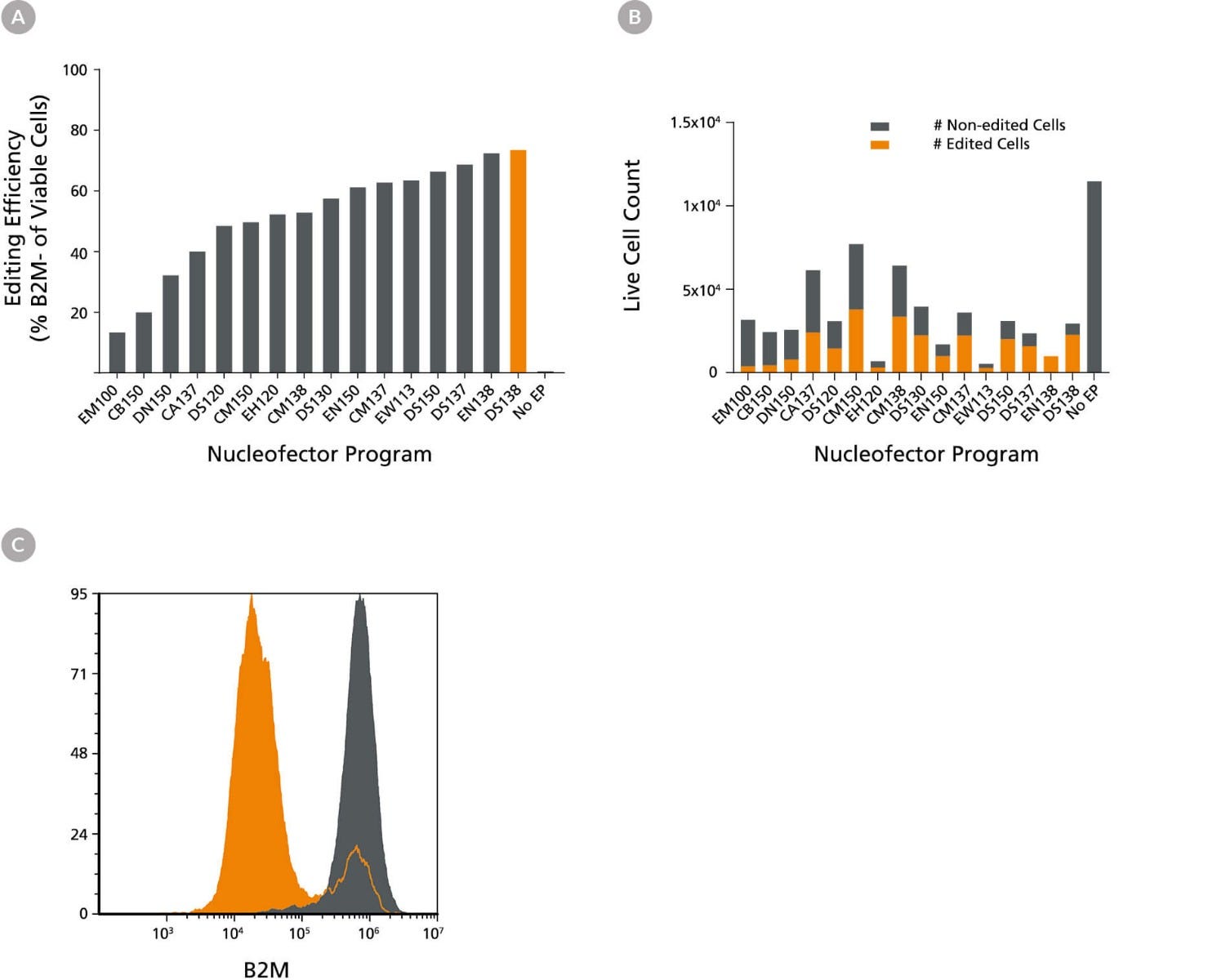
Figure 2. Optimization of Liver Organoid-Derived Cell RNP Delivery Using the 4D- Nucleofector™ System
Liver organoids were derived from a healthy control and cultured in HepatiCult™ Organoid Growth Medium (Human). RNP complexes containing ArciTect™ Cas9 Nuclease and ArciTect™ sgRNA targeting the B2M gene were delivered using the indicated 4D-Nucleofector™ program, and editing efficiency was monitored by flow cytometry to detect surface expression of B2M. (A) Editing efficiency was measured 7 days after electroporation by flow cytometry to detect functional knockout of B2M using the ArciTect™ Human CRISPR Optimization Kit (Cat # 100-0472). (B) The total number of edited cells (orange bars) and non-edited liver organoid cells (grey bars) was calculated by multiplying editing efficiency by post-electroporation recovery; this indicated which electroporation programs resulted in the best overall editing efficiency and cell recovery. (C) Representative histogram of B2M flow cytometry data from RNP-electroporated (RNP, orange; DS-138) and non-electroporated (No EP; grey) liver organoid cells 7 days after delivery of the CRISPR-Cas9 RNP complexes containing sgRNA targeting B2M.
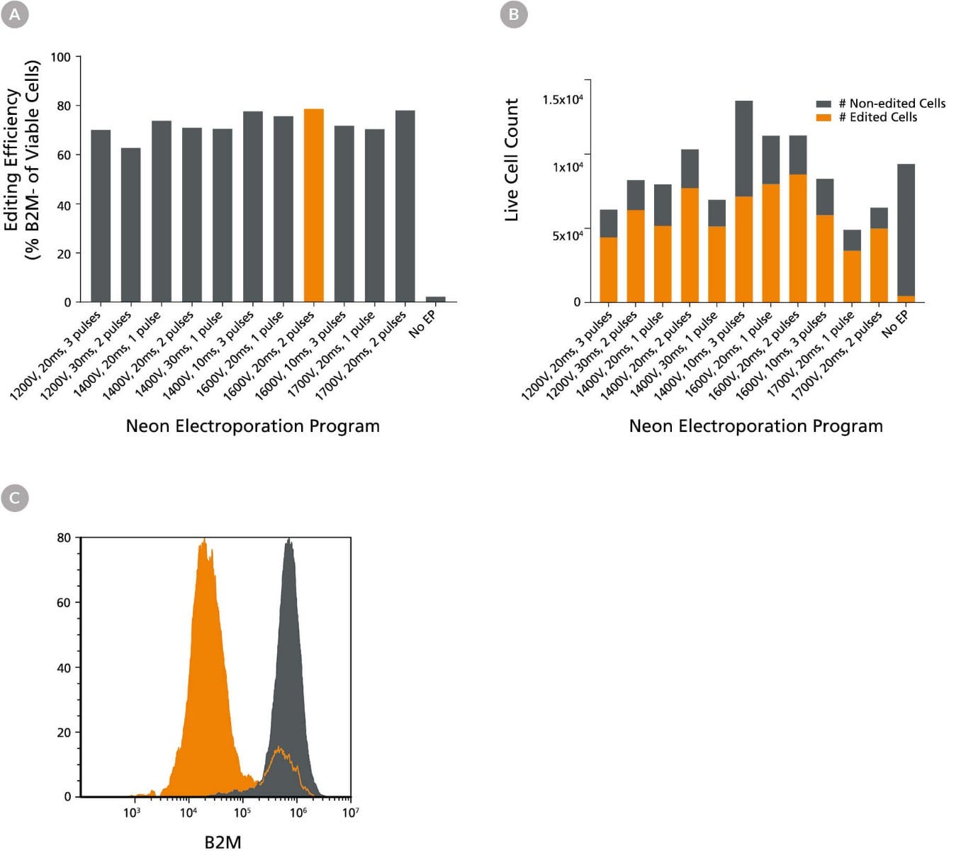
Figure 3. Optimization of Liver Organoid-Derived Cell RNP Delivery Using the Neon® Transfection System 10 μL Kit
Liver organoids were derived from a healthy control and cultured in HepatiCult™ Organoid Growth Medium (Human). RNP complexes containing ArciTect™ Cas9 Nuclease and ArciTect™ sgRNA targeting the B2M gene were delivered using the indicated electroporation program with the Neon® Transfection System, and editing efficiency was monitored by flow cytometry to detect surface expression of B2M. (A) Editing efficiency was measured 7 days after electroporation by flow cytometry to detect functional knockout of B2M using the ArciTect™ Human CRISPR Optimization Kit (Cat # 100-0472). (B) The total number of edited cells (orange bars) and non-edited liver organoid cells (grey bars) was calculated by multiplying editing efficiency by post-electroporation recovery; this indicated which electroporation programs resulted in the best overall editing efficiency and cell recovery. (C) Representative histogram of B2M flow cytometry data from RNP-electroporated (RNP, orange; 1600v, 20ms, 2 pulses) and non-electroporated (No EP; grey) liver organoid cells 7 days after delivery of the CRISPR-Cas9 RNP complexes containing sgRNA targeting B2M.
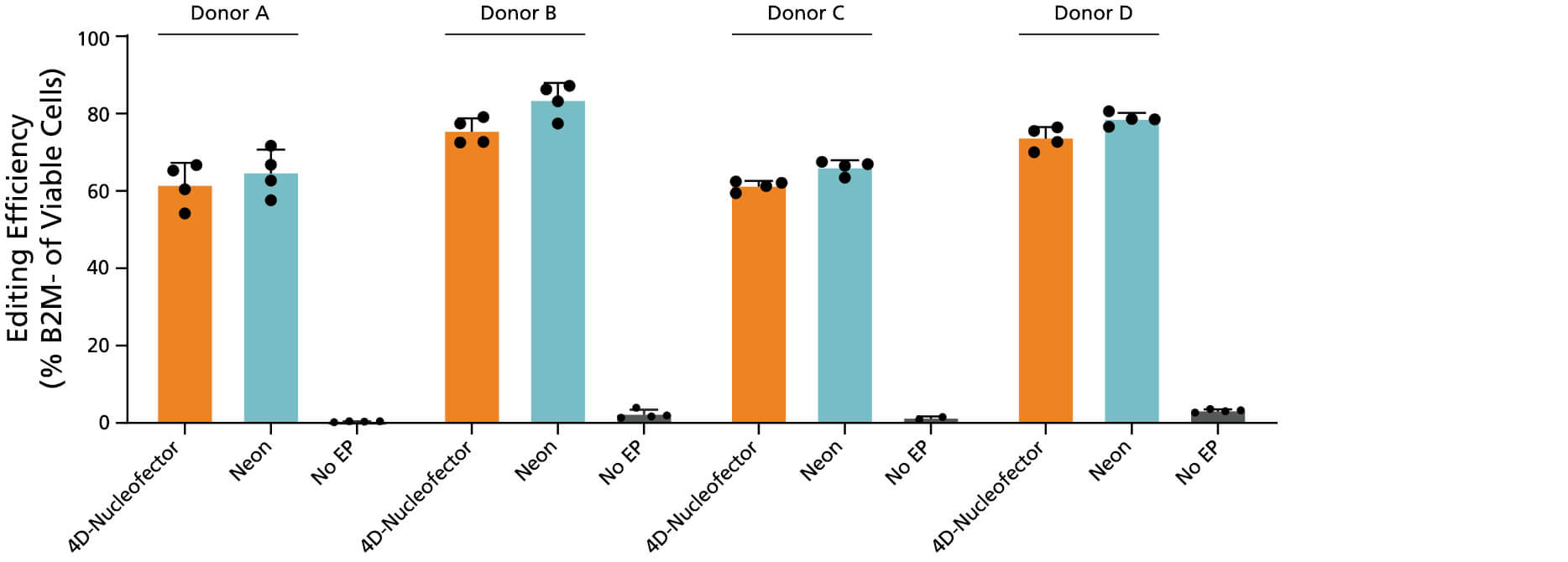
Figure 4. Optimized RNP Delivery Electroporation Programs for Liver Organoid-Derived Cells using the 4D-Nucleofector™ or Neon® Transfection Systems
Liver organoids were derived from four healthy donor lines and cultured in HepatiCult™ Organoid Growth Medium. RNP complexes containing ArciTect™ Cas9 Nuclease and ArciTect™ sgRNA targeting the B2M gene were delivered into the cells using either the 4D-Nucleofector™ System (program code: DS-138) or the Neon® Transfection System (1600 V, 20 ms, 2 pulses), and editing efficiency was monitored by flow cytometry to detect B2M knockout.

Figure 5. Expansion of Edited Liver Organoids in HepatiCult™ Organoid Growth Medium Using an Optimized Single-Cell Dissociation Protocol
Liver organoids were edited with an RNP complex containing ArciTect™ Cas9 Nuclease and ArciTect™ sgRNA targeting the B2M using the indicated electroporation programs with the Neon® Transfection System. Edited liver organoids were dissociated into single cells using Animal Component-Free Cell Dissociation Kit (Catalog # 05426). Cells were plated at a density of 6.67 x 10⁴ cells per Matrigel® dome and cultured in HepatiCult™ Organoid Growth Medium (Human) for 7 days before harvesting for downstream analysis.
Explore These Resources
Request Pricing
Thank you for your interest in this product. Please provide us with your contact information and your local representative will contact you with a customized quote. Where appropriate, they can also assist you with a(n):
Estimated delivery time for your area
Product sample or exclusive offer
In-lab demonstration
