RosetteSep™ Human Total Lymphocyte Enrichment Cocktail

Immunodensity negative selection cocktail
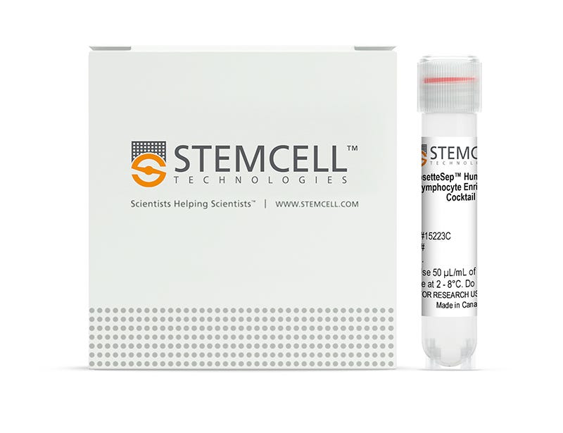
New look, same high quality and support! You may notice that your instrument or reagent packaging looks slightly different from images displayed on the website, or from previous orders. We are updating our look but rest assured, the products themselves and how you should use them have not changed. Learn more

RosetteSep™ Human Total Lymphocyte Enrichment Cocktail

Immunodensity negative selection cocktail

From: 240 USD
Catalog #
(Select a product)
Immunodensity negative selection cocktail
Add to Wish List

Product Advantages


  • Fast and easy-to-use

  • Requires no special equipment or training

  • Untouched, viable cells

  • Can be combined with SepMate™ for consistent, high-throughput sample processing

What's Included

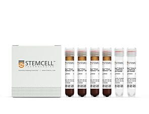
  • RosetteSep™ Human Total Lymphocyte Enrichment Cocktail (Catalog #15223)
    • RosetteSep™ Human Total Lymphocyte Enrichment Cocktail, 2 mL
  • RosetteSep™ Human Total Lymphocyte Enrichment Cocktail (Catalog #15263)
    • RosetteSep™ Human Total Lymphocyte Enrichment Cocktail, 5 x 2 mL
Products for Your Protocol

Overview

The RosetteSep™ Human Total Lymphocyte Enrichment Cocktail is designed to enrich lymphocytes from whole blood by negative selection. Unwanted cells are targeted for removal with Tetrameric Antibody Complexes recognizing CD16, CD36, CD66b and glycophorin A on red blood cells (RBCs). When centrifuged over a buoyant density medium such as RosetteSep™ DM-L (Catalog #15705) or Lymphoprep™ (Catalog #07801), the unwanted cells pellet along with the RBCs. The purified lymphocytes are present as a highly enriched population at the interface between the plasma and the buoyant density medium.
Subtype
Cell Isolation Kits
Cell Type
Lymphocytes
Species
Human
Sample Source
Buffy Coat, Whole Blood
Selection Method
Negative
Application
Cell Isolation
Brand
RosetteSep
Area of Interest
HLA, Immunology

Data Figures

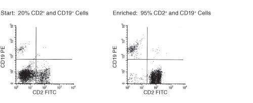
FACS Profile Results Using RosetteSep™ Human Total Lymphocyte Enrichment Cocktail

Figure 1. FACS Profile Results Using RosetteSep™ Human Total Lymphocyte Enrichment Cocktail

Starting with fresh peripheral blood, the CD2+ and CD19+ cell content of the enriched fraction is typically 94 ± 2%.

Protocols and Documentation

Find supporting information and directions for use in the Product Information Sheet or explore additional protocols below.

Document Type
Product Name
Catalog #
Lot #
Language
Catalog #
15223, 15263
Lot #
All
Language
English
Document Type
Safety Data Sheet
Catalog #
15223, 15263
Lot #
All
Language
English

Applications

This product is designed for use in the following research area(s) as part of the highlighted workflow stage(s). Explore these workflows to learn more about the other products we offer to support each research area.

Resources and Publications

Frequently Asked Questions

What is RosetteSep™?

RosetteSep™ is a rapid cell separation procedure for the isolation of purified cells directly from whole blood, without columns or magnets.

How does RosetteSep™ work?

The antibody cocktail crosslinks unwanted cells to red blood cells (RBCs), forming rosettes. The unwanted cells then pellet with the free RBCs when centrifuged over a density centrifugation medium (e.g. Ficoll-Paque™ PLUS, Lymphoprep™).

What factors affect cell recovery?

The temperature of the reagents can affect cell recovery. All reagents should be at room temperature (sample, density centrifugation medium, PBS, centrifuge) before performing the isolations. Layering can also affect recovery so be sure to carefully layer the sample to avoid mixing with the density centrifugation medium as much as possible. Be sure to collect the entire enriched culture without disturbing the RBC pellet. A small amount of density centrifugation medium can be collected without worry.

Which cell samples can RosetteSep™ be used with?

RosetteSep™ can be used with leukapheresis samples, bone marrow or buffy coat, as long as: the concentration of cells does not exceed 5 x 10⁷ per mL (can dilute if necessary); and there are at least 100 RBCs for every nucleated cell (RBCs can be added if necessary).

Can RosetteSep™ be used with previously frozen or cultured cells?

Yes. Cells should be re-suspended at 2 - 5 x 10⁷ cells / mL in PBS + 2% FBS. Fresh whole blood should be added at 250 µL per mL of sample, as a source of red cells.

Can RosetteSep™ be used to enrich progenitors from cord blood?

Yes. Sometimes cord blood contains immature nucleated red cells that have a lower density than mature RBCs. These immature red cells do not pellet over Ficoll™, which can lead to a higher RBC contamination than peripheral blood separations.

Does RosetteSep™ work with mouse cells?

No, but we have developed EasySep™, a magnetic-based cell isolation system which works with mouse and other non-human species.

Which anticoagulant should be used with RosetteSep™?

Peripheral blood should be collected in heparinized Vacutainers. Cord blood should be collected in ACD.

Should the anticoagulant be washed off before using RosetteSep™?

No, the antibody cocktail can be added directly to the sample.

Publications (3)

Root cause analysis of limitations of virtual crossmatch for kidney allocation to highly-sensitized patients. Jani V et al. Human immunology 2016 NOV

Abstract

Efficient allocation of deceased donor organs depends upon effective prediction of immunologic compatibility based on donor HLA genotype and recipient alloantibody profile, referred to as virtual crossmatching (VCXM). VCXM has demonstrated utility in predicting compatibility, though there is reduced efficacy for patients highly sensitized against allogeneic HLA antigens. The recently revised deceased donor kidney allocation system (KAS) has increased transplantation for this group, but with an increased burden for histocompatibility testing and organ sharing. Given the limitations of VCXM, we hypothesized that increased organ offers for highly-sensitized patients could result in a concomitant increase in offers rejected due to unexpectedly positive crossmatch. Review of 645 crossmatches performed for deceased donor kidney transplantation at our center did not reveal a significant increase in positive crossmatches following KAS implementation. Positive crossmatches not predicted by VCXM were concentrated among highly-sensitized patients. Root cause analysis of VCXM failures identified technical limitations of anti-HLA antibody testing as the most significant contributor to VCXM error. Contributions of technical limitations including additive/synergistic antibody effects, prozone phenomenon, and antigens not represented in standard testing panels, were evaluated by retrospective testing. These data provide insight into the limitations of VCXM, particularly those affecting allocation of kidneys to highly-sensitized patients.
TAT-BH4 and TAT-Bcl-xL peptides protect against sepsis-induced lymphocyte apoptosis in vivo. Hotchkiss RS et al. Journal of immunology (Baltimore, Md. : 1950) 2006 MAY

Abstract

Apoptosis is a key pathogenic mechanism in sepsis that induces extensive death of lymphocytes and dendritic cells, thereby contributing to the immunosuppression that characterizes the septic disorder. Numerous animal studies indicate that prevention of apoptosis in sepsis improves survival and may represent a potential therapy for this highly lethal disorder. Recently, novel cell-penetrating peptide constructs such as HIV-1 TAT basic domain and related peptides have been developed to deliver bioactive cargoes and peptides into cells. In the present study, we investigated the effects of sepsis-induced apoptosis in Bcl-x(L) transgenic mice and in wild-type mice treated with an antiapoptotic TAT-Bcl-x(L) fusion protein and TAT-BH4 peptide. Lymphocytes from Bcl-x(L) transgenic mice were resistant to sepsis-induced apoptosis, and these mice had a approximately 3-fold improvement in survival. TAT-Bcl-x(L) and TAT-BH4 prevented Escherichia coli-induced human lymphocyte apoptosis ex vivo and markedly decreased lymphocyte apoptosis in an in vivo mouse model of sepsis. In conclusion, TAT-conjugated antiapoptotic Bcl-2-like peptides may offer a novel therapy to prevent apoptosis in sepsis and improve survival.
Accelerated lymphocyte death in sepsis occurs by both the death receptor and mitochondrial pathways. Hotchkiss RS et al. Journal of immunology (Baltimore, Md. : 1950) 2005 APR

Abstract

Patients with sepsis are immune compromised, as evidenced by their failure to clear their primary infection and their propensity to develop secondary infections with pathogens that are often not particularly virulent in normal healthy individuals. A potential mechanism for immunosuppression in sepsis is lymphocyte apoptosis, which may occur by either a death receptor or a mitochondrial-mediated pathway. A prospective study of blood samples from 71 patients with sepsis, 55 nonseptic patients, and 6 healthy volunteers was undertaken to quantitate lymphocyte apoptosis and determine cell death pathways and mechanisms of apoptosis. Apoptosis was evaluated by flow cytometry and Western blotting. Lymphocyte apoptosis was increased in CD4 and CD8 T cells, B cells (CD20), and NK cells (CD56) in septic vs nonseptic patients. Samples taken sequentially from 10 patients with sepsis showed that the degree of CD3 T cell apoptosis correlated with the activity of his/her sepsis. In septic patients, apoptotic lymphocytes were positive for active caspases 8 and 9, consistent with death occurring by both mitochondrial-mediated and receptor-mediated pathways. In support of the concept that both death pathways were operative, lymphocyte apoptosis occurred in cells with markedly decreased Bcl-2 (an inhibitor of mitochondrial-mediated apoptosis) as well as cells with normal concentrations of Bcl-2. In conclusion, apoptosis occurs in a broad range of lymphocyte subsets in patients with sepsis and correlates with the activity of the disease. Lymphocyte loss occurs by both death receptor and mitochondrial-mediated apoptosis, suggesting that there may be multiple triggers for lymphocyte apoptosis.
New look, same high quality and support! You may notice that your instrument or reagent packaging looks slightly different from images displayed on the website, or from previous orders. We are updating our look but rest assured, the products themselves and how you should use them have not changed. Learn more