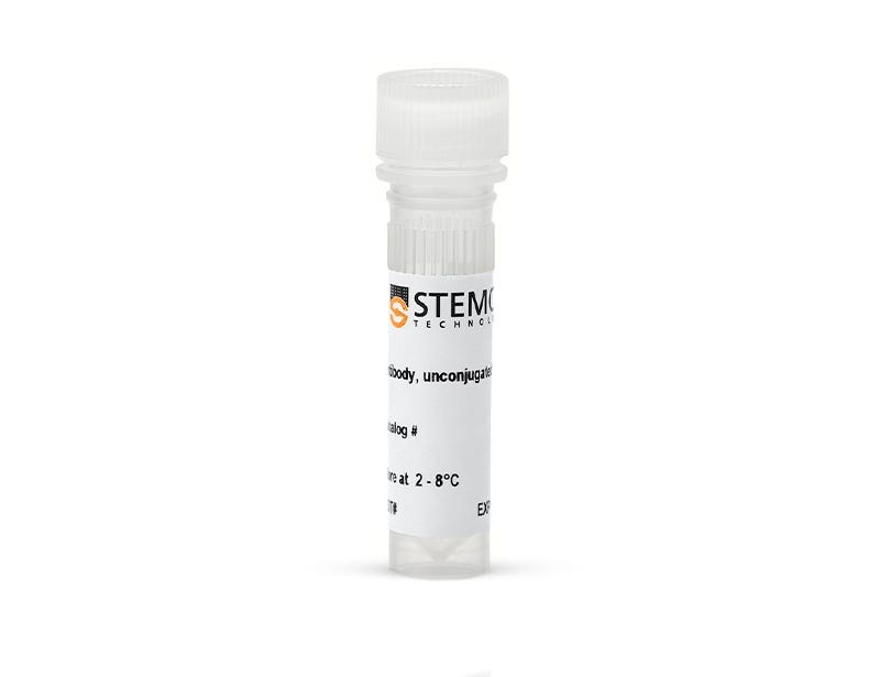
Anti-Human CD9 Antibody, Clone HI9a

Mouse monoclonal antibody against human, rhesus, cynomolgus CD9, unconjugated

Anti-Human CD9 Antibody, Clone HI9a

Mouse monoclonal antibody against human, rhesus, cynomolgus CD9, unconjugated

From: 359 USD
Catalog #
(Select a product)
Mouse monoclonal antibody against human, rhesus, cynomolgus CD9, unconjugated
Add to Wish List

Overview

The HI9a antibody reacts with an extracellular epitope on human CD9, a 24 kDa single-chain type III transmembrane glycoprotein. CD9 is broadly expressed, including on the surface of monocytes, developing B cells, activated T cells, granulocytes, epithelial cells, endothelial cells, and platelets (mainly the α-granules) and is also a marker characteristic of extracellular vesicles.

CD9 belongs to the tetraspanin family of proteins, which contain four transmembrane domains, two extracellular loops, and short cytoplasmic N- and C-termini. It is involved in mediating several cellular processes, including angiogenesis, by promoting cell adhesion, migration, and signal transduction, and is involved in cell fusion processes linked to fertilization, osteoclastogenesis, and myogenesis.
Subtype
Primary Antibodies
Target Antigen
CD9
Alternative Names
DRAP-24
Reactive Species
African Green Monkey, Baboon, Capuchin Monkey, Chimpanzee, Cynomolgus, Human, Rhesus
Conjugation
Unconjugated
Host Species
Mouse
Cell Type
Other
Application
Flow Cytometry, Western Blotting
Area of Interest
Extracellular Vesicle Research
Clone
HI9a
Gene ID
928
Isotype
IgG1, kappa

Data Figures

Data showing western blot analysis of extracellular vesicles isolated from mesenchymal stromal cell-conditioned medium with Anti-Human CD9 Antibody, Clone HI9a and flow cytometry analysis of human peripheral blood mononuclear cells labeled with Anti-Human CD9 Antibody, Clone HI9a.

Figure 1. Analysis of Anti-Human CD9 Antibody, Clone HI9a with Western Blot and Flow Cytometry

Extracellular vesicles (EVs) were isolated from mesenchymal stromal cell (MSC)-conditioned medium using a 2 mL EV size exclusion chromatography column and analyzed by (A) western blot with Anti-Human CD9 Antibody, Clone HI9a. Lanes (left to right) were loaded with isolated fractions 9, 10, and 11, respectively. Further validation was performed by flow cytometry analysis of human peripheral blood mononuclear cells (PBMCs; viable lymphocytes gated for analysis) stimulated with phorbol myristate acetate (PMA) and ionomycin and then (B) labeled with Anti-Human CD9 Antibody, Clone HI9a, followed by a goat anti-mouse IgG1 antibody, APC (filled histogram), or Anti-Dextran Antibody, Clone DX1 (Catalog #60026) as an isotype control, followed by a goat anti-mouse IgG1 antibody, APC (solid line histogram), or (C) labeled with Anti-Dextran Antibody, Clone DX1 (Catalog #60026) as an isotype control, followed by a goat anti-mouse IgG1 antibody, APC, or (D) labeled with Anti-Human CD9 Antibody, Clone HI9a, followed by a goat anti-mouse IgG1 antibody, APC.

Protocols and Documentation

Find supporting information and directions for use in the Product Information Sheet or explore additional protocols below.

Document Type
Product Name
Catalog #
Lot #
Language
Catalog #
100-0138
Lot #
All
Language
English
Document Type
Safety Data Sheet
Catalog #
100-0138
Lot #
All
Language
English

Applications

This product is designed for use in the following research area(s) as part of the highlighted workflow stage(s). Explore these workflows to learn more about the other products we offer to support each research area.