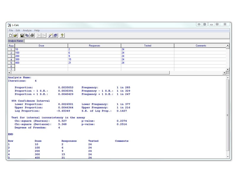
L-Calc™ Software
Software for limiting dilution analysis
Request Pricing
Thank you for your interest in this product. Please provide us with your contact information and your local representative will contact you with a customized quote. Where appropriate, they can also assist you with a(n):
Estimated delivery time for your area
Product sample or exclusive offer
In-lab demonstration
By submitting this form, you are providing your consent to STEMCELL Technologies Canada Inc. and its subsidiaries and affiliates (“STEMCELL”) to collect and use your information, and send you newsletters and emails in accordance with our privacy policy. Please contact us with any questions that you may have. You can unsubscribe or change your email preferences at any time.
Overview
Applications
This product is designed for use in the following research area(s) as part of the highlighted workflow stage(s). Explore these workflows to learn more about the other products we offer to support each research area.
Resources and Publications
Educational Materials (2)


Publications (19)
p53-TP53-Induced Glycolysis Regulator Mediated Glycolytic Suppression Attenuates DNA Damage and Genomic Instability in Fanconi Anemia Hematopoietic Stem Cells.
Stem cells (Dayton, Ohio) 2019 jul
Abstract
Emerging evidence has shown that resting quiescent hematopoietic stem cells (HSCs) prefer to utilize anaerobic glycolysis rather than mitochondrial respiration for energy production. Compelling evidence has also revealed that altered metabolic energetics in HSCs underlies the onset of certain blood diseases; however, the mechanisms responsible for energetic reprogramming remain elusive. We recently found that Fanconi anemia (FA) HSCs in their resting state are more dependent on mitochondrial respiration for energy metabolism than on glycolysis. In the present study, we investigated the role of deficient glycolysis in FA HSC maintenance. We observed significantly reduced glucose consumption, lactate production, and ATP production in HSCs but not in the less primitive multipotent progenitors or restricted hematopoietic progenitors of Fanca-/- and Fancc-/- mice compared with that of wild-type mice, which was associated with an overactivated p53 and TP53-induced glycolysis regulator, the TIGAR-mediated metabolic axis. We utilized Fanca-/- HSCs deficient for p53 to show that the p53-TIGAR axis suppressed glycolysis in FA HSCs, leading to enhanced pentose phosphate pathway and cellular antioxidant function and, consequently, reduced DNA damage and attenuated HSC exhaustion. Furthermore, by using Fanca-/- HSCs carrying the separation-of-function mutant p53R172P transgene that selectively impairs the p53 function in apoptosis but not cell-cycle control, we demonstrated that the cell-cycle function of p53 was not required for glycolytic suppression in FA HSCs. Finally, ectopic expression of the glycolytic rate-limiting enzyme PFKFB3 specifically antagonized p53-TIGAR-mediated metabolic reprogramming in FA HSCs. Together, our results suggest that p53-TIGAR metabolic axis-mediated glycolytic suppression may play a compensatory role in attenuating DNA damage and proliferative exhaustion in FA HSCs. Stem Cells 2019;37:937-947.
Metformin blunts muscle hypertrophy in response to progressive resistance exercise training in older adults: A randomized, double-blind, placebo-controlled, multicenter trial: The MASTERS trial.
Aging cell 2019 dec
Abstract
Progressive resistance exercise training (PRT) is the most effective known intervention for combating aging skeletal muscle atrophy. However, the hypertrophic response to PRT is variable, and this may be due to muscle inflammation susceptibility. Metformin reduces inflammation, so we hypothesized that metformin would augment the muscle response to PRT in healthy women and men aged 65 and older. In a randomized, double-blind trial, participants received 1,700 mg/day metformin (N = 46) or placebo (N = 48) throughout the study, and all subjects performed 14 weeks of supervised PRT. Although responses to PRT varied, placebo gained more lean body mass (p = .003) and thigh muscle mass (p {\textless} .001) than metformin. CT scan showed that increases in thigh muscle area (p = .005) and density (p = .020) were greater in placebo versus metformin. There was a trend for blunted strength gains in metformin that did not reach statistical significance. Analyses of vastus lateralis muscle biopsies showed that metformin did not affect fiber hypertrophy, or increases in satellite cell or macrophage abundance with PRT. However, placebo had decreased type I fiber percentage while metformin did not (p = .007). Metformin led to an increase in AMPK signaling, and a trend for blunted increases in mTORC1 signaling in response to PRT. These results underscore the benefits of PRT in older adults, but metformin negatively impacts the hypertrophic response to resistance training in healthy older individuals. ClinicalTrials.gov Identifier: NCT02308228.
Revision of the Human Hematopoietic Tree: Granulocyte Subtypes Derive from Distinct Hematopoietic Lineages
Cell Reports 2013 5
Abstract
The classical model of hematopoiesis predicts a dichotomous lineage restriction of multipotent hematopoietic progenitors (MPPs) into common lymphoid progenitors (CLPs) and common myeloid progenitors (CMPs). However, this idea has been challenged by the identification of lymphoid progenitors retaining partial myeloid potential (e.g., LMPPs), implying that granulocytes can arise within both the classical lymphoid and the myeloid branches. Here, we resolve this issue by using cell-surface CD133 expression to discriminate functional progenitor populations. We show that eosinophilic and basophilic granulocytes as well as erythrocytes and megakaryocytes derive from a common erythro-myeloid progenitor (EMP), whereas neutrophilic granulocytes arise independently within a lympho-myeloid branch with long-term progenitor function. These findings challenge the concept of a CMP and restore dichotomy to the classical hematopoietic model.
Related Products
Related Products


Quality Statement:
PRODUCTS ARE FOR RESEARCH USE ONLY AND NOT INTENDED FOR HUMAN OR ANIMAL DIAGNOSTIC OR THERAPEUTIC USES UNLESS OTHERWISE STATED. FOR ADDITIONAL INFORMATION ON QUALITY AT STEMCELL, REFER TO WWW.STEMCELL.COM/COMPLIANCE.
PRODUCTS ARE FOR RESEARCH USE ONLY AND NOT INTENDED FOR HUMAN OR ANIMAL DIAGNOSTIC OR THERAPEUTIC USES UNLESS OTHERWISE STATED. FOR ADDITIONAL INFORMATION ON QUALITY AT STEMCELL, REFER TO WWW.STEMCELL.COM/COMPLIANCE.
